Vats Procedure — Explained by Medical Evidence, Not Myths

The vats procedure uses a tiny camera and slim instruments through small chest incisions. It can be used to diagnose disease, remove tissue, drain fluid, or treat selected lung conditions.
Key Takeaways
- The vats procedure uses a tiny camera and slim instruments through small chest incisions.
- It can be used to diagnose disease, remove tissue, drain fluid, or treat selected lung conditions.
- Compared with open surgery, VATS often means less pain, shorter hospital stay, and quicker recovery for suitable patients.
- Not everyone is a candidate; the best approach depends on the condition, anatomy, and overall health.
- Recovery usually includes breathing exercises, walking, pain control, and follow-up imaging or pathology review.
The vats procedure, also called video-assisted thoracoscopic surgery, is a minimally invasive operation that allows surgeons to diagnose or treat problems inside the chest through small incisions. It is commonly used for certain lung, pleural, and chest conditions, often with less pain and faster recovery than traditional open chest surgery.
Overview: what the vats procedure is
The vats procedure is short for video-assisted thoracoscopic surgery. It is a type of minimally invasive chest surgery in which a surgeon inserts a small camera and fine instruments through a few small incisions between the ribs. The camera shows the inside of the chest on a monitor, helping the surgeon examine, biopsy, or treat tissues without making the larger opening used in traditional thoracotomy.
In simple terms, the vats procedure is a way to operate inside the chest with less disruption to muscles and ribs. It may be used for diagnosis, such as taking a biopsy from the lung or pleura, or for treatment, such as removing a small lung nodule, treating recurrent collapsed lung, or draining pleural fluid. The exact operation depends on the reason for surgery.
Although VATS is often associated with lung surgery, it is not limited to one disease. It can help manage problems involving the lungs, pleura, mediastinum, and chest wall. For some patients, it offers a balance between precision and recovery comfort, but the final decision is always individualized after specialist assessment.
Why it is done and which conditions it can help
Doctors may recommend a vats procedure when imaging, symptoms, or previous test results suggest that a problem inside the chest needs a closer look or active treatment. One common use is obtaining a tissue sample when scans alone cannot confirm a diagnosis. In other cases, the goal is to remove an abnormal area or treat symptoms caused by fluid, air, or inflammation.
Common reasons for VATS include evaluating or removing lung nodules, treating recurrent pneumothorax, draining pleural effusions, taking pleural or lung biopsies, and removing selected early-stage tumors. It may also be used in some infections, mediastinal masses, or for procedures such as pleurodesis. Depending on the condition, a surgeon may remove a very small piece of tissue, a wedge of lung, or a larger lung section.
Examples of conditions that may lead to this surgery include lung cancer under evaluation or treatment planning, persistent or unexplained pleural fluid, and selected cases of pneumothorax. In some patients, VATS is also part of the pathway for lung cancer treatment when surgery is an appropriate option.
- Diagnostic biopsy of lung, pleura, or lymph nodes
- Removal of small lung nodules or abnormal tissue
- Treatment of recurrent collapsed lung
- Drainage of fluid or treatment of pleural disease
- Selected operations for tumors in the chest
How the procedure is performed

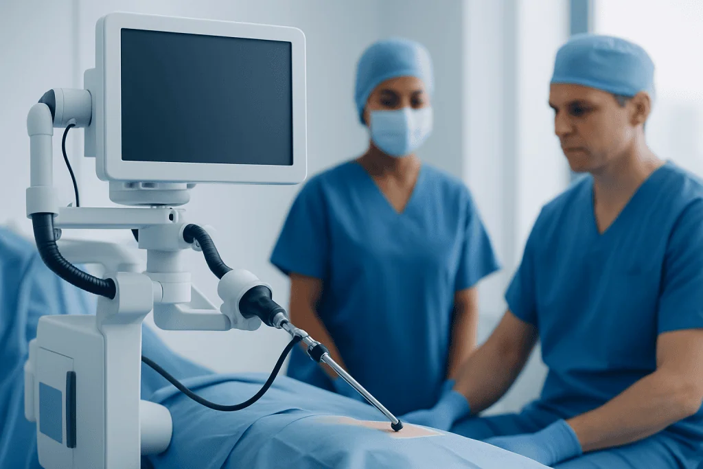
The vats procedure is usually performed under general anesthesia, so the patient is asleep and does not feel pain during surgery. The surgeon makes small incisions, often two to four, on the side of the chest. A thoracoscope, which is a thin tube with a camera, is inserted through one incision, while surgical instruments are passed through the others.
During the operation, the surgeon examines the target area and carries out the planned procedure. This may include taking biopsies, removing blebs that cause recurrent lung collapse, draining fluid, or removing a lung segment or lobe in selected cases. At the end of surgery, a chest tube is often left in place temporarily to drain air or fluid and help the lung re-expand.
Some procedures begin as VATS but may need to be converted to open surgery if visibility is limited, bleeding occurs, or the anatomy is more complex than expected. This does not mean the operation has failed; it means the surgical team is choosing the safest approach. In experienced centers, minimally invasive thoracic techniques may also overlap with other advanced options, including selected robotic surgery approaches when appropriate.
Benefits, limitations, and possible risks
A major advantage of the vats procedure is that it usually avoids the larger incision and greater tissue disruption of open chest surgery. For many patients, this can mean less postoperative pain, less blood loss, a shorter hospital stay, and a faster return to daily activities. Smaller incisions may also reduce the risk of some wound-related complications.
However, minimally invasive does not mean minor or risk-free. VATS is still a real operation inside the chest and requires careful preoperative planning. It may not be the best choice if the disease is very extensive, if there is dense scarring from prior infection or surgery, or if the patient has medical issues that increase anesthesia or surgical risk.
Possible complications can include bleeding, infection, air leak, pneumonia, irregular heart rhythm, pain, reactions to anesthesia, or the need for another procedure. The exact risk profile depends on what is being treated, how much tissue is removed, and the person’s overall health, especially lung and heart function. Doctors explain these points in detail before surgery so patients can make an informed decision.
Preparation, diagnosis, and who may be a candidate
Before recommending a vats procedure, doctors first confirm why surgery is needed and whether the patient is likely to benefit from it. Evaluation often includes a physical examination, chest imaging such as X-ray or CT, blood tests, and sometimes pulmonary function testing. If a suspicious growth is present, additional tests may be needed to understand its size, location, and whether surgery is the right next step.
Cardiac health, breathing reserve, smoking history, prior chest surgery, and current medications all matter. Blood thinners may need special planning. Patients are usually advised not to eat or drink for a period before surgery and may be asked to stop smoking, as this can improve healing and lower respiratory complications.
Candidate selection is important because the safest and most effective procedure differs from person to person. Some people are well suited to a minimally invasive approach, while others may benefit more from a different operation or a nonsurgical plan. If a biopsy or pathology review points to cancer, the next steps may involve a broader oncology pathway and, in selected cases, thoracic surgery as part of treatment planning.
Recovery after VATS surgery
Recovery after a vats procedure varies with the type of surgery performed. A short diagnostic procedure may allow discharge within a relatively short time, while a lung resection usually requires a longer hospital stay. Most patients can expect monitoring in the hospital, pain management, breathing exercises, early walking, and chest tube care until it is safe for the tube to be removed.
It is common to feel tired, sore around the incisions, and somewhat limited in deep breathing or activity at first. Good pain control is important because it helps with coughing, movement, and prevention of complications such as pneumonia. The care team may recommend an incentive spirometer, gentle activity, and gradual return to normal routines.
At home, patients are generally advised to walk regularly, avoid heavy lifting until cleared, and watch for warning signs such as fever, worsening shortness of breath, or increasing wound redness. Follow-up visits are important to review healing, imaging, and any pathology results. If the procedure was done for a tumor or another serious condition, the follow-up plan may include input from pulmonology, oncology, or surgery specialists.
Self-care, prevention, and when to seek medical care
There is no single way to prevent every condition that leads to a vats procedure, but some steps support lung and chest health. Avoiding smoking, staying physically active within personal limits, keeping vaccinations up to date when appropriate, and seeking evaluation for persistent respiratory symptoms can all help reduce delays in diagnosis and improve surgical readiness if an operation becomes necessary.
After surgery, self-care focuses on healing and protecting lung function. Patients should follow discharge instructions carefully, take medicines only as prescribed, perform breathing exercises, stay hydrated unless told otherwise, and attend follow-up appointments. If a person has an underlying lung condition, long-term care with a pulmonologist may help prevent future flare-ups or complications.
Medical care should be sought promptly if there is chest pain that is new or worsening, increasing shortness of breath, fever, persistent cough with concerning mucus, heavy bleeding from an incision, severe swelling, or symptoms that suddenly change. A healthcare professional should also be contacted if pain is not controlled well enough to allow deep breathing and movement. Near the end of the treatment journey, some patients may choose centers with multidisciplinary thoracic expertise; Acibadem International’s specialists in JCI-accredited hospitals diagnose and treat chest conditions for international patients.
Frequently asked questions
Is the vats procedure a major surgery?
Yes, it is still a surgical procedure performed under anesthesia, even though it is minimally invasive. The incisions are smaller than in open chest surgery, but the operation still involves important structures in the chest and needs specialist care.
How long does recovery from VATS surgery take?
Recovery depends on why the surgery was done and how much tissue was treated or removed. Some people recover fairly quickly after a biopsy, while others need more time after a lung resection, with gradual improvement over days to weeks.
What is the difference between VATS and open thoracotomy?
VATS uses small incisions, a camera, and long instruments to work inside the chest. Open thoracotomy uses a larger incision and wider opening of the chest, which may be necessary for more complex cases or if safety requires conversion during surgery.
Will a chest tube be needed after the vats procedure?
In many cases, yes. A chest tube is commonly placed after surgery to drain air or fluid and help the lung expand properly, and it is usually removed once the surgical team confirms it is no longer needed.
Can the vats procedure be used to diagnose cancer?
Yes, VATS is often used to obtain tissue samples from the lung, pleura, or lymph nodes when a diagnosis is uncertain. The final diagnosis depends on pathology results, which guide the next treatment steps.
Is the vats procedure always better than open surgery?
Not always. VATS offers advantages for many suitable patients, but the best surgical method depends on the exact condition, anatomy, and overall health. A surgeon chooses the approach that provides the safest and most effective treatment.
References
- American College of Chest Physicians
- National Cancer Institute
- National Heart, Lung, and Blood Institute
- Society of Thoracic Surgeons
- MedlinePlus
This article is for general information only and is not a substitute for professional medical advice. Please consult a qualified doctor about your individual situation.
Oncology care in Turkey — second opinion and treatment plan
JCI-accredited · board-certified surgeons · reply within 24h
Add us as a Preferred Source to see more of our trusted health content across Google Search, AI Overviews and Discover.