Percutaneous Closure
Percutaneous closure is a catheter-based cardiology procedure used to close selected structural heart openings, such as atrial septal defects or patent foramen ovale, without open surgery.
Quick answer
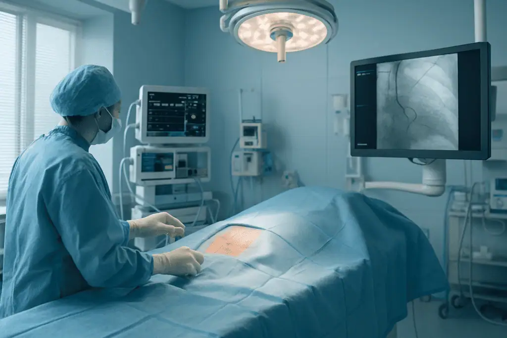
Percutaneous closure is a catheter-based procedure that seals selected openings inside the heart, most commonly a patent foramen ovale (PFO) or an atrial septal defect (ASD). A folded implant is guided through a vein to the heart, opened across the defect and left in place while the body's own tissue grows over it. Most patients avoid open-heart surgery and many stay only one night in hospital.
PFO, ASD and What Percutaneous Closure Does
Percutaneous closure is a catheter-based procedure that seals selected openings inside the heart without open surgery. It is used most often for a patent foramen ovale — a PFO — and for certain atrial septal defects, known as ASDs. A folded implant travels through a vein to the heart, opens across the defect and stays there permanently while your own tissue grows over it. Closure is considered when an opening places measurable strain on the heart, or when a PFO is judged to have contributed to a stroke and other causes have been assessed.
People arrive at this diagnosis by very different routes. Many adults learn they have a PFO only after a stroke evaluation or detailed heart imaging, when the finding comes as a complete surprise. Others are diagnosed with an ASD after years of subtle symptoms: shortness of breath on exertion, reduced exercise capacity, palpitations or unexplained fatigue. Parents may hear that a child has a structural heart opening during evaluation of a murmur, and immediately wonder about growth, activity, long-term heart function and whether surgery will ever be needed.
Here is the point worth holding onto from the start: the decision to close an opening is never based on its presence alone. Many small PFOs never need treatment. Some ASDs are monitored for years before any intervention is recommended. The questions that actually matter are clinical ones. Is the opening placing extra volume on the right side of the heart? Is it associated with symptoms, rhythm disturbance or stroke risk? Is the anatomy suitable for a catheter-based device? Answering these takes detailed imaging, careful interpretation and a cardiology team experienced in structural heart work.
This page explains what percutaneous closure involves, who genuinely benefits, how the procedure is performed step by step, what recovery looks like and what influences the long-term result — including the situations in which closure is not the right answer.
What does PFO mean?
PFO stands for patent foramen ovale. The foramen ovale is a natural flap-like passage between the two upper chambers of the heart, the atria. Before birth, every baby has one: it allows oxygen-rich blood from the placenta to bypass the lungs, which are not yet in use. After birth, pressure changes in the heart normally press the flap shut, and in most people it seals over time. In some people it never seals completely. “Patent” simply means “open” — a PFO is a foramen ovale that stayed open into adult life. It is a remnant of normal fetal anatomy, not a disease in itself.
Is PFO a serious heart condition?
For most people, no. A PFO usually causes no symptoms, does not weaken the heart muscle and is discovered by chance, if at all. Many people carry one for an entire lifetime without knowing. The condition becomes clinically relevant in a narrower set of circumstances — chiefly when a person has an ischaemic stroke or transient ischaemic attack with no other identifiable cause, and the PFO is suspected of having allowed a clot to cross from the venous side of the circulation to the arterial side. That mechanism is called paradoxical embolism. Whether a specific PFO is “serious” therefore depends less on the opening itself and more on the clinical story around it.
Can you live a long life with a PFO?
Yes. A PFO on its own does not shorten life expectancy, restrict exercise or require lifestyle changes for the great majority of people who have one. Most never need treatment of any kind. Where a PFO has been linked to a stroke, the focus shifts to preventing a recurrence — through medication, closure or both, depending on individual assessment — but even then the aim is to manage a specific risk, not to treat a failing heart. A PFO diagnosis alone is not a reason for alarm; it is a reason for a considered conversation with a cardiologist.
What Percutaneous Closure Is
Percutaneous closure is a minimally invasive cardiology procedure used to close selected openings or abnormal communications within the heart. The word percutaneous means “through the skin”. Instead of opening the chest, the cardiologist inserts thin, flexible tubes called catheters through a blood vessel — most commonly the femoral vein in the groin — and guides them to the heart under live imaging. A specially designed closure device is delivered through the catheter and positioned across the opening. Over the following months, your own tissue grows over the device and incorporates it into the heart’s internal wall. Procedures of this kind sit within the broader field of structural heart treatment, which deals with defects in the heart’s walls, valves and chambers rather than its arteries or rhythm.
The procedure is not the same for every patient, because the two main indications serve different purposes. ASD closure aims to eliminate a significant left-to-right shunt and relieve long-term strain on the right heart. PFO closure is usually considered in a different context — after a neurological event and a detailed search for other causes. Not every opening can be closed with a catheter: some ASDs are too large, sit too close to important structures, or lack adequate rims of tissue to anchor a device safely. Certain complex defects need surgical correction instead. High-quality imaging and multidisciplinary discussion exist precisely to sort these cases apart.
What is percutaneous closure of ASD?
Percutaneous closure of ASD is the sealing of an atrial septal defect — an opening in the wall between the two upper chambers of the heart — using a catheter-delivered device rather than surgery. The type treated this way is almost always a secundum ASD, which sits in the central part of the atrial septum. When significant, an ASD lets extra blood pass from the left atrium to the right atrium with every heartbeat, increasing blood flow through the right heart and lungs. Over years, that extra volume enlarges the right chambers and can set the stage for rhythm problems and raised lung pressures. ASDs belong to the family of congenital heart diseases, meaning they are present from birth, though many are only recognised in adulthood.
What is PFO closure?
PFO closure is the sealing of a patent foramen ovale with a catheter-delivered device, usually to reduce the risk of a further stroke in carefully selected patients. It is generally considered after a cryptogenic ischaemic stroke — one with no definite cause found despite appropriate testing — when cardiologists and neurologists together judge that the PFO plausibly allowed a clot to cross to the arterial circulation. In selected patients, clinical trials have shown that PFO closure combined with appropriate medical therapy can reduce the risk of recurrent stroke compared with medical therapy alone, though individual benefit varies. PFO closure is not performed simply because a PFO exists, and it is not a standard answer for migraines, dizziness or nonspecific symptoms.
How do an ASD and a PFO differ?
Clinics and search results often group these conditions together under “ASD PFO heart” headings, but the two openings are structurally and clinically different. An ASD is a true deficiency of tissue — a hole in the septum that allows continuous abnormal flow, typically from left to right, loading the right heart with extra volume. A PFO is not a hole in that sense; it is a flap valve that failed to seal, and it usually allows flow only under certain pressure conditions, such as coughing or straining. An ASD threatens the heart itself over time. A PFO rarely troubles the heart at all — its significance lies in what it may allow to pass through. This difference shapes everything: who is treated, why, and what success means afterwards.
What is percutaneous left atrial appendage closure?
Percutaneous left atrial appendage closure is a different procedure from PFO or ASD closure, although it uses similar catheter techniques. It seals off the left atrial appendage — a small pouch on the left atrium where blood clots tend to form in people with atrial fibrillation — with the aim of reducing stroke risk in selected patients who cannot take long-term blood thinners. If your diagnosis involves atrial fibrillation rather than a septal opening, the relevant treatment pathway sits within heart rhythm disorders rather than septal defect care. The two procedures are often confused because both are “percutaneous closures” performed for stroke prevention, but they address entirely different structures for entirely different reasons.
Who May Need Percutaneous Closure
Patients reach this treatment at different points in life. Some are children whose murmur prompted an echocardiogram. Some are adults who lived for decades with an undetected ASD and only developed symptoms in middle age. Others learn about a PFO after a stroke, a transient ischaemic attack or an evaluation for unexplained neurological symptoms. In every case, the treatment decision starts with the same question: is this opening responsible for current problems or future risk?
The symptoms of a clinically significant ASD are often subtle enough to be dismissed for years. You may notice reduced stamina, breathlessness during exertion, easy fatigue, palpitations or a sense of irregular heartbeat. Some patients develop leg swelling, recurrent respiratory infections or right-sided heart enlargement visible on imaging. In adults, an untreated significant ASD can be associated with atrial arrhythmias, pulmonary hypertension and progressive strain on right heart function — changes that, left long enough, overlap with the territory of heart failure. Importantly, some patients feel entirely well despite measurable changes in the heart, which is why imaging findings often carry as much weight as symptoms.
Does a PFO need to be fixed?
Usually not. Most people with a PFO have no symptoms and no indication for closure; the opening can simply be left alone. The calculation changes when a patient has had a stroke or transient ischaemic attack and no other clear cause is found after appropriate evaluation. Cardiologists and neurologists then weigh whether the PFO likely played a role. Factors that influence the decision include the size of the shunt, the presence of an atrial septal aneurysm — a floppy, mobile section of the septum — the patient’s age, the stroke pattern on brain imaging, and other vascular risk factors. Closure is recommended only when the expected benefit outweighs the risks of the procedure and of carrying a permanent implant.
How is a PFO or ASD diagnosed?
Diagnosis usually involves several complementary tests, each answering a different question. A transthoracic echocardiogram — an ultrasound performed on the chest — is typically the first-line test to assess heart structure and function. A transoesophageal echocardiogram, performed with a probe passed into the oesophagus, gives far more detailed images of the atrial septum and nearby structures, and often decides whether catheter closure is anatomically feasible. Bubble contrast echocardiography, in which agitated saline is injected into a vein while the heart is imaged, helps identify right-to-left shunting and is particularly useful in PFO evaluation. Cardiac CT or cardiac MRI may be used in selected cases to clarify anatomy or measure right heart size and blood flow. Electrocardiography and rhythm monitoring are added when palpitations or atrial arrhythmias are suspected. In stroke-related cases, neurological evaluation, vascular imaging and blood clotting assessment usually form part of the same pathway.
You may be considered a candidate when the opening is anatomically suitable, the clinical indication is strong, and the expected benefit clearly exceeds the combined risk of the procedure and long-term device implantation. Pre-procedure assessment also takes in pregnancy plans, current medications, allergies, kidney function, bleeding risk, infection history and any previous cardiac interventions.
Conditions and Indications Percutaneous Closure Can Address
Percutaneous closure is used most commonly for selected atrial septal defects and patent foramen ovale. It can also be applied in other specialised structural situations, depending on anatomy, device availability and the experience of the treating team. The governing principle never changes: the opening must be appropriate for catheter-based treatment, and closing it must serve a clear medical purpose.
For a secundum ASD, closure may be recommended when a significant shunt is enlarging the right atrium and right ventricle, increasing pulmonary blood flow, or producing symptoms attributable to the defect. The goal is to stop the abnormal flow, allow the right heart to remodel where it still can, and reduce the risk of future complications. In children, closure can prevent long-term strain before symptoms ever appear. In adults, closure may improve exercise tolerance and slow the progression of right heart enlargement — although how much a heart recovers depends on the patient’s age, how long the defect has been loading the heart, and whether pulmonary hypertension or arrhythmias have already taken hold.
For a PFO, the established indication is a history of cryptogenic ischaemic stroke after thorough evaluation. “Cryptogenic” means no definite cause was found despite appropriate testing — it does not mean testing was skipped. When that criterion is met and the anatomy supports the mechanism, closure alongside appropriate medical therapy is a recognised option for reducing recurrence risk. When it is not met — when the stroke has another explanation, or there has been no stroke at all — closing the PFO adds procedural risk without a corresponding benefit.
Other structural openings are occasionally evaluated for catheter closure: certain residual leaks after previous cardiac surgery, or selected congenital communications. These cases are individualised and usually reviewed by a multidisciplinary heart team or congenital cardiology board. The plan depends on the exact location of the defect, the surrounding tissue, the pressures inside the heart and whether other cardiac conditions coexist.
There are also clear situations where percutaneous closure is not appropriate. If the defect is very large, if there is insufficient tissue to anchor a device, if pulmonary vascular disease is advanced, if active infection is present, or if the patient needs open surgery for another reason anyway, then surgical or medical management is the safer route. A well-designed treatment plan identifies not only who benefits from closure, but also — just as importantly — when not to close.
How Percutaneous Closure Is Performed
The pathway begins well before the day of the procedure. The first step is a review of your medical records and imaging — echocardiography reports, cardiac images, neurological records, discharge summaries and medication lists from previous care. If the existing images are incomplete or too old, additional imaging is arranged. The aim at this stage is to confirm the diagnosis, judge suitability for catheter closure and select the most appropriate device size and strategy.
Before the procedure, the care team reviews your medications, allergies, previous reactions to anaesthesia or contrast agents, bleeding history and any implanted devices. Blood tests and an electrocardiogram are commonly performed. If you take blood thinners or antiplatelet medication, any adjustment around the procedure is decided and directed by your treating doctor — never something to change on your own. When the closure relates to stroke prevention, coordination between cardiology and neurology is particularly important. For children, paediatric cardiology, anaesthesia and family counselling are built into the plan from the start.
On the day itself, you are admitted to a cardiac catheterisation or structural heart unit — an environment equipped for live imaging, haemodynamic monitoring and sterile catheter-based intervention. Anaesthesia ranges from local anaesthetic with sedation to full general anaesthesia, depending on your age, the type of closure and the imaging method. Transoesophageal echocardiography during the procedure usually calls for deeper sedation or general anaesthesia; intracardiac echocardiography, which images from inside the heart itself, can allow some procedures to be done without an oesophageal probe.
The procedure then follows a defined sequence:
- 1. Vascular access. The cardiologist punctures a vein — usually the femoral vein in the groin — through a small opening in the skin, and introduces a sheath.
- 2. Reaching the heart. A catheter is advanced through the venous system into the right atrium under fluoroscopic and ultrasound guidance, with pressure measurements taken where needed.
- 3. Crossing the defect. The physician identifies the opening and passes a guidewire across it. In ASD closure, a sizing step may measure the effective diameter of the defect. In PFO closure, the tunnel-like anatomy and any atrial septal aneurysm are assessed.
- 4. Delivering the device. The closure device is compressed inside a delivery catheter and advanced to the heart. It typically has two discs that open on either side of the septum, clamping across the opening to create a secure seal.
- 5. Checking before release. The cardiologist confirms the device is stable and that nearby structures — valves, veins — are not obstructed. If the position is not ideal, the device can usually be recaptured and repositioned before final release.
- 6. Release and removal. Once imaging confirms a good result, the device is released, the catheters are withdrawn, and the puncture site is closed.
After release, pressure is applied to the puncture site to reduce bleeding; some centres use dedicated vascular closure techniques depending on the access site and patient factors. You are then monitored in a recovery area, where heart rhythm, blood pressure, oxygen levels and the groin site are checked. Many patients stay in hospital overnight, though the length of stay depends on the indication, your age, the anaesthesia used, the access site and your overall condition. Before discharge, an echocardiogram or equivalent assessment usually confirms the device position and checks for residual shunting or fluid around the heart.
The technology in the catheterisation laboratory is not there for show. Echocardiography provides real-time visualisation of the heart’s internal structures; fluoroscopy guides catheter and device movement; haemodynamic monitoring lets the team read pressures and flow patterns as they work. The value of these tools is practical: they allow the physician to plan precisely, work through small access points, and verify the result before the procedure ends. Procedure times vary with anatomy — a straightforward PFO closure can be relatively short, while an ASD closure or complex anatomy takes longer — and you should also allow time for preparation, anaesthesia and post-procedure monitoring.
How dangerous is percutaneous transcatheter closure?
In experienced hands and properly selected patients, percutaneous transcatheter closure is generally considered a low-risk procedure — considerably less invasive than open-heart surgery — but no cardiac intervention is free of risk. The complications the team prepares for include bleeding or bruising at the access site, device malposition or, rarely, device embolisation requiring retrieval, heart rhythm disturbances, fluid accumulation around the heart (pericardial effusion), clot formation on the device, and residual shunting through or around it. Allergic reactions to contrast or device materials are uncommon but recognised. The honest framing is this: the risk of the procedure has to be weighed against the risk of leaving a significant defect open, and that balance differs for every patient. It is exactly the calculation your cardiologist works through before recommending closure — and it is why closure is declined for patients in whom the balance does not favour it.
What is a Perclose device?
Perclose is the brand name of a suture-based vascular closure device used to close the puncture in the blood vessel at the end of a catheter procedure — it does not close the PFO or ASD itself. The distinction trips up many patients researching their treatment. The implant that seals the opening inside the heart is a septal occluder, a double-disc device that stays in the heart permanently. A perclose device, by contrast, delivers a stitch to the vessel wall at the access site, most often when an artery has been used or when a larger sheath needs secure closure. In many septal closure procedures, which use the femoral vein, the puncture is managed with manual pressure or a temporary suture instead. If you see both terms in your paperwork, they refer to two different steps of the same procedure: one closes the heart defect, the other closes the doorway the catheter came through.
Aftercare and Recovery Timeline
After discharge, most patients take antiplatelet medication for a defined period; the drug, dose and duration are set by the treating physician according to the reason for closure, and any changes belong to that conversation, not to guesswork at home. Temporary antibiotic precautions before certain dental or medical procedures may be advised while tissue grows over the device. Strenuous activity and heavy lifting are restricted for a short time — mainly to protect the access site and allow early healing, not because the heart itself is fragile. Follow-up echocardiography is scheduled to assess device position, heart chamber size and any residual flow. If closure followed a stroke, neurological follow-up and ongoing vascular risk management remain just as important as the cardiac checks.
Recovery varies by patient, but the timeline below reflects what many people can generally expect after an uncomplicated percutaneous closure.
| Time Period | What Patients Can Expect |
|---|---|
| Day 1 | Monitoring in the recovery area or hospital room, checks of heart rhythm and the groin access site, and gradual return to eating and walking as advised. |
| First Week | Mild soreness or bruising at the puncture site may occur. Most patients avoid heavy lifting, intense exercise and long travel until cleared by the physician. |
| First Month | Daily activities usually become easier. Follow-up medication, often antiplatelet therapy, is continued as prescribed. A follow-up visit or echocardiogram may be scheduled. |
| Three to Six Months | Tissue gradually grows over the device. Additional imaging may assess device position, residual shunt and changes in heart chamber size. |
| Longer Term | Patients may continue periodic cardiology follow-up, especially after ASD closure, prior stroke, arrhythmia or congenital heart disease. Long-term medication needs depend on the individual situation. |
Two points deserve emphasis. First, the device is permanent: once tissue has grown over it, it becomes part of the heart’s internal wall and does not need replacement. Second, follow-up is not a formality. Imaging over the first months confirms that the device sits well, that any small residual leak is closing as tissue grows, and that the heart chambers are responding as expected. Your care team will tell you which symptoms are worth reporting between scheduled visits.
Why Acting Early Matters
Not every heart opening needs urgent treatment, and some patients are appropriately monitored for years. But when a defect is clinically significant and closure has been recommended, delay allows preventable changes to progress. In ASD, ongoing excess flow to the right heart gradually enlarges the right atrium and right ventricle. Over time this feeds reduced exercise capacity, atrial arrhythmias and rising pressures in the pulmonary circulation. Closure performed before advanced changes develop gives the heart its best chance of favourable remodelling and reduces the likelihood of long-term complications.
In adults with a longstanding ASD, timing can be decisive. Once pulmonary hypertension becomes severe or irreversible, closure may no longer help — and in certain circumstances may be unsafe. This is why careful measurement of pulmonary pressures and vascular resistance is essential whenever the diagnosis is made later in life. Waiting until symptoms are severe can narrow the range of safe treatment options rather than simply postponing a decision.
For PFO-related stroke, the calculation cuts both ways. Delay may mean continued exposure to recurrent neurological risk in selected patients. At the same time, rushing to close a PFO before the evaluation is complete can be genuinely inappropriate — if the stroke has another mechanism, the procedure treats the wrong problem. The balanced approach is prompt but thorough: neurological workup, cardiac imaging, rhythm evaluation and a proper comparison of medical therapy against closure. Acting early means getting the right assessment before another event occurs, not skipping steps to reach a procedure date.
Symptoms can also change the calculation. Worsening breathlessness, fainting, chest discomfort, new palpitations or swelling in the legs are findings that can shift a case from routine monitoring to more urgent evaluation, because they may signal progressive right heart strain or an emerging rhythm problem.
Benefits of Percutaneous Closure
When the indication is appropriate and the anatomy suitable, percutaneous closure offers several clinical and practical advantages over open surgery and over leaving a significant defect untreated.
| Benefit | What It Means for You |
|---|---|
| Less invasive than open surgery | The procedure is performed through a blood vessel using catheters, avoiding a chest incision and cardiopulmonary bypass in suitable cases. |
| Correction of abnormal blood flow | For significant ASD, closure can reduce excess flow to the right side of the heart and lungs, helping protect heart function over time. |
| Potential reduction in recurrent stroke risk for selected PFO patients | In carefully evaluated patients with PFO-associated cryptogenic stroke, closure may reduce the likelihood of another event when combined with appropriate medical care. |
| Shorter recovery compared with open surgery | Many patients return to normal daily activities relatively quickly, although heavy exercise and travel plans should follow physician guidance. |
| Real-time imaging during treatment | Echocardiography and fluoroscopic guidance help the cardiologist position the device accurately and confirm the result before completing the procedure. |
| Individualised planning | Treatment is based on your anatomy, symptoms, imaging findings, neurological history and overall health rather than a one-size-fits-all approach. |
Factors That Influence Outcomes
A good result begins long before the device is deployed: it begins with selecting the right patient for the right treatment. The type of defect, its size and shape, the quality of the surrounding tissue and its relationship to nearby structures all determine whether catheter closure is appropriate. In ASD closure, adequate rims of tissue are needed for the device to sit securely. In PFO closure, features such as a large shunt or an atrial septal aneurysm influence both the treatment decision and the choice of device. The device must also sit clear of the heart’s valves — one reason imaging is scrutinised so carefully, and why patients with coexisting heart valve diseases need a broader assessment before any closure plan is fixed.
Timing matters. Patients with a significant ASD generally do better when closure happens before advanced right heart enlargement, persistent atrial arrhythmias or severe pulmonary vascular disease has developed. In older adults, the heart can still improve after closure, but the response tends to be less complete when structural changes have been present for decades. Some patients need additional testing to establish whether closure remains safe at all, particularly where pulmonary hypertension is suspected.
For PFO closure, the outcome rests heavily on correct diagnosis. The procedure is most meaningful when the PFO is a plausible contributor to a cryptogenic stroke after other causes have been ruled out. If the stroke was actually caused by atrial fibrillation, carotid artery disease, small vessel disease or another defined mechanism, closing the PFO leaves the true risk untouched. This is why cardiology and neurology must work the case together rather than in sequence.
Operator experience and imaging quality carry real weight. Structural heart procedures demand precise catheter manipulation, careful device sizing and — just as importantly — the judgement to recognise when the anatomy is not suitable and to stop. Real-time imaging reduces uncertainty, but only when interpreted by clinicians familiar with congenital and structural anatomy. The team must also be ready to manage uncommon complications: device malposition, vascular bleeding, arrhythmia, pericardial effusion, thrombus formation or residual shunt.
Your own profile shapes recovery too. Age, kidney function, bleeding risk, medication adherence, dental health, clotting disorders, smoking, diabetes, hypertension and prior stroke history can all influence the care plan. Following the instructions on antiplatelet therapy, activity restrictions, infection precautions and follow-up imaging is part of achieving a safe result — the procedure is one afternoon; the aftercare is a partnership over months.
Finally, honesty about limits: in most patients percutaneous closure is highly effective at sealing the intended opening, but no procedure is free of risk. Some patients keep a small residual leak that improves as tissue grows over the device; others need continued monitoring. Atrial arrhythmias can occur, particularly in patients whose atria were already enlarged or rhythm-prone. A transparent discussion of expected benefit, risks and alternatives is not an add-on to informed consent — it is the substance of it.
How Acibadem Organises Percutaneous Closure Care
Patients weighing up percutaneous closure usually need more than a procedure date. They need confidence that the diagnosis has been reviewed carefully, that the recommended treatment aligns with current evidence, and that the hospital can support them through every stage of care. Acibadem’s cardiovascular programmes are organised around that sequence: diagnosis, treatment planning, procedure, recovery and follow-up form one coordinated pathway rather than separate appointments.
Evaluation is multidisciplinary. Cardiologists work alongside cardiac imaging specialists, anaesthesiologists, neurologists, paediatric cardiologists or cardiovascular surgeons as each case requires. Complex situations — congenital heart disease, prior stroke, pulmonary hypertension, rhythm disorders or anatomy that may challenge catheter closure — can be reviewed through multidisciplinary boards before a plan is fixed. The point of this structure is that a recommendation for closure emerges from discussion, not from a single opinion.
Imaging sits at the centre of the pathway. Modern echocardiographic and catheter-based diagnostics are used to assess the atrial septum, chamber sizes, blood flow and device suitability. The purpose is not merely to confirm that an opening exists, but to answer the questions that actually decide treatment: Does this opening explain the patient’s condition? Is closure likely to help? Can it be done safely through a catheter? What should be monitored afterwards?
Continuity after discharge is treated as part of the treatment, not an afterthought. Before leaving hospital, patients receive procedure details, medication instructions, follow-up recommendations and imaging reports prepared for use by their own cardiologist or neurologist — including device information, the planned duration of antiplatelet therapy and the recommended timing of follow-up echocardiography. For patients who need long-term congenital heart follow-up or ongoing secondary stroke prevention, this documentation is what allows local care to pick up seamlessly where the procedure left off. And because most percutaneous closures are planned but structural heart work demands readiness, the care team can broaden the evaluation if additional testing changes the diagnosis or if a different treatment turns out to be more appropriate — rather than forcing every patient down a single procedural pathway.
Deciding Whether Percutaneous Closure Is Right for You
If you have been told you may need percutaneous closure, the useful next step is a careful review of your diagnosis, symptoms, imaging and overall health — not an immediate procedure date. Some patients are ideal candidates for catheter-based closure. Others are better served by monitoring, medication, rhythm evaluation, further neurological workup or surgical consultation. A considered second opinion can clarify these choices and protect you from both unnecessary delay and unnecessary intervention. The right centre is the one that explains whether closure is necessary, not just whether it is possible.
Percutaneous closure is an important treatment for selected structural heart openings: significant atrial septal defects, and PFO in carefully evaluated stroke cases. Performed for the right indication, it corrects abnormal blood flow, reduces specific future risks and lets many patients recover with a far shorter hospital stay than open surgery would involve. The quality of the result rests on the same four pillars throughout: precise diagnosis, appropriate patient selection, experienced procedural care and consistent follow-up.
Whatever you decide, the presence of a PFO or a small ASD is rarely an emergency and never a reason to make a rushed choice. Take the time to understand what your imaging shows, what the opening is — and is not — doing to your heart, and what each pathway realistically offers. That understanding, more than any single test or procedure, is what turns an unsettling diagnosis into a manageable plan.
Preparation
- Before the procedure, patients usually have echocardiography, ECG, blood tests, and imaging to confirm suitability. Blood thinners and regular medications are reviewed by the cardiology team. Fasting is typically required for several hours before the procedure.
Aftercare
- After percutaneous closure, heart rhythm, puncture site, and vital signs are monitored closely. Patients may need antiplatelet medication and follow-up echocardiography. Strenuous activity is usually avoided for a short period as advised by the cardiologist.
Turkey vs UK, Germany & USA
Percutaneous closure is a minimally invasive catheter-based approach used for selected structural heart openings, such as atrial septal defect or patent foramen ovale. Costs and the overall patient experience vary by country, hospital setting, specialist expertise, device selection and the level of international patient support required.
The comparison below highlights practical factors that can influence the cost and experience of percutaneous closure in Turkey, the UK, Germany and the USA.
| Factor | Turkey | UK | Germany | USA |
|---|---|---|---|---|
| Price drivers | Costs are influenced by hospital category, cardiologist expertise, imaging needs, device type, anaesthesia and length of stay. Package-style planning is common for international patients. | Costs vary between public and private pathways, consultant fees, diagnostic imaging, device costs and hospital stay. Private care may offer more predictable scheduling. | Costs depend on hospital type, specialist team, imaging, device choice, catheterisation laboratory resources and inpatient care. | Costs are strongly influenced by hospital billing systems, physician fees, device charges, imaging, anaesthesia, facility fees and insurance arrangements. |
| Hospital and specialist factors | International hospitals may provide experienced interventional cardiology teams, modern catheterisation laboratories and coordinated pre-travel review. | Care may be provided in specialist cardiac centres with structured referral pathways and multidisciplinary review. | Care is often delivered in established cardiac centres with strong diagnostic and interventional infrastructure. | Care may be available in high-volume cardiac centres, with costs and administrative processes varying widely by provider and insurer. |
| Accreditation and quality signals | Patients can look for JCI-accredited hospitals, cardiac intensive care capability and clear international patient protocols. | Patients may consider national regulatory oversight, cardiac unit experience and consultant credentials. | Patients may consider hospital certification, cardiac centre experience and specialist training background. | Patients may consider hospital accreditation, cardiac programme reputation and insurer network status. |
| Waiting time and scheduling | International patient departments may help coordinate consultations, imaging review and procedure dates in a planned itinerary. | Timing depends on the pathway used, referral urgency, private availability and local capacity. | Scheduling depends on referral process, cardiac centre availability and the need for additional tests. | Timing depends on specialist access, insurance approval, hospital availability and pre-authorisation requirements. |
| Travel and language logistics | Support may include airport transfer guidance, interpreters, accommodation coordination and medical record translation assistance. | International patients may need to arrange travel, accommodation, language support and payment administration separately, depending on provider. | International patient offices may assist, although translation and administrative arrangements vary by hospital. | Travel, accommodation, language support and insurance administration can add complexity, especially for self-paying international patients. |
| What a package may include | A package may include specialist consultation, pre-procedure tests, catheter procedure, closure device, hospital stay, standard medications and follow-up planning, depending on the case. | Private packages may include selected consultations and hospital services, but device, imaging and follow-up coverage should be clarified. | Packages or estimates may include hospital and physician components, with separate details for diagnostics, device and aftercare. | Estimates may be itemised across facility, physician, anaesthesia, imaging and device components, with coverage depending on payer arrangements. |
What affects your final cost
- Type and size of the heart opening and whether percutaneous closure is technically suitable.
- Choice of closure device and catheterisation laboratory requirements.
- Pre-procedure imaging, such as echocardiography or cardiac imaging requested by the specialist.
- Need for general anaesthesia, sedation, intensive monitoring or additional hospital stay.
- Cardiologist, anaesthesiology and hospital facility fees.
- Whether travel, accommodation, interpreter support and post-discharge follow-up are included.
Compare your options
Percutaneous closure is not a single fixed treatment; the best option depends on the anatomy of the opening, symptoms, stroke or cardiac risk profile, imaging results and overall health. Suitability is decided by a specialist after clinical assessment.
| Option | What it is | Typical use | Key considerations |
|---|---|---|---|
| Percutaneous atrial septal defect closure | A catheter-based procedure that places a closure device across a suitable atrial septal defect. | Used for selected atrial septal defects when the anatomy supports device placement and closure is clinically indicated. | Requires detailed imaging, assessment of defect rims, heart chamber effects and pulmonary pressure. Not all defects are suitable for catheter closure. |
| Percutaneous patent foramen ovale closure | A device is delivered through a catheter to seal a patent foramen ovale when closure is considered appropriate. | May be considered for selected patients after specialist evaluation, often involving cardiology and neurology input when stroke prevention is relevant. | Decision-making depends on clinical history, imaging findings, competing risk factors and medication considerations. |
| Percutaneous patent ductus arteriosus closure | A catheter-based device or coil is used to close a persistent vessel connection when suitable. | Used in selected patients where the duct size and anatomy allow safe catheter closure. | Specialist assessment considers duct anatomy, heart strain, vascular access and imaging findings. |
| Surgical closure | An operation to repair the opening directly when catheter closure is not appropriate. | Used when the defect is too large, anatomically unsuitable, associated with other cardiac problems or requires direct repair. | May involve a longer recovery and different hospital resources, but can be the safer or more definitive option for certain anatomies. |
| Medical monitoring and follow-up | Regular specialist review, imaging and medication where closure is not recommended or not urgent. | Used when the opening is small, low risk, asymptomatic or when procedure risks outweigh expected benefit. | Follow-up plans should be individualised, and symptoms or imaging changes may alter future recommendations. |
General information only — not medical or financial advice. Final costs depend on the factors above and your individual case; request a free, personalised quote.
Frequently Asked Questions
What affects the cost of percutaneous closure?
The final cost depends on the diagnosis, anatomy of the opening, closure device, imaging requirements, anaesthesia, hospital stay, specialist fees and whether travel-related support is included. A personalised medical review is needed before a reliable quote can be prepared.
How can I get a personalised quote from Acibadem?
You can request a complimentary consultation by sharing recent medical reports, echocardiography results, imaging files and a summary of symptoms or previous events. The cardiology team reviews suitability and the international patient team can prepare a tailored treatment plan and estimate.
Is the closure device included in the treatment package?
Device inclusion depends on the hospital proposal and the device selected for your anatomy. Before confirming treatment, ask whether the estimate includes the device, catheterisation laboratory use, anaesthesia, hospital stay, standard medications and follow-up consultation.
Why might I need more tests before receiving a final quote?
Percutaneous closure depends heavily on detailed anatomy. Additional echocardiography or cardiac imaging may be required to confirm whether catheter closure is suitable, which device may be used and whether any other heart condition needs attention.
Does international travel change the overall cost?
Yes. Flights, accommodation, interpreter needs, companion travel, transfers and post-discharge follow-up arrangements can affect the total budget. International patient coordinators can help clarify which services are included and which are separate.
Is percutaneous closure always cheaper than surgery?
Not necessarily. Although catheter-based treatment may reduce some recovery-related needs in suitable patients, device costs, imaging and specialist resources can be significant. The appropriate option should be chosen for safety and clinical suitability, not cost alone.
Medically reviewed by the Acıbadem International Medical Board — August 30, 2026
See our medical review board →
Update history
- PublishedJune 8, 2026
- Medical review approvedAugust 30, 2026
- Last content updateAugust 30, 2026
Trusted care for international patients
Doctors Performing This Treatment

Prof. Dr. Cahide Soydaş Çınar
Cardiology
Prof. Dr. Metin Gürsürer
Cardiology
Prof. Dr. Elif Eroğlu Büyüköner
Cardiology
Prof. Dr. Ahmet Akyol
Cardiology
Prof. Dr. Ahmet Karabulut
Cardiology
Prof. Dr. Mustafa Hakan Dinçkal
Cardiology
Prof. Dr. Sinan Dağdelen
Cardiology
Prof. Dr. Ahmet Kaya Bilge
Cardiology
Prof. Dr. Duhan Fatih Bayrak
Cardiology
Prof. Dr. Barış Kılıçaslan
Cardiology
Prof. Dr. Aleks Değirmencioğlu
Cardiology
Prof. Dr. Alper Özkan
Cardiology
Prof. Dr. Gültekin Karakuş
Cardiology
Prof. Dr. Refik Erdim
Cardiology
Prof. Dr. Mert İlker Hayıroğlu
Cardiology
Prof. Dr. Osman Bilgin Timuralp
Cardiology
Prof. Dr. Ethem Kumbay
Cardiology
Prof. Dr. Ali Aydınlar
Cardiology
Prof. Dr. Ender Semiz
Cardiology
Prof. Dr. Ercüment Yılmaz
Cardiology
Prof. Dr. Bekir Sıtkı Cebeci
Cardiology
Prof. Dr. Haldun Akgöz
Cardiology
Prof. Dr. Ergün Seyfeli
Cardiology
Prof. Dr. Mustafa Kemal Batur
CardiologyMedical Units
Available at These Hospitals