Mediastinal Mass: A Complete Medical Overview

A mediastinal mass is a growth or abnormal tissue in the space between the lungs. Some mediastinal masses cause no symptoms and are found incidentally on imaging.
Key Takeaways
- A mediastinal mass is a growth or abnormal tissue in the space between the lungs.
- Some mediastinal masses cause no symptoms and are found incidentally on imaging.
- Causes include cysts, enlarged lymph nodes, thymic tumors, thyroid tissue, germ cell tumors, and lymphoma.
- Diagnosis often relies on CT or MRI scans and sometimes a biopsy to identify the exact type.
- Treatment depends on the cause and may include monitoring, surgery, medicines, chemotherapy, or radiation therapy.
- New chest symptoms, breathing difficulty, or signs of pressure on nearby organs should be assessed by a doctor.
A mediastinal mass is an abnormal area of tissue in the mediastinum, the central part of the chest between the lungs. It may be benign or malignant, and proper evaluation usually includes imaging, possible biopsy, and treatment tailored to the cause.
What Is a Mediastinal Mass?
A mediastinal mass is an abnormal growth, lump, or enlarged tissue located in the mediastinum, the area in the middle of the chest between the lungs. This space contains important structures such as the heart, major blood vessels, trachea, esophagus, thymus, lymph nodes, and nerves. A mediastinal mass is not a diagnosis by itself; it is a descriptive term that means something abnormal has been found in that region.
Some mediastinal masses are harmless and slow-growing, such as certain cysts. Others may be infections, inflammatory conditions, or cancers. Because many different tissues are present in the mediastinum, the possible causes are broad, and the next step is usually to define exactly what type of mass it is.
Doctors often classify mediastinal masses by where they are located: the front (anterior), middle, or back (posterior) mediastinum. This location helps narrow the likely causes. For example, thymic tumors, thyroid-related growths, and some germ cell tumors are more common in the front part, while nerve-related tumors are more often found in the back part.
Many people learn they have a mediastinal mass after a chest X-ray or CT scan done for another reason. Even when the word “mass” sounds worrying, it does not automatically mean cancer. Careful imaging and, when needed, tissue sampling help determine whether the finding is benign or malignant and what treatment, if any, is needed.
Possible Symptoms and How It May Affect the Body
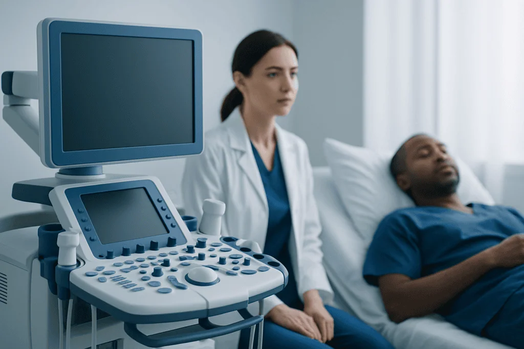
A mediastinal mass may cause no symptoms at all, especially when it is small. When symptoms do occur, they often happen because the mass presses on nearby structures in the chest. The trachea, lungs, esophagus, nerves, and blood vessels are all close together in the mediastinum, so even a noncancerous mass can create noticeable problems if it grows.
Common symptoms can include chest pain or pressure, shortness of breath, cough, hoarseness, wheezing, trouble swallowing, or a feeling of fullness in the chest. Some people also notice swelling in the face, neck, or arms if blood flow through a large vein is affected. These symptoms do not confirm a specific cause, but they are important clues during medical evaluation.
In some cases, symptoms are related to the type of underlying disease rather than pressure alone. For example, lymphoma may be associated with fever, night sweats, or unexplained weight loss. Thymic conditions can sometimes occur with autoimmune disorders such as myasthenia gravis, which may cause muscle weakness, drooping eyelids, or fatigue.
- Chest discomfort or pressure
- Persistent cough or shortness of breath
- Hoarseness or wheezing
- Difficulty swallowing
- Swelling of the face or neck
- Fever, night sweats, or weight loss in some cases
Common Causes and Risk Factors

The causes of a mediastinal mass vary by age, location in the mediastinum, and the tissue it comes from. In adults, common causes include thymoma and other thymic tumors, enlarged lymph nodes, thyroid enlargement extending into the chest, cysts, germ cell tumors, and lymphoma. In children, nerve-related tumors and congenital cysts are relatively more common.
Benign causes include bronchogenic cysts, pericardial cysts, some thyroid goiters, and certain noncancerous nerve sheath tumors. Infections and inflammatory disorders can also enlarge lymph nodes or create masses. On the malignant side, possibilities include lymphoma, thymic carcinoma, metastatic cancer, germ cell malignancies, and some primary lung-related or nerve-related tumors.
Risk factors depend on the exact diagnosis rather than on the mass itself. A personal history of cancer may raise concern for metastatic disease. Certain autoimmune conditions are associated with thymic abnormalities. Smoking does not cause every mediastinal mass, but it can increase the risk of some chest cancers and may complicate breathing symptoms. Family history and age can also influence which causes are more likely.
Because the list of possibilities is wide, doctors focus on pattern recognition rather than assumptions. The patient’s age, symptoms, imaging appearance, blood tests, and occasionally biopsy results are used together to identify whether the mass is likely benign, malignant, infectious, or inflammatory.
How a Mediastinal Mass Is Diagnosed
Diagnosis usually begins with a medical history, physical examination, and chest imaging. A chest X-ray may first show a widened area or shadow in the mediastinum, but a CT scan is often the main test used to look more closely. CT helps define the size, exact location, shape, and relationship of the mass to nearby structures. In some situations, MRI can provide additional detail, especially for nerve-related tumors or tissue characterization.
Depending on what imaging suggests, doctors may order blood tests. These can look for signs of infection, inflammation, thyroid disease, or tumor markers linked to certain germ cell tumors. If lymphoma is suspected, or if there are symptoms suggesting an autoimmune condition, additional targeted testing may be recommended. PET-CT may sometimes be used to assess metabolic activity and look for disease elsewhere in the body.
A biopsy is often needed when imaging alone cannot reliably identify the mass or when cancer is suspected. Tissue can be obtained in several ways, such as needle biopsy guided by CT or ultrasound, bronchoscopy with endobronchial ultrasound, mediastinoscopy, or surgery. The choice depends on the safest and most accurate route to the mass.
The goal of diagnosis is not only to label the mass but also to determine its stage and whether nearby organs are involved. This information directly affects treatment planning. If there is concern for tumors such as lung cancer or lymphoma, a coordinated evaluation by radiology, pathology, thoracic surgery, pulmonology, and oncology can be especially helpful.
Treatment Options and What They Depend On
Mediastinal mass treatment depends entirely on the cause. Some benign cysts or small, stable masses may only need observation with follow-up imaging. Others need active treatment because they are causing symptoms, growing, or have features that raise concern for cancer. The treatment plan is guided by the diagnosis, location, size, and whether nearby structures are affected.
Surgery may be recommended for many localized masses, especially thymic tumors, some cysts, thyroid-related growths, or nerve sheath tumors. If the mass can be safely removed, surgery can both confirm the diagnosis and treat the condition. In appropriate cases, minimally invasive approaches may be possible through robotic surgery or other thoracic techniques, depending on the patient and the center’s expertise.
If the mass is cancerous, treatment may include surgery, chemotherapy, radiation therapy, or a combination of these. Lymphoma, for example, is often treated primarily with systemic therapy rather than surgery. Some thymic or germ cell tumors may need a combined plan involving several specialties.
Supportive care is also important. Breathing symptoms, cough, pain, swallowing difficulty, or complications from compression of nearby structures may need prompt attention while the main diagnosis is being clarified. Near the end of the care pathway, some patients benefit from review by a thoracic surgery team experienced in chest surgery procedures when an operation is being considered.
Prevention, Monitoring, and Self-care
There is no single way to prevent a mediastinal mass because it is a broad finding rather than one disease. However, general health steps can support lung and chest health overall. Avoiding tobacco, keeping up with routine medical care, and seeking attention for persistent chest symptoms may help conditions be identified earlier.
For people who have a known benign mediastinal mass, follow-up imaging may be recommended to monitor for growth or change. It is important to attend scheduled scans and specialist visits, even if there are no symptoms. Monitoring plans vary, and a doctor will usually explain how often imaging is needed and what changes would lead to treatment.
Self-care cannot replace medical treatment, but it can support comfort and recovery. Patients may feel better by staying physically active within their limits, eating well, reporting new symptoms promptly, and following instructions before and after tests or procedures. Questions about work, exercise, travel, or surgery recovery are best discussed directly with the treating team.
For international patients who need further assessment, Acibadem International’s multidisciplinary specialists and JCI-accredited hospitals diagnose and treat mediastinal conditions using coordinated imaging, pathology, thoracic surgery, and oncology care.
When to Seek Medical Care
Medical advice should be sought if there is a persistent cough, unexplained chest pain, shortness of breath, hoarseness, trouble swallowing, or swelling of the face or neck. These symptoms do not always mean a serious condition, but they do deserve evaluation, especially when they are new, worsening, or not improving.
Urgent assessment is especially important if breathing becomes difficult, chest pressure is severe, swallowing becomes hard, or facial and neck swelling develops suddenly. These signs can suggest that a mass is pressing on important structures in the chest. Immediate care is also needed for coughing up blood, fainting, or severe weakness.
Anyone told they have a mediastinal mass on imaging should follow up with a qualified doctor, even if they feel well. The most useful next step is usually a clear diagnostic plan rather than delay. Early clarification can reduce uncertainty and help avoid unnecessary treatment or missed disease.
Frequently asked questions
Is a mediastinal mass always cancer?
No. A mediastinal mass can be benign, malignant, inflammatory, infectious, or cystic. Imaging and sometimes a biopsy are needed to determine exactly what it is.
Can a mediastinal mass cause no symptoms?
Yes. Many mediastinal masses are found incidentally during imaging for another reason. Symptoms are more likely when the mass is large or presses on nearby structures such as the airway or esophagus.
What tests are used to evaluate a mediastinal mass?
Common tests include chest X-ray, CT scan, and sometimes MRI or PET-CT. Blood tests and a biopsy may also be needed to confirm the diagnosis and guide treatment.
Does every mediastinal mass need surgery?
No. Some masses can be monitored, while others are treated with medication, chemotherapy, radiation therapy, or a combination of treatments. Surgery is mainly used when removal is appropriate or when tissue is needed for diagnosis.
What are the common symptoms of a mediastinal mass?
Possible symptoms include chest discomfort, cough, shortness of breath, wheezing, hoarseness, and trouble swallowing. Some people may also develop facial or neck swelling if nearby veins are compressed.
How serious is a mediastinal mass?
The seriousness depends on the cause, size, location, and whether it affects nearby organs. Some are harmless and stable, while others require prompt treatment, which is why specialist evaluation is important.
References
- American Cancer Society
- National Cancer Institute
- American College of Radiology
- National Comprehensive Cancer Network
- Merck Manual Consumer Version
This article is for general information only and is not a substitute for professional medical advice. Please consult a qualified doctor about your individual situation.
Oncology care in Turkey — second opinion and treatment plan
JCI-accredited · board-certified surgeons · reply within 24h
Add us as a Preferred Source to see more of our trusted health content across Google Search, AI Overviews and Discover.

More from the Health Library

Helichrysum: A Complete Medical Overview

Vein Ablation: A Complete Medical Overview

Meaning of pac in medical: A Complete Medical Guide for Patients

Olive Oil Cholesterol: An Evidence-Based Guide for Patients
Breathalyzer Test: An Evidence-Based Guide for Patients