Eye Ball: A Complete Medical Overview
The eye ball includes several structures that work together to support clear vision. Pain, sudden vision changes, flashes, floaters, or injury should not be ignored.
Key Takeaways
- The eye ball includes several structures that work together to support clear vision.
- Pain, sudden vision changes, flashes, floaters, or injury should not be ignored.
- Common eye ball problems include dryness, infection, trauma, pressure-related disorders, and retinal conditions.
- Regular eye examinations can detect problems before vision is affected.
- Protective eyewear, chronic disease control, and healthy habits support long-term eye health.
The eye ball is the rounded organ that captures light, focuses images, and sends visual information to the brain. Understanding its parts, normal function, and common warning signs can help people protect vision and know when medical care is needed.
Overview: what the eye ball is and what it does
The eye ball is the globe-shaped organ that makes vision possible. It receives light from the environment, focuses that light through clear structures at the front of the eye, and converts it into nerve signals that travel to the brain. In simple terms, the eye ball acts like a highly specialized optical system connected to the nervous system.
Although the eye appears small from the outside, it contains many delicate parts that need to work in balance. The clear cornea and lens focus light, the iris controls how much light enters, the retina detects images, and the optic nerve carries visual information to the brain. The white outer wall, called the sclera, helps protect the eye and maintain its shape.
The term “eye ball” is often used broadly, but clinicians may distinguish between the globe itself and nearby structures such as the eyelids, tear glands, and eye muscles. This distinction matters because symptoms such as redness, pain, swelling, or blurred vision can come from the globe, the surface of the eye, or surrounding tissues.
A person does not usually need to think about these processes during daily life. However, knowing the basics of how the eye ball works can make it easier to recognize symptoms early and seek appropriate care before a minor problem becomes more serious.
Main parts of the eye ball
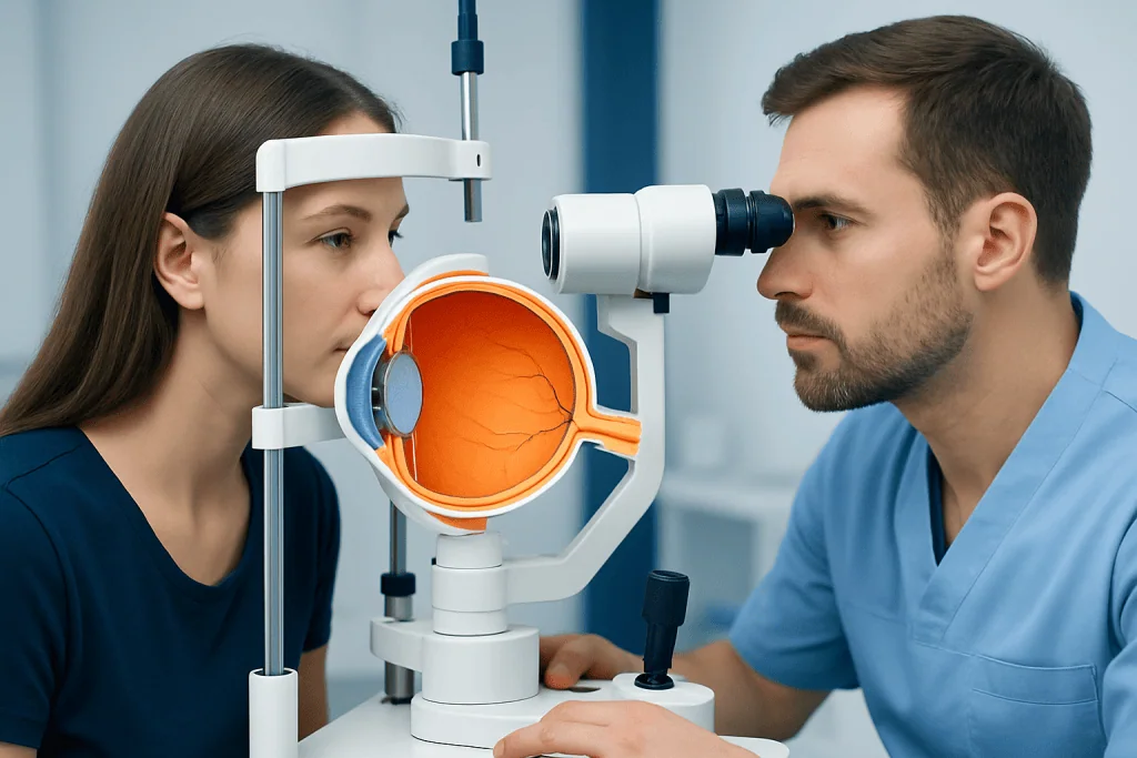
The front of the eye ball includes the cornea, a clear curved surface that begins focusing incoming light. Behind it is the anterior chamber, which contains fluid that nourishes the eye and helps maintain pressure. The iris gives the eye its color, while the central opening in the iris, the pupil, changes size to regulate light entry.
Behind the pupil sits the lens, which fine-tunes focus so a person can see at different distances. With age, the lens can become less flexible or cloudy. This can lead to common conditions such as cataract, in which the lens loses clarity and vision becomes dim, blurry, or glare-prone.
The back portion of the eye ball is filled largely by the vitreous, a gel-like substance that helps the globe keep its shape. Lining the inside back wall is the retina, a thin layer of light-sensitive tissue. The central part of the retina, the macula, is especially important for reading, recognizing faces, and seeing fine detail.
The optic nerve leaves the back of the eye and carries signals to the brain, where images are interpreted. Damage anywhere along this pathway may affect sight. Eye specialists may also assess the pressure inside the eye because elevated pressure can be associated with glaucoma, a condition that can gradually harm the optic nerve.
Common symptoms that may involve the eye ball
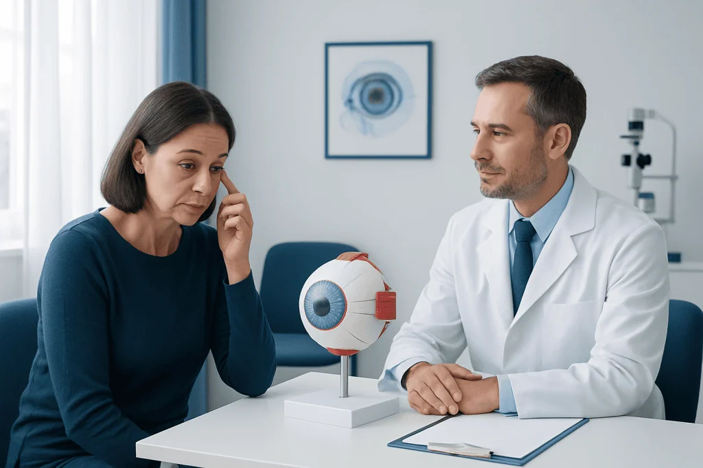
Symptoms linked to the eye ball can range from mild irritation to urgent warning signs. Common complaints include redness, watering, dryness, itching, burning, blurred vision, light sensitivity, and the sensation that something is in the eye. These symptoms may come from the eye’s surface, deeper structures, or nearby tissues.
Eye pain deserves attention because it can have many causes. Mild discomfort may occur with dryness, eye strain, or surface irritation. More significant pain may be associated with infection, inflammation, pressure changes inside the eye, trauma, or a scratched cornea. Pain combined with vision loss, nausea, or marked redness should be assessed promptly.
Some visual symptoms can suggest a problem in the back of the eye rather than on the surface. Sudden flashes of light, new floaters, a curtain-like shadow, or rapid vision loss may indicate a retinal problem that requires urgent evaluation. Gradual changes such as increasing blur, difficulty seeing at night, or halos around lights may reflect lens or pressure-related conditions.
Not every symptom signals a serious disease, but the pattern matters. Symptoms in one eye only, symptoms after injury, or symptoms that are sudden, severe, or worsening are more concerning than brief, mild irritation that improves with rest. A proper eye examination helps determine whether the issue is routine or urgent.
Causes and risk factors affecting the eye ball
Eye ball problems can result from infection, inflammation, injury, pressure changes, aging, or chronic health conditions. Surface irritation may be caused by dry air, prolonged screen use, allergies, contact lens overuse, or foreign bodies such as dust. Infections can involve the conjunctiva, cornea, or deeper eye tissues and may produce redness, discharge, pain, and sensitivity to light.
Injuries are another important cause. A blow to the eye, chemical splash, penetrating trauma, or even vigorous rubbing can damage delicate structures. Sports, workplace hazards, and home accidents increase risk, especially when protective eyewear is not used. Contact lens misuse can also raise the risk of corneal injury and infection.
Several medical conditions can affect the eye ball over time. Diabetes can damage blood vessels in the retina, while high blood pressure may also affect retinal circulation. Autoimmune diseases may cause inflammation inside the eye, and thyroid disorders can influence tissues around the eyes. Age is a strong risk factor for conditions involving the lens, vitreous, retina, and optic nerve.
Family history also matters. Some disorders, especially glaucoma and certain retinal diseases, may occur more often in families. Smoking, uncontrolled blood sugar, long-term ultraviolet exposure, and not having regular eye examinations can increase the chance that a problem develops or progresses unnoticed.
How doctors examine and diagnose eye ball problems
Diagnosis begins with a careful history. A doctor will usually ask when symptoms began, whether one or both eyes are affected, whether there was injury or chemical exposure, and whether the person has pain, discharge, flashes, floaters, headaches, or vision loss. Medical conditions such as diabetes, hypertension, and autoimmune disease are also relevant.
The examination may include visual acuity testing, assessment of the pupils, and measurement of eye pressure. A slit-lamp examination allows the clinician to inspect the cornea, lens, iris, and front chamber in detail. Fluorescein dye may be used to look for scratches or damage on the eye’s surface. If the back of the eye needs evaluation, dilating drops may be used so the retina and optic nerve can be examined.
Additional testing depends on the suspected cause. Imaging of the retina or optic nerve may help monitor structural changes. Visual field testing can assess peripheral vision when glaucoma is a concern. Ultrasound, optical coherence tomography, or other specialized tests may be used when the view into the eye is limited or when retinal disease is suspected.
Because symptoms can overlap, self-diagnosis is not always reliable. For example, red eye may come from a simple irritation, but it can also reflect a corneal ulcer, uveitis, or acute pressure-related problem. Prompt professional assessment is the safest way to identify the cause and begin the right treatment.
Treatment options for eye ball conditions
Treatment depends entirely on the underlying problem. Mild dryness or irritation may improve with environmental adjustments, breaks from screen use, and lubricating drops recommended by a clinician. Allergic symptoms may be treated differently from infection, and bacterial problems require a different approach from viral or inflammatory conditions. It is important not to use leftover eye drops or someone else’s medication.
When an infection, inflammation, or injury affects the eye ball, treatment may include prescription drops, protective measures, pain control, or close follow-up. Chemical injuries need immediate first aid and urgent medical evaluation. Contact lens users are often advised to stop lens wear until the eye has healed and a doctor confirms that it is safe to resume.
Some conditions involve the internal structures of the eye and may need procedures or surgery. Lens-related vision loss may be treated with cataract surgery. Pressure-related optic nerve disease may require medication, laser therapy, or glaucoma surgery in selected cases. Retinal tears or detachments may need timely treatment to help preserve vision.
If vision changes are severe or the diagnosis is uncertain, referral to an ophthalmologist is often needed. In complex cases, a multidisciplinary approach may be helpful. Near the end of the care pathway, patients seeking evaluation abroad may choose centers such as Acibadem International, where multidisciplinary specialists in JCI-accredited hospitals diagnose and treat eye conditions for international patients, including advanced retinal detachment surgery when appropriate.
Protecting the eye ball: prevention and self-care
Many eye ball problems cannot be completely prevented, but healthy habits can lower risk. Wearing sunglasses with ultraviolet protection may help protect the eyes from long-term sun exposure. Protective eyewear is strongly recommended for certain sports, construction work, laboratory settings, and home projects involving chemicals or flying particles.
Screen habits also matter. Long periods of digital device use can reduce blinking and worsen dryness or eye strain. Taking regular visual breaks, blinking consciously, and adjusting screen position and room lighting may help. People who wear contact lenses should follow hygiene instructions carefully, avoid sleeping in lenses unless specifically approved, and never clean lenses with tap water.
General health supports eye health. Good control of diabetes, blood pressure, and cholesterol can help protect retinal blood vessels. Not smoking, staying physically active, eating a balanced diet rich in fruits and vegetables, and attending routine eye examinations are practical steps that support long-term vision.
Self-care has limits. Home measures may ease mild irritation, but they do not replace a professional assessment for persistent symptoms, eye pain, trauma, or any sudden change in vision. Seeking timely care is one of the most effective ways to protect the eye ball from avoidable damage.
When to seek medical care
Medical care should be sought promptly if a person develops sudden blurred vision, partial vision loss, flashes of light, a sudden increase in floaters, severe pain, marked redness, or light sensitivity. These symptoms can signal problems that need urgent treatment. Any chemical splash or eye injury should also be treated as a medical priority.
Evaluation is also important for symptoms that do not improve within a short time, recur frequently, or affect daily activities such as reading, driving, or work. Redness with discharge, pain in a contact lens wearer, or swelling around the eye should not be ignored. Children, older adults, and people with diabetes or immune system problems may need earlier assessment.
A routine eye examination is still useful even when symptoms are absent. Some important conditions, including glaucoma and early retinal disease, may cause little or no warning at first. Regular checkups can identify changes early, when treatment may be more effective and vision is often easier to preserve.
Frequently asked questions
What is the eye ball made of?
The eye ball is made of several connected structures, including the cornea, iris, pupil, lens, vitreous, retina, sclera, and optic nerve. Each part has a specific job in focusing light, detecting images, and sending signals to the brain.
Why does the eye ball sometimes hurt?
Eye ball pain can happen for many reasons, including dryness, eye strain, allergies, scratches on the cornea, infection, inflammation, or increased pressure inside the eye. Pain that is severe, sudden, or associated with vision changes should be assessed by a doctor promptly.
Can eye ball problems cause blurred vision?
Yes. Blurred vision can come from surface problems such as dryness, but it can also result from lens changes, retinal disease, corneal injury, or optic nerve problems. Because the causes vary widely, persistent or sudden blurred vision should be evaluated professionally.
Are flashes and floaters in the eye ball dangerous?
Flashes and floaters are not always dangerous, especially if they are mild and stable. However, a sudden increase in floaters, repeated flashes, or a shadow over part of the vision can suggest a retinal tear or detachment and needs urgent eye care.
How can a person keep the eye ball healthy?
Useful steps include wearing protective eyewear, using UV-protective sunglasses, limiting contact lens risks, managing chronic conditions such as diabetes, and having regular eye exams. Healthy lifestyle habits, including not smoking and eating a balanced diet, also support eye health.
When should someone see a doctor for a red eye?
A red eye should be checked if it is painful, affects vision, follows an injury, occurs in a contact lens wearer, or does not improve quickly. Redness with discharge, light sensitivity, or swelling may also point to a condition that needs medical treatment.
This article is for general information only and is not a substitute for professional medical advice. Please consult a qualified doctor about your individual situation.
Explore treatments in Turkey — costs, top hospitals & a free quote
JCI-accredited · board-certified surgeons · reply within 24h
Add us as a Preferred Source to see more of our trusted health content across Google Search, AI Overviews and Discover.

More from the Health Library

Pudendal Nerve: An Evidence-Based Guide for Patients
Psychotic Episode: What Patients Need to Know
Benztropine: A Complete Medical Overview

Chamomile Tea — Explained by Medical Evidence, Not Myths

Ideomotor Apraxia: A Complete Medical Overview