Pericholecystic Fluid — Explained by Medical Evidence, Not Myths

Pericholecystic fluid is an imaging finding, not a diagnosis on its own. It is often associated with acute gallbladder inflammation, especially when paired with pain, fever, or gallstones.
Key Takeaways
- Pericholecystic fluid is an imaging finding, not a diagnosis on its own.
- It is often associated with acute gallbladder inflammation, especially when paired with pain, fever, or gallstones.
- Doctors interpret it together with symptoms, blood tests, and imaging features such as gallbladder wall thickening.
- Treatment depends on the cause and may range from observation to antibiotics, drainage, or surgery.
- Urgent medical care is important if severe right upper abdominal pain, fever, vomiting, or jaundice develops.
Pericholecystic fluid means there is fluid around the gallbladder, usually detected on ultrasound or CT imaging. It is not a disease by itself, but a sign that may point to gallbladder inflammation, infection, or another abdominal problem that needs medical interpretation in context.
Overview: what pericholecystic fluid means
Pericholecystic fluid is fluid that collects around the gallbladder. In many cases, it is found during an ultrasound or CT scan performed because a person has abdominal pain, nausea, fever, or abnormal liver-related blood tests. The term describes where the fluid is seen, but it does not by itself explain why it is there.
Medical interpretation depends on the whole picture. A small rim of fluid around the gallbladder may suggest inflammation of the gallbladder, especially when combined with gallstones, gallbladder wall thickening, or tenderness during ultrasound. In other situations, the fluid may reflect another illness such as liver disease, heart failure, generalized fluid retention, or inflammation nearby in the abdomen.
This is why pericholecystic fluid should be understood as an evidence-based imaging sign rather than a stand-alone diagnosis. Some people need prompt treatment, while others need careful follow-up and evaluation of the underlying cause. The meaning changes depending on symptoms, examination findings, blood work, and the full imaging report.
Symptoms and how it may present
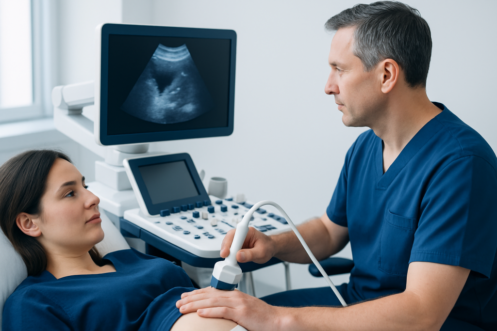
Pericholecystic fluid itself does not cause unique symptoms that a person can feel. Instead, symptoms come from the condition causing the fluid. When the gallbladder is inflamed, a person may have pain in the right upper abdomen, pain after eating fatty meals, nausea, vomiting, loss of appetite, fever, or pain that radiates to the right shoulder or back.
Some people have milder symptoms, such as a sense of fullness, indigestion, bloating, or intermittent discomfort. Others may have more concerning signs, including jaundice, chills, or severe tenderness in the upper abdomen. In older adults and people with diabetes, symptoms can sometimes be less typical, which may delay recognition.
Doctors often look for a pattern rather than a single complaint. Symptoms become more meaningful when they occur along with imaging features of gallstones or gallbladder inflammation. If the fluid is due to a non-gallbladder problem, symptoms may point elsewhere, such as swelling in the legs, shortness of breath, or more generalized abdominal discomfort.
- Right upper abdominal pain
- Nausea or vomiting
- Fever or chills
- Pain after meals, especially fatty foods
- Jaundice in some cases
Common causes and risk factors

The most common clinically important cause of pericholecystic fluid is acute cholecystitis, which is inflammation of the gallbladder, usually triggered by a gallstone blocking the cystic duct. This blockage can lead to swelling, irritation, and sometimes infection. On imaging, doctors may see fluid around the gallbladder along with gallstones, a thickened gallbladder wall, and tenderness over the gallbladder area.
Not all pericholecystic fluid means acute cholecystitis. It can also appear with liver cirrhosis, hepatitis, pancreatitis, low blood protein, heart failure, kidney disease, trauma, or widespread inflammation in the abdomen. In these settings, the fluid may reflect leakage or congestion rather than a primary gallbladder infection.
Risk factors depend on the underlying cause. For gallbladder disease, risk may be higher in people with gallstones, obesity, rapid weight loss, pregnancy, increasing age, diabetes, or certain blood and metabolic conditions. Risk can also rise in people who are seriously ill, hospitalized, fasting for long periods, or receiving intensive care, because the gallbladder may become inflamed even without stones in a condition called acalculous cholecystitis.
How doctors confirm the cause
Diagnosis starts with a medical history and physical examination. A clinician asks where the pain is, when it began, whether it worsens after meals, and whether fever, vomiting, or jaundice are present. During the examination, tenderness in the right upper abdomen can support suspicion of gallbladder disease, but symptoms alone are not enough to identify the cause of the fluid.
Ultrasound is often the first imaging test because it can show gallstones, wall thickening, bile duct enlargement, and fluid around the gallbladder. CT scanning may be used when the diagnosis is unclear or when doctors need a broader view of the abdomen. In some cases, a hepatobiliary scan may help assess whether the gallbladder is functioning or blocked.
Blood tests are also important. Doctors may check white blood cell count, liver enzymes, bilirubin, pancreatic enzymes, kidney function, and markers of inflammation. A diagnosis of acute cholecystitis is strongest when symptoms, examination findings, blood tests, and imaging all point in the same direction. This combined approach helps avoid overinterpreting a single imaging term such as pericholecystic fluid.
Treatment options depend on the underlying problem
There is no single treatment for pericholecystic fluid because treatment is aimed at the condition causing it. If acute cholecystitis is confirmed or strongly suspected, treatment may include hospital observation, intravenous fluids, pain control, antibiotics when infection is likely, and a plan to remove the gallbladder when appropriate. If the fluid is due to another illness, care focuses on that condition instead.
When gallbladder disease is the cause, surgery to remove the gallbladder is a common definitive treatment. Many patients are treated with minimally invasive laparoscopic gallbladder removal, especially if symptoms are ongoing or complications are a concern. If a person is too unwell for immediate surgery, doctors may first stabilize the condition or consider temporary drainage.
Additional procedures may be needed if imaging or blood tests suggest a blocked bile duct, stone migration, or infection extending beyond the gallbladder. In those situations, a specialist may recommend ERCP to evaluate and relieve bile duct obstruction. In selected cases, further gastroenterology evaluation helps clarify whether the imaging finding reflects gallbladder disease or another digestive disorder.
Treatment decisions are individualized. The urgency depends on pain severity, fever, laboratory findings, overall health, and whether complications such as perforation, abscess, or sepsis are suspected. For international patients, Acibadem International’s multidisciplinary specialists and JCI-accredited hospitals diagnose and treat gallbladder conditions using coordinated medical, imaging, and surgical care.
Prevention and self-care
Pericholecystic fluid cannot be prevented directly because it is a sign rather than a disease. Prevention focuses on reducing the risk of gallstones and supporting prompt evaluation of abdominal symptoms. Maintaining a balanced weight, avoiding very rapid weight loss, staying physically active, and following a nutritious eating pattern may help lower the risk of some gallbladder problems.
If a person already knows they have gallstones, keeping regular medical follow-up can be useful, especially if episodes of pain are recurring. People with diabetes, liver disease, or serious chronic illness should mention new abdominal pain early, as symptoms can progress more quietly. Self-treating persistent right upper abdominal pain with repeated antacids or pain relievers may delay proper diagnosis.
After treatment for gallbladder disease, the care team may advise gradual return to meals, hydration, and follow-up testing if needed. People should follow the specific instructions given by their doctor, especially after a procedure or surgery. Home care supports recovery, but it does not replace evaluation when symptoms suggest active inflammation or infection.
When to seek medical care
Medical care should be sought promptly if right upper abdominal pain is severe, lasts more than a few hours, or is accompanied by fever, repeated vomiting, jaundice, chills, faintness, or worsening weakness. These symptoms can suggest a gallbladder attack, infection, bile duct blockage, or another urgent abdominal condition.
Even when pain is not severe, a recent imaging report that mentions pericholecystic fluid should be reviewed with a qualified doctor who can explain what it means in context. This is especially important if there are abnormal blood tests, known gallstones, or a history of liver, pancreatic, kidney, or heart disease.
Emergency evaluation is important if a person has confusion, very low blood pressure, inability to keep fluids down, shortness of breath, or rapidly worsening abdominal swelling. These are not typical minor symptoms and should not be ignored. Early assessment can help identify whether the fluid is part of gallbladder inflammation or a different problem requiring treatment.
Frequently asked questions
Is pericholecystic fluid the same as gallbladder disease?
No. Pericholecystic fluid is an imaging finding that describes fluid around the gallbladder. It may occur with gallbladder disease, but it can also be related to other conditions such as liver disease, pancreatitis, or generalized fluid retention.
Does pericholecystic fluid always mean surgery is needed?
Not always. Surgery may be recommended if the fluid is part of acute cholecystitis or another gallbladder problem that is unlikely to settle safely on its own. If the fluid comes from a different cause, treatment may focus on that underlying condition instead.
Can pericholecystic fluid be found without gallstones?
Yes. Although gallstones are a common cause, pericholecystic fluid can also appear in acalculous cholecystitis, liver disease, heart failure, kidney problems, trauma, and other abdominal illnesses. That is why doctors interpret the finding together with symptoms and tests.
How serious is pericholecystic fluid on an ultrasound?
Its significance varies. In some cases it supports a diagnosis of active gallbladder inflammation and needs urgent treatment, while in others it is a less specific sign that requires follow-up and broader evaluation. The seriousness depends on the full clinical picture, not the term alone.
What tests are usually done after this finding?
Doctors often review the ultrasound carefully and may order blood tests for infection, liver function, and pancreatic enzymes. Depending on symptoms, they may also use CT imaging or a hepatobiliary scan to better understand whether the gallbladder is inflamed or blocked.
Can pericholecystic fluid go away on its own?
It can, but only if the underlying cause resolves. For example, fluid related to temporary inflammation or systemic fluid overload may improve as the main problem is treated. It should not be assumed to be harmless without medical review.
References
- National Institute of Diabetes and Digestive and Kidney Diseases
- American College of Gastroenterology
- American College of Radiology
- Society of American Gastrointestinal and Endoscopic Surgeons
- Merck Manual Professional Edition
This article is for general information only and is not a substitute for professional medical advice. Please consult a qualified doctor about your individual situation.
Explore treatments in Turkey — costs, top hospitals & a free quote
JCI-accredited · board-certified surgeons · reply within 24h
Add us as a Preferred Source to see more of our trusted health content across Google Search, AI Overviews and Discover.

More from the Health Library

Hair Growth Serum: What Patients Need to Know
Cupping therapy: A Complete Medical Guide for Patients

Code Blue — Explained by Medical Evidence, Not Myths

Clitor Big: A Complete Medical Overview

Pruritus of the Neck: Common Causes of Itching

Candesartan: What Patients Need to Know
Infectious Diseases & Clinical Microbiology Specialists at Acibadem

Dr. Hakan Kutlu
Infectious Diseases & Clinical Microbiology
Dr. Hande Aygün
Infectious Diseases & Clinical Microbiology
Asst. Prof. Dr. Hülya Kuşoğlu
Infectious Diseases & Clinical Microbiology
Prof. Dr. İftahar Köksal
Infectious Diseases & Clinical Microbiology