Intraocular Pressure: A Complete Medical Overview
Intraocular pressure refers to the fluid pressure inside the eye. High eye pressure may increase the risk of glaucoma, but it does not always cause symptoms.
Key Takeaways
- Intraocular pressure refers to the fluid pressure inside the eye.
- High eye pressure may increase the risk of glaucoma, but it does not always cause symptoms.
- A simple eye exam can measure pressure and assess the optic nerve and visual field.
- Treatment may include monitoring, eye drops, laser procedures, or surgery depending on the cause and risk.
- Regular eye checks are especially important for people with risk factors such as age, family history, or steroid use.
Intraocular pressure is the pressure created by fluid inside the eye. It is an important part of eye health because pressure that is too high, and sometimes too low, can affect the optic nerve and vision, although many people have no early symptoms.
Overview: what intraocular pressure means
Intraocular pressure is the pressure of the fluid inside the eye. In simple terms, it reflects the balance between how much aqueous humor, a clear fluid in the front part of the eye, is produced and how well it drains away. This pressure helps the eye keep its shape and function normally.
A normal reading does not guarantee perfect eye health, and a high reading does not always mean vision loss will occur. However, when intraocular pressure is consistently higher than the eye can safely tolerate, it may damage the optic nerve over time. This is why eye pressure is closely linked with glaucoma, a group of conditions that can gradually affect vision.
Eye pressure is measured in millimeters of mercury, often written as mmHg. While there is a commonly accepted normal range, each person’s eye may tolerate pressure differently. For that reason, doctors look at pressure together with optic nerve appearance, corneal thickness, visual field testing, and overall eye health rather than relying on one number alone.
How the eye regulates pressure
The eye constantly makes aqueous humor behind the iris. This fluid flows through the pupil into the front chamber of the eye and then drains through specialized channels, mainly at the angle where the cornea and iris meet. When production and drainage are balanced, intraocular pressure remains stable.
If the drainage system becomes less efficient, pressure can rise. If fluid leaks or production becomes too low, pressure can fall. Both situations matter, although high intraocular pressure is far more common in routine eye care.
Pressure is not fixed throughout the day. It may change slightly from morning to evening and can be influenced by body position, medications, and certain eye conditions. Because of this, a doctor may repeat measurements over time or combine them with imaging and optic nerve evaluation to understand the bigger picture.
Symptoms and what people may notice
One of the most important facts about intraocular pressure is that it often causes no symptoms in the early stages. Many people with ocular hypertension, meaning pressure higher than normal without clear optic nerve damage, feel completely well. This is why routine eye examinations are so valuable.
When symptoms do occur, they depend on the cause and how quickly pressure changes. Gradual pressure elevation may be silent, while a sudden and marked increase can lead to more noticeable problems. Possible symptoms can include:
- Blurred vision
- Eye pain or pressure sensation
- Headache
- Halos around lights
- Redness of the eye
- Nausea or vomiting when pressure rises suddenly
- Progressive loss of side vision in untreated glaucoma
Low intraocular pressure is less common but can also affect vision. It may cause blurred vision, discomfort, or visual distortion, especially after surgery or injury. Any sudden change in vision or significant eye pain should be assessed promptly by a qualified doctor.
Causes and risk factors for high or low eye pressure
High intraocular pressure usually happens when the eye’s drainage system does not remove fluid efficiently. This may occur on its own or as part of another eye condition. Some people develop elevated pressure without immediate nerve damage, while others are more vulnerable and may develop glaucoma even at pressures that are not very high.
Common causes or contributors to elevated pressure include aging changes in the drainage angle, family history of glaucoma, long-term corticosteroid use, previous eye injury, certain inflammatory eye diseases, and some structural features of the eye. Conditions that block the drainage angle can cause sudden pressure elevation and need urgent attention.
Risk factors that may increase the chance of pressure-related optic nerve damage include older age, thinner corneas, diabetes, severe nearsightedness or farsightedness in some cases, and African, Asian, or Hispanic ancestry depending on the glaucoma type. Related eye problems may include cataract or retinal disorders, although these do not automatically cause high pressure.
Low intraocular pressure may result from eye surgery, trauma, inflammation, dehydration of the eye tissues, or leakage of fluid from the eye. Because both high and low pressure can threaten visual function, the cause should be identified rather than focusing on the pressure number alone.
How doctors diagnose intraocular pressure problems
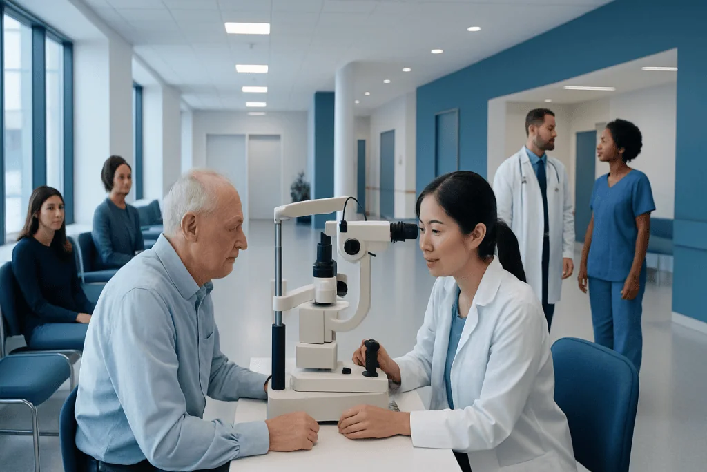
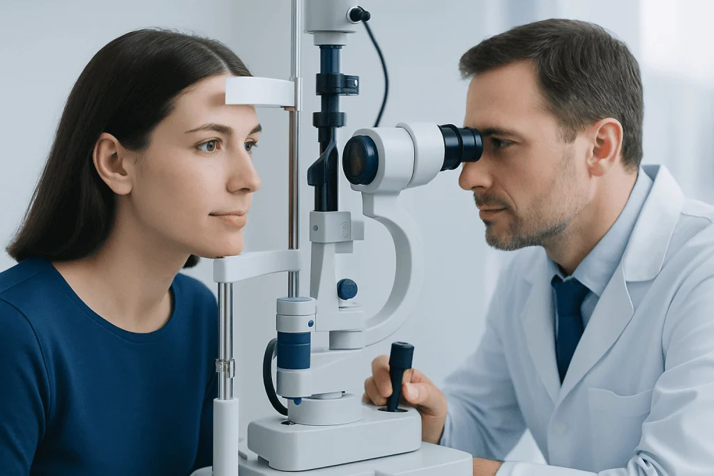
Diagnosis begins with a comprehensive eye examination. The pressure itself is often measured with tonometry, which may be done using a slit-lamp device after numbing eye drops or with non-contact methods that briefly puff air onto the eye. These tests are quick and are commonly performed during routine ophthalmology visits.
Because pressure alone does not tell the whole story, the doctor usually examines the drainage angle, optic nerve, and retina. Corneal thickness may also be measured because it can influence pressure readings. A thick or thin cornea can make the number appear higher or lower than the true pressure inside the eye.
Additional tests may include visual field testing to look for side-vision loss and imaging of the optic nerve and retinal nerve fiber layer. If pressure is elevated or glaucoma is suspected, these tests help determine whether there is early nerve damage and guide treatment decisions. In some patients, evaluation may also include advanced imaging or combined care with specialists in eye examination and eye check-up services.
Treatment options and long-term management
Treatment depends on the pressure level, the underlying cause, and whether there is evidence of optic nerve damage. Not every person with elevated intraocular pressure needs immediate treatment. In lower-risk cases, careful monitoring may be enough, especially if the optic nerve and visual field remain stable over time.
When treatment is recommended, eye drops are often the first step. Different medications work by either reducing fluid production or improving drainage. The choice of medicine depends on the individual’s pressure goals, general health, side effects, and ability to use drops regularly as prescribed.
If drops are not enough or are not suitable, laser treatment may help improve drainage or reduce fluid production. In some cases, surgery is advised to create a new drainage pathway or enhance the eye’s natural outflow. Patients with established glaucoma may also need ongoing follow-up and procedures tailored to the stage of disease, including options discussed in glaucoma treatment.
Low eye pressure is managed by treating the cause, such as inflammation, wound leakage, or a postoperative issue. The aim is to stabilize the eye and protect vision. Near the end of the care pathway, some patients may benefit from multidisciplinary assessment; Acibadem International’s specialists in JCI-accredited hospitals diagnose and treat eye conditions for international patients when coordinated care is needed.
Prevention, monitoring, and self-care
Intraocular pressure cannot always be prevented, but regular eye examinations can help detect changes early, before vision is affected. This is particularly important for people over 40, anyone with a family history of glaucoma, people using steroid medications, and those with diabetes or previous eye injury.
Good self-care supports overall eye health, even though lifestyle measures do not replace medical treatment. Patients should use prescribed eye drops exactly as directed, attend follow-up visits, and tell their doctor about any side effects or difficulties using medication. Stopping treatment without medical advice can allow pressure to rise again.
Helpful habits may include protecting the eyes from injury, managing chronic health conditions, and reporting any new visual symptoms promptly. People should also mention all medicines and supplements they take, since some treatments can affect eye pressure or interact with other eye conditions. If pressure changes are related to another eye problem, treatment such as cataract surgery may sometimes be part of the broader plan.
When to seek medical care
People should arrange an eye examination if they have a family history of glaucoma, have been told they have high eye pressure, use steroid medicines for a long time, or notice gradual changes in vision. Even without symptoms, routine care is the best way to identify pressure-related problems early.
Prompt medical attention is important for sudden eye pain, rapidly blurred vision, halos around lights, redness, severe headache with eye symptoms, nausea, or vomiting. These symptoms can occur with a sudden rise in intraocular pressure and should not be ignored.
Anyone who has had recent eye surgery or an eye injury and then develops discomfort, worsening vision, or unusual sensitivity should contact their eye doctor. Early assessment can help prevent complications and protect sight.
Frequently asked questions
What is a normal intraocular pressure reading?
Intraocular pressure is often considered normal when it falls within a typical range used in eye care, but there is no single perfect number for everyone. Some people develop optic nerve damage at pressures within the usual range, while others tolerate slightly higher readings without harm.
Does high intraocular pressure always mean glaucoma?
No. High intraocular pressure can occur without visible optic nerve damage, a condition often called ocular hypertension. Glaucoma is diagnosed when there is pressure-related or other optic nerve damage, often supported by changes on imaging or visual field testing.
Can intraocular pressure cause symptoms?
Often it does not cause symptoms in the early stages, which is why regular eye exams are so important. Sudden or severe pressure elevation may cause eye pain, blurred vision, halos, redness, headache, or nausea and needs prompt assessment.
How is intraocular pressure measured?
Doctors measure eye pressure with tonometry during an eye exam. The test may involve a gentle instrument touching the eye after numbing drops or a puff of air, and it is usually quick and well tolerated.
Can intraocular pressure be lowered naturally?
Healthy habits support overall eye health, but they do not replace proven medical treatment for high eye pressure. If a doctor prescribes monitoring, eye drops, laser treatment, or surgery, following that plan is the safest way to protect vision.
Who should have regular eye pressure checks?
Regular checks are especially important for adults over 40, people with a family history of glaucoma, steroid users, and those with diabetes or previous eye injury. A doctor can recommend how often testing is needed based on age, risk factors, and previous exam findings.
References
- American Academy of Ophthalmology
- National Eye Institute
- Glaucoma Research Foundation
- Mayo Clinic
- World Health Organization
This article is for general information only and is not a substitute for professional medical advice. Please consult a qualified doctor about your individual situation.
Explore treatments in Turkey — costs, top hospitals & a free quote
JCI-accredited · board-certified surgeons · reply within 24h
Add us as a Preferred Source to see more of our trusted health content across Google Search, AI Overviews and Discover.

More from the Health Library



Related Specialists

Dyt. Şeyda Sıla Bilgili Tokgöz
Nutrition & Diet
Dr. Jirayr Ağazaryan
General Surgery
Prof. Dr. Gülfer Mehtap Yazicioğlu
Pediatric Allergy
Prof. Dr. Ceyda Erel Kirişoğlu
Pulmonology