

Synechiae — Explained by Medical Evidence, Not Myths

Synechiae are adhesions between tissues that are normally separate. They most often affect the eye or the uterus, but the term can be used in other parts of the body.
Key Takeaways
- Synechiae are adhesions between tissues that are normally separate.
- They most often affect the eye or the uterus, but the term can be used in other parts of the body.
- Symptoms vary by location and may include eye pain, blurred vision, menstrual changes, or fertility problems.
- Treatment focuses on the cause, the severity of symptoms, and whether organ function is affected.
- Early evaluation can help prevent complications such as glaucoma, vision damage, or reproductive problems.
Synechiae are abnormal bands of tissue that cause nearby structures to stick together, most commonly in the eye or inside the uterus. The term describes a physical adhesion rather than a single disease, so symptoms, causes, and treatment depend on where the synechiae form.
What synechiae means in medical terms
Synechiae are abnormal adhesions, meaning bands of tissue that make two surfaces stick together when they would normally remain separate. The word is descriptive rather than diagnostic: it tells doctors what has happened anatomically, but not yet why it happened. In practice, synechiae are most often discussed in ophthalmology, where the iris can adhere to nearby eye structures, and in gynecology, where scar tissue can form inside the uterus.
This distinction matters because many people search for “synechiae” expecting it to name one disease. Instead, it is a finding that can result from inflammation, injury, surgery, infection, or healing after a procedure. Some synechiae cause no symptoms and are found incidentally, while others interfere with normal function and need treatment.
In the eye, synechiae may block fluid flow, distort the pupil, or raise the risk of pressure-related complications. In the uterus, adhesions may change menstrual bleeding, contribute to infertility, or increase the risk of pregnancy complications. A careful diagnosis helps identify both the location of the adhesion and the underlying condition that led to it.
Where synechiae occur and how they differ
Eye synechiae are usually described as either posterior or anterior. Posterior synechiae occur when the iris sticks to the lens behind it. Anterior synechiae occur when the iris adheres to the cornea or drainage angle at the front of the eye. These forms can develop after inflammation such as uveitis, after trauma, or sometimes after eye surgery.
Uterine synechiae refer to adhesions inside the uterine cavity. When extensive, they may be part of Asherman syndrome, a condition linked to scarring of the endometrium. These adhesions can form after procedures involving the uterus, such as dilation and curettage, after infection, or less commonly after other types of uterine injury.
Doctors may also use the term in other settings, such as nasal or abdominal adhesions, but eye and uterine synechiae are the most clinically recognized forms in general patient education. Because the word covers more than one body system, medical care is guided by symptoms, examination findings, and the specialty involved.
Symptoms and possible effects on health

Symptoms depend on the location and size of the adhesions. In the eye, synechiae may cause eye redness, pain, light sensitivity, blurred vision, an irregularly shaped pupil, or a feeling that vision has changed suddenly. Some people have no obvious symptoms at first, and the adhesion is discovered during an eye examination performed for another reason.
If eye synechiae affect fluid drainage or pupil movement, they can contribute to increased eye pressure and glaucoma-related damage. For that reason, symptoms such as severe eye pain, halos around lights, rapidly worsening blurred vision, or headache with nausea should not be ignored. Prompt ophthalmic evaluation is important because pressure-related complications can harm vision.
In uterine synechiae, symptoms can include lighter periods than usual, absent periods, pelvic discomfort, repeated miscarriage, difficulty becoming pregnant, or menstrual pain caused by trapped blood flow. However, some people with mild adhesions still have regular cycles and only learn about the problem during an infertility workup or after imaging of the uterus.
Symptoms alone cannot confirm synechiae. Similar complaints may occur with glaucoma, infection, hormonal disorders, fibroids, endometriosis, or other eye and gynecologic conditions. That is why diagnosis depends on a targeted exam and appropriate imaging or procedures rather than self-diagnosis.
Why synechiae develop: causes and risk factors
Synechiae usually develop when inflammation or injury causes tissues to heal abnormally and stick together. In the eye, one of the most common drivers is uveitis, an inflammatory condition involving the middle layer of the eye. Trauma, previous eye surgery, some infections, and uncontrolled inflammation can also contribute. In some cases, synechiae are part of a broader eye disorder and appear alongside other findings.
In the uterus, adhesions are more likely after procedures that affect the lining of the womb, especially if the endometrium has been injured. This can happen after pregnancy-related procedures, miscarriage management, removal of retained tissue, or other intrauterine surgery. Infection of the uterine lining, though less common in some settings, is another recognized cause.
Not everyone with inflammation or surgery develops synechiae. Risk can be influenced by the extent of tissue damage, repeated procedures, delayed treatment of inflammation, and the body’s own healing response. This is one reason follow-up after surgery or inflammatory illness is important even when a person initially feels well.
It can help to think of synechiae as a healing-related complication rather than a contagious disease. They do not spread from person to person. The main concern is whether the adhesion interferes with normal anatomy, blocks movement or drainage, or reflects an untreated underlying condition that still needs care.
How synechiae are diagnosed
Diagnosis begins with the history: symptoms, timing, previous surgery, injury, infection, pregnancy history, and any inflammatory disease can all provide important clues. Because synechiae are structural adhesions, direct visualization is often central to diagnosis. The exact tests depend on whether doctors suspect an eye or uterine form.
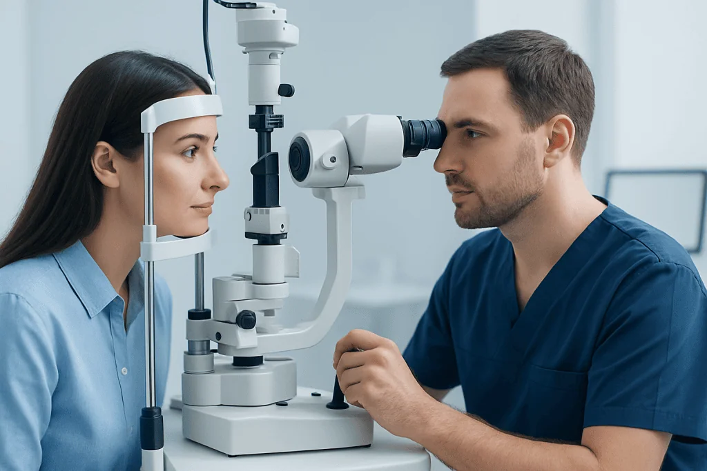
For eye synechiae, an ophthalmologist typically uses a slit-lamp examination to inspect the front structures of the eye in detail. Eye pressure may be measured, and the pupil and drainage angle may be assessed to look for signs of blockage or inflammation. If needed, the specialist may evaluate for associated conditions such as uveitis that can trigger adhesions.
For uterine synechiae, diagnosis may involve transvaginal ultrasound, saline infusion sonography, hysterosalpingography, or hysteroscopy. Hysteroscopy is especially useful because it allows a direct view of the uterine cavity and can sometimes move straight into treatment planning. When infertility, miscarriage, or menstrual changes are present, doctors may also look for hormonal or structural causes beyond adhesions alone.
Because the word “synechiae” can sound technical and broad, patients may find it helpful to ask three simple questions during diagnosis: where is the adhesion, what likely caused it, and is it affecting function? Those answers usually shape the next steps more clearly than the term itself.
Treatment options and what recovery may involve
Treatment depends on location, severity, symptoms, and the underlying cause. Some mild synechiae that do not affect function may simply be monitored. When treatment is needed, the goal is usually to reduce inflammation, restore normal anatomy as much as possible, and prevent recurrence.
Eye synechiae are commonly treated by addressing the inflammation that caused them. This may involve anti-inflammatory eye drops and dilating drops that help prevent or reduce iris adhesions in selected cases. If the synechiae contribute to increased eye pressure or structural problems, treatment may also include care for glaucoma or other related disorders. In certain situations, specialists may recommend glaucoma treatment or uveitis treatment as part of the broader plan.
Uterine synechiae may be treated hysteroscopically, especially when they affect menstruation, fertility, or pregnancy outcomes. During hysteroscopy, the doctor can directly evaluate the uterine cavity and, when appropriate, divide adhesions to help restore the normal shape of the uterus. Follow-up may include repeat imaging or hysteroscopy, depending on the extent of scarring and the treatment goals.
Recovery and long-term outlook vary. Many people do well when synechiae are recognized early and the underlying problem is controlled. However, recurrence can happen, especially if inflammation remains active or scar tissue was extensive to begin with. Ongoing follow-up helps the care team monitor healing and adjust treatment if needed.
Prevention, self-care, and protecting function
Not all synechiae can be prevented, but timely care lowers risk. In the eye, rapid treatment of redness, pain, light sensitivity, or diagnosed inflammation can reduce the chance that adhesions will form or worsen. Attending follow-up visits after eye surgery or trauma is also important because some changes are visible to the doctor before they cause clear symptoms.
In gynecologic care, prevention centers on careful management of uterine procedures and appropriate follow-up after miscarriage, postpartum complications, infection, or intrauterine surgery. People who notice a major change in menstruation after a uterine procedure should mention it to a doctor rather than waiting for the problem to resolve on its own.
Self-care cannot remove established synechiae. Home remedies, supplements, or online “scar-clearing” claims should not replace medical evaluation, especially when vision, menstrual health, or fertility is affected. The safest step is to seek an evidence-based assessment from the right specialist.
For international patients who need coordinated evaluation, Acibadem International’s multidisciplinary specialists and JCI-accredited hospitals diagnose and treat conditions related to synechiae, including advanced ophthalmic and gynecologic problems, with plans tailored to the individual patient.
When to seek medical care
Medical care is advisable if symptoms suggest that synechiae may be affecting vision, menstruation, or reproductive health. An eye specialist should assess persistent eye redness, pain, light sensitivity, sudden blurred vision, or an irregular pupil. Urgent care is especially important if eye symptoms are severe or come on quickly.
A gynecologist should evaluate periods that become much lighter or stop after a uterine procedure, unexplained pelvic pain, repeated miscarriages, or difficulty becoming pregnant. These symptoms do not always mean uterine synechiae, but they do warrant professional assessment. Early diagnosis may make treatment more straightforward and help protect long-term function.
Patients should also seek follow-up if they have already been treated for synechiae and symptoms return. Recurrence, ongoing inflammation, or incomplete resolution can require a change in management. A qualified doctor can explain the likely cause, the available options, and whether any further testing is needed.
Frequently asked questions
Are synechiae serious?
They can be, but seriousness depends on where they are and whether they affect normal function. Eye synechiae may raise the risk of pressure problems or vision damage, while uterine synechiae may affect menstruation or fertility. Many cases can be managed effectively once properly diagnosed.
Can synechiae go away on their own?
Established synechiae usually do not simply disappear on their own. Mild cases may be monitored if they are not causing problems, but treatment is often needed when function is affected. The underlying inflammation or injury also needs attention to reduce further adhesions.
What is the difference between synechiae and Asherman syndrome?
Synechiae is a general term for adhesions between tissues. Asherman syndrome refers specifically to scar tissue and adhesions inside the uterus, often when they are extensive enough to cause symptoms such as menstrual changes or infertility. In other words, Asherman syndrome is one type of uterine synechiae pattern.
Do synechiae always cause infertility?
No. Some uterine synechiae are mild and may not prevent pregnancy. Fertility effects depend on how much of the uterine cavity is involved and whether the endometrial lining has been significantly damaged.
How are eye synechiae treated?
Treatment usually focuses first on the cause, especially inflammation such as uveitis. Doctors may use eye drops to reduce inflammation and sometimes to help keep the pupil moving freely. If eye pressure or drainage is affected, additional glaucoma-related treatment may be needed.
Can surgery cause synechiae?
Yes. Synechiae can develop after surgery because healing tissue may form adhesions, especially if inflammation is significant. This is why follow-up after eye procedures or uterine surgery is important, even if recovery seems smooth at first.
References
- American Academy of Ophthalmology
- American College of Obstetricians and Gynecologists
- National Eye Institute
- Royal College of Obstetricians and Gynaecologists
- Mayo Clinic
This article is for general information only and is not a substitute for professional medical advice. Please consult a qualified doctor about your individual situation.
Explore treatments in Turkey — costs, top hospitals & a free quote
JCI-accredited · board-certified surgeons · reply within 24h
Add us as a Preferred Source to see more of our trusted health content across Google Search, AI Overviews and Discover.

More from the Health Library



Related Specialists

Assoc. Prof. Dr. Önder Doksöz
Pediatric Cardiology
Asst. Prof. Dr. Neval Yurttutan Uyar
Clinical Laboratory
Assoc. Prof. Dr. Özge Vural Topuz
Nuclear Medicine
Dr. Kumsal Kurucu
Acibadem Life Clinical Service