Shoulder MRI: A Complete Medical Overview
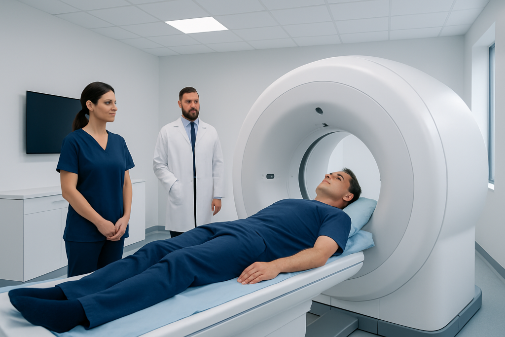
A shoulder MRI uses magnetic fields and radio waves rather than X-rays or radiation. It is particularly useful for evaluating the rotator cuff, labrum, cartilage, ligaments and muscles.
Key Takeaways
- A shoulder MRI uses magnetic fields and radio waves rather than X-rays or radiation.
- It is particularly useful for evaluating the rotator cuff, labrum, cartilage, ligaments and muscles.
- The examination is painless, although remaining still in the scanner can be challenging for some people.
- Not every shoulder problem requires an MRI; a clinician selects imaging based on symptoms, examination findings and medical history.
- MRI results should be interpreted alongside a physical examination and, when needed, other imaging tests.
A shoulder MRI is a detailed imaging examination used to assess the bones, joints, tendons, muscles, cartilage and other soft tissues of the shoulder. It can help clinicians investigate persistent pain, weakness, instability, reduced movement or suspected injury without using ionizing radiation.
Shoulder MRI overview
A shoulder MRI, or magnetic resonance imaging scan, is a noninvasive test that creates highly detailed pictures of the shoulder. It uses a powerful magnetic field and radio waves to show structures that may not be clearly visible on standard X-rays, including tendons, muscles, ligaments, cartilage, nerves and fluid around the joint. It does not use ionizing radiation.
The shoulder is a complex joint made up of the upper arm bone, shoulder blade and collarbone. It also depends on the rotator cuff, a group of muscles and tendons that helps lift and rotate the arm. Because several structures work together in a small area, shoulder pain can have many possible causes. An MRI can provide useful information when symptoms, physical examination findings or initial imaging suggest a soft-tissue injury or a more detailed assessment is needed.
A shoulder MRI is not automatically required for every painful shoulder. Many common problems improve with appropriate rest, activity modification, physiotherapy and other conservative care. The referring clinician considers the person’s symptoms, how the problem began, examination findings, prior treatment and whether the scan result is likely to guide care.
What a shoulder MRI can show
One of the main uses of shoulder MRI is assessment of the rotator cuff. The scan may show tendon inflammation, degeneration, partial-thickness tears or full-thickness tears. It can also show changes in the associated muscles, such as shrinkage or fatty change, which may be relevant when planning treatment for a longstanding tear.
MRI can evaluate the labrum, a ring of cartilage that helps deepen and stabilize the shoulder socket. Labral injuries may occur after a dislocation, repetitive overhead activity or trauma. The scan may also identify cartilage loss, joint inflammation, fluid accumulation, bursitis, ligament injury, bone bruising, stress-related changes and some fractures that are difficult to see on plain X-rays.
In selected situations, a clinician may request an MR arthrogram rather than a routine shoulder MRI. For this test, contrast material is placed into the shoulder joint before imaging, which can improve visualization of certain labral tears, subtle instability-related injuries or some partial tendon tears. Whether this is appropriate depends on the clinical question and the individual’s history.
- Rotator cuff tears, tendinopathy and impingement-related changes
- Labral tears and signs of shoulder instability
- Bursitis, joint inflammation and fluid collections
- Cartilage damage and osteoarthritis-related changes
- Occult fractures, bone marrow changes and selected tumors or infections
Why a clinician may recommend the scan

A clinician may recommend a shoulder MRI when pain persists despite initial care, especially if it affects sleep, work, sport or daily activities. It may also be considered after a fall, direct blow, sudden pulling injury or shoulder dislocation. Weakness when lifting the arm, repeated episodes of the shoulder slipping out of place, or a marked loss of movement can also prompt further evaluation.
MRI is often used after X-rays when the concern involves soft tissues. X-rays are valuable for showing bone alignment, many fractures and arthritis, but they do not provide the same detail of tendons and cartilage. Ultrasound is another useful test for some rotator cuff conditions and can assess moving structures in real time; however, MRI may offer a broader view of the entire shoulder and deeper structures.
The reason for imaging should be clear before the appointment. A detailed symptom history helps the radiologist and treating clinician focus on the most relevant possibilities. Information such as the location of pain, whether symptoms began gradually or suddenly, prior injuries, dislocations, surgery and any history of cancer or infection should be shared with the healthcare team.
What happens during a shoulder MRI
Before the examination, the imaging team asks about medical conditions, prior operations, implanted devices and possible metal fragments. People are asked to remove metal objects such as jewelry, watches, hearing aids, removable dental appliances and clothing with metallic parts. The team provides instructions about clothing and personal belongings.
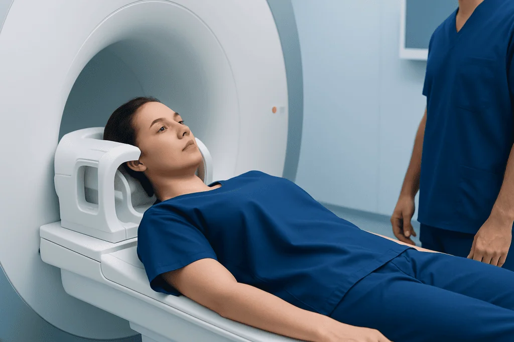
For the scan, the person lies on a movable table, usually on their back, with the arm positioned comfortably at the side or in a suitable position for imaging. A special device called a coil is placed around the shoulder to capture detailed images. The table moves into the MRI scanner, and the person must remain still while several image sequences are obtained.
The machine makes rhythmic knocking, tapping or humming sounds during scanning. Ear protection is provided. Staff can see and communicate with the person throughout the procedure, and an alert device is usually available if assistance is needed. The imaging itself is painless, although holding one position may cause temporary discomfort in an already painful shoulder.
Scan duration varies according to the protocol and whether contrast is used, but many shoulder MRI examinations take roughly 20 to 45 minutes. After a standard MRI, people can generally resume normal activities immediately unless they have received medication for anxiety or sedation, in which case they need to follow the care team’s instructions about transport and activities.
Contrast, safety and preparation
Most shoulder MRI scans do not require contrast. When contrast is needed, it may be administered into a vein or, for an MR arthrogram, into the joint. MRI contrast agents commonly contain gadolinium. Serious allergic reactions are uncommon, but patients should tell the team about previous contrast reactions, kidney disease, pregnancy, breastfeeding or other relevant health concerns.
MRI is considered safe for many people because it does not use radiation. However, the strong magnet means certain implants or metal objects require careful review. Some pacemakers, neurostimulators, cochlear implants, aneurysm clips, infusion pumps and older surgical devices may not be compatible with MRI. Many modern implants can be scanned under specific conditions, so it is important not to assume that an implant prevents MRI without checking its details.
Preparation is usually simple. Unless instructed otherwise, people can eat and drink normally before a routine shoulder MRI. Bringing previous imaging reports or providing information about where prior scans were performed may help comparison. Anyone with claustrophobia, severe pain when lying flat or difficulty remaining still should notify the imaging center in advance; practical measures, relaxation support or, in selected cases, medication may be arranged.
Understanding the results and next steps
A radiologist, a doctor specially trained to interpret medical images, reviews the MRI images and prepares a report. The report describes relevant findings, such as tendon changes, tears, joint degeneration, inflammation or bone abnormalities. The referring clinician then discusses what those findings mean in the context of the person’s symptoms and physical examination.
An MRI finding does not always explain pain or require treatment. For example, tendon wear and small tears can sometimes be present in people without symptoms, especially with increasing age. Conversely, a person may have significant pain even when imaging changes are mild. This is why treatment decisions should not be based on a scan alone.
Depending on the diagnosis, next steps may include activity adjustments, physiotherapy, pain management, an injection when clinically appropriate, follow-up assessment or referral to an orthopedic specialist. Surgery is generally considered only when there is a clear indication, such as certain acute injuries, ongoing functional limitation, recurrent instability or symptoms that have not improved with suitable nonsurgical care. Acibadem International’s multidisciplinary specialists and JCI-accredited hospitals can assess shoulder conditions and discuss appropriate treatment pathways for international patients.
When to seek medical care
Medical assessment is appropriate for shoulder pain that persists, repeatedly returns or limits normal sleep, self-care, work or sporting activities. It is also sensible to seek advice after an injury if there is weakness, reduced range of movement, a visible change in shoulder shape, a sensation of instability or difficulty using the arm. A clinician can determine whether an MRI or another test is needed.
Urgent medical care is important after a serious injury when the shoulder appears dislocated, the arm cannot be moved, there is severe swelling or deformity, or the hand becomes numb, pale, cool or weak. Prompt evaluation is also needed for shoulder pain accompanied by fever, redness, warmth, feeling unwell, unexplained weight loss, chest pain, shortness of breath or sudden sweating, as these symptoms may require assessment beyond the shoulder itself.
While waiting for medical advice after a minor injury, avoiding activities that sharply worsen pain and using a supported, comfortable arm position may help. People should avoid trying to force a suspected dislocation back into place or beginning strenuous strengthening exercises before the injury has been assessed.
Frequently asked questions
What does a shoulder MRI show?
A shoulder MRI can show the rotator cuff tendons, muscles, labrum, cartilage, ligaments, joint fluid and bones in detailed cross-sectional images. It may help identify tears, inflammation, degeneration, instability-related injury, cartilage damage or hidden bone injury. Results are interpreted together with symptoms and a physical examination.
Is a shoulder MRI painful?
The MRI scan itself is painless. However, lying still with a painful shoulder in one position may be uncomfortable, and the scanner makes loud sounds. The imaging team can help with positioning and provides hearing protection.
How long does a shoulder MRI take?
A routine shoulder MRI often takes about 20 to 45 minutes, although the exact time varies by scan protocol and equipment. An examination using contrast or an MR arthrogram may take longer because of additional preparation. The imaging center can provide more specific timing before the appointment.
Does a shoulder MRI use radiation?
No. MRI uses magnetic fields and radio waves, not X-rays or ionizing radiation. This makes it different from CT scans and conventional X-rays, although MRI still requires safety screening because of the strong magnet.
Do people need contrast for a shoulder MRI?
Many shoulder MRI scans are completed without contrast. Contrast may be recommended when the clinician needs more detail about certain labral injuries, inflammation, infection, tumors or postoperative changes. The decision depends on the reason for the examination and the person’s medical history.
Can a shoulder MRI detect a rotator cuff tear?
Yes. MRI is commonly used to assess rotator cuff tendons and can show many partial and full-thickness tears, as well as inflammation and muscle changes. The scan helps describe the injury, but treatment depends on symptoms, function, tear features and individual goals.
References
- American College of Radiology
- Radiological Society of North America
- National Institute of Arthritis and Musculoskeletal and Skin Diseases
- NHS
- Mayo Clinic
This article is for general information only and is not a substitute for professional medical advice. Please consult a qualified doctor about your individual situation.
Joint and spine care in Turkey — expert assessment & treatment
JCI-accredited · board-certified surgeons · reply within 24h
Add us as a Preferred Source to see more of our trusted health content across Google Search, AI Overviews and Discover.

More from the Health Library



Related Specialists

Assoc. Prof. Dr. Fatih Büyükcam
Emergency Service
Fzt. Alperen Mustafa Oğuz
Physical Medicine & Rehabilitation
Fzt. Ali Demir
Pediatric Rehabilitation
Dr. Süleyman Büber
Clinical Laboratory