Slit Lamp Exam — Explained by Medical Evidence, Not Myths
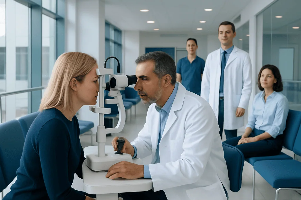
A slit lamp exam is usually quick, noninvasive, and painless. It allows detailed assessment of the eyelids, cornea, iris, lens, and sometimes the retina and optic nerve.
Key Takeaways
- A slit lamp exam is usually quick, noninvasive, and painless.
- It allows detailed assessment of the eyelids, cornea, iris, lens, and sometimes the retina and optic nerve.
- Eye drops may be used to numb the eye, add dye, or widen the pupils depending on what needs to be examined.
- The test can help evaluate symptoms such as redness, pain, blurred vision, light sensitivity, or a sensation of something in the eye.
- Temporary blurred near vision and light sensitivity can occur after pupil-dilating drops.
- Sudden vision changes, severe eye pain, or an eye injury require prompt medical assessment.
A slit lamp exam is a close-up examination of the eye using a bright, narrow beam of light and a microscope. It helps an ophthalmologist or optometrist assess eye structures, identify changes that may need treatment, and monitor known eye conditions.
Slit Lamp Exam: an evidence-based overview
A slit lamp exam is an eye examination performed with a binocular microscope and an adjustable, high-intensity beam of light. The focused beam creates an illuminated “optical section” through transparent tissues of the eye, allowing the clinician to view layers that cannot be assessed as clearly with a standard light alone. It is a routine part of many comprehensive eye examinations and is not a surgery, scan, or treatment.
The examination can reveal signs of irritation, infection, injury, dryness, inflammation, cataract, and other eye changes. It may also support the assessment of conditions affecting the eye pressure, retina, or optic nerve when combined with other tests. A normal result is reassuring, but the meaning of any finding depends on symptoms, eye history, visual testing, and the rest of the examination.
Despite its name, the slit lamp does not touch the eye during the basic examination. The person sits at the instrument, resting their chin and forehead on supports while the clinician directs the light and looks through the microscope. Additional lenses, eye drops, or gentle contact techniques may be used in selected situations.
What parts of the eye does a slit lamp examine?
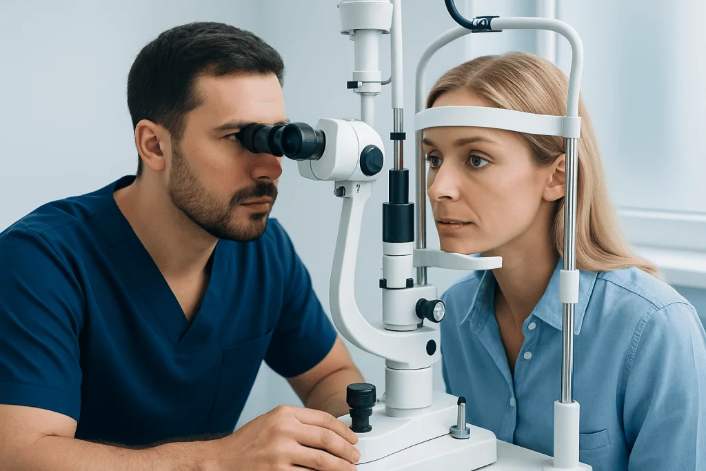
The slit lamp is particularly useful for examining the front, or anterior, portion of the eye. This includes the eyelids and eyelashes, the conjunctiva (the clear membrane over the white of the eye), the tear film, and the cornea. The clinician can also inspect the anterior chamber, which is the fluid-filled space behind the cornea, as well as the iris, pupil, and natural lens.
These structures can show subtle findings such as corneal scratches, contact lens-related changes, inflammation, abnormal cells in the eye fluid, deposits on the cornea, or early lens clouding. Looking at these features closely helps clinicians distinguish between conditions that can feel similar, such as dry eye, allergy, infection, and inflammation.
With pupil-dilating drops and a special hand-held or contact lens, the slit lamp can also help examine the vitreous, retina, macula, and optic nerve at the back of the eye. This does not replace every retinal test, but it is an important way to look for changes linked with diabetes, high blood pressure, retinal tears, and optic nerve disorders.
Why might an eye specialist recommend this test?
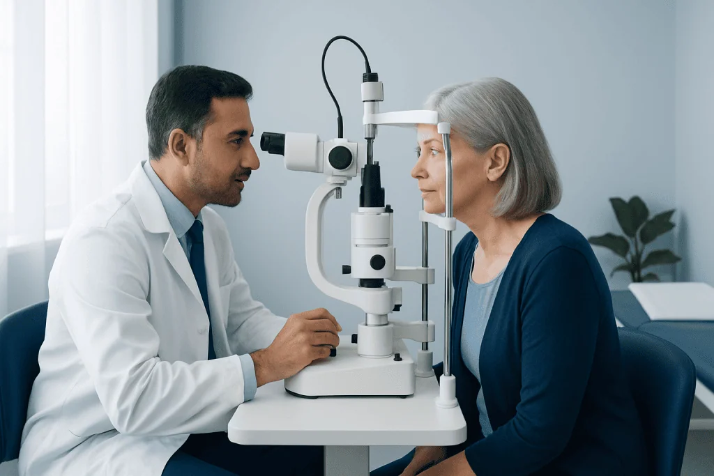
A slit lamp exam may be recommended during a routine eye check, before or after certain eye procedures, or when a person has new eye symptoms. It is commonly used to investigate red eye, irritation, tearing, discharge, itchiness, light sensitivity, blurry vision, floaters, flashes, or a feeling that there is grit in the eye. It also helps assess an eye injury, including possible foreign material on the eye surface.
For people with known eye conditions, the test can help monitor response to care and identify complications. For example, clinicians may use it when following cataracts, dry eye disease, corneal disease, uveitis, contact lens concerns, or glaucoma risk. A slit lamp examination is only one component of glaucoma assessment; eye pressure measurement, optic nerve evaluation, visual field testing, and imaging may also be appropriate.
The test is also useful when medication or a general health condition may affect the eyes. Diabetes, autoimmune disorders, skin conditions involving the eyelids, and some medicines can be associated with eye-surface or internal eye changes. The specialist will decide which additional tests are useful based on the person’s needs rather than using the same testing plan for everyone.
What happens during a slit lamp exam?
Before the examination, the clinician may ask about symptoms, current medicines, contact lens use, allergies, past eye injuries, surgery, and relevant health conditions. If the person wears contact lenses, they may be asked to remove them. Glasses are usually removed for the examination, although they should be brought to the appointment if vision testing is also planned.
The person sits facing the device and places their chin on a chin rest and forehead against a bar. The clinician adjusts the height and focus, then asks the person to look in different directions while examining each eye. The light can feel bright, but the standard examination should not be painful. Blinking normally between instructions is generally fine.
Depending on the reason for the visit, the clinician may place drops in the eyes. Fluorescein dye can highlight damage to the cornea or problems with the tear film under blue light. Numbing drops may be used before certain measurements or before removing a small surface foreign body. Dilating drops enlarge the pupil so the back of the eye can be examined more thoroughly.
Some examinations include gonioscopy, in which a special lens is gently placed on a numbed eye to view the drainage angle where the cornea and iris meet. This is often done when assessing glaucoma or a narrow angle. It may feel like mild pressure or awareness of the lens, but it should not cause sharp pain.
Understanding results and common findings
In many cases, the clinician can discuss initial findings immediately after the slit lamp exam. Results may be described as normal, or they may show a change that needs observation, treatment, further testing, or referral. A finding on a slit lamp exam is not always a diagnosis by itself; it must be considered alongside vision measurements, symptoms, and the medical history.
Common findings include dry or irritated eye surfaces, blepharitis affecting the eyelid margins, allergic changes, conjunctivitis, corneal staining, cataract changes, or signs of inflammation inside the eye. Some findings are minor and respond to simple care, while others require more urgent assessment. The clinician should explain what was seen, what it may mean, and the next appropriate step.
A dilated examination may detect changes in the retina or optic nerve that are not noticeable to the person at an early stage. If an abnormality is suspected, additional tests may include eye pressure measurement, retinal photography, optical coherence tomography, visual field testing, ultrasound, or blood tests. Further tests are chosen to clarify the cause rather than because the slit lamp examination itself is harmful or inconclusive.
Preparation, safety, and aftercare
Most people do not need special preparation for a slit lamp exam. It can be helpful to bring a list of medicines and eye drops, details of any past eye surgery, and the name of the contact lens products used. A person should tell the clinician if they are pregnant, have had a reaction to eye drops, have narrow-angle glaucoma, or have a history of eye injury or infection.
The examination has a very low risk of complications. Fluorescein dye may briefly make tears appear yellow and can temporarily stain soft contact lenses, which is why lenses may need to remain out for a period advised by the clinician. Numbing drops should only be used in the clinical setting unless specifically prescribed; repeated unsupervised use can damage the cornea.
Pupil-dilating drops can cause light sensitivity and blurred near vision for several hours, and occasionally longer depending on the drops used and the individual. Sunglasses may improve comfort outdoors. Because vision can be temporarily affected, it is sensible to ask in advance whether driving or detailed visual work may be difficult after the appointment and to arrange assistance if needed.
Rarely, dilation can trigger a sudden rise in eye pressure in people with anatomically narrow drainage angles. This is uncommon, but severe eye pain, headache, nausea, halos around lights, or marked redness after dilation should be assessed urgently. The clinician will consider relevant risk factors before using drops and can answer questions about their purpose.
When to seek medical care
A slit lamp exam should be arranged promptly for persistent red eye, increasing discomfort, unexplained blurred vision, light sensitivity, discharge, or a sensation of a foreign body that does not settle. People who wear contact lenses should seek timely assessment for a painful red eye, as certain corneal infections can progress quickly and should not be treated with leftover eye drops or by continuing lens wear.
Urgent medical care is appropriate for sudden loss or major reduction of vision, severe eye pain, a chemical splash, a penetrating eye injury, new flashes with many floaters, or a curtain-like shadow across vision. After a chemical exposure, the eye should be rinsed immediately with clean lukewarm water or saline while urgent help is arranged; rinsing should not be delayed to search for eye drops or other products.
Acibadem International’s multidisciplinary specialists and JCI-accredited hospitals can evaluate eye symptoms and conditions for international patients when diagnostic assessment or treatment is needed. For any ongoing concern, an ophthalmologist can explain whether a slit lamp exam, dilation, or another eye test is appropriate.
Frequently asked questions
Is a slit lamp exam painful?
A standard slit lamp exam is not painful because the microscope and light remain outside the eye. The light may feel bright, and the person may need to hold their eyes open briefly. If special drops or a contact lens are needed, the clinician explains the process and usually uses numbing drops first.
How long does a slit lamp exam take?
The basic examination often takes only a few minutes as part of a wider eye appointment. It can take longer if dilation, eye pressure checks, imaging, or other tests are needed. The total visit length depends on the reason for the examination and the findings.
Will eyes be dilated for every slit lamp exam?
No. A clinician may examine the front of the eye thoroughly without dilating the pupils. Dilation is used when a clearer view of the retina, macula, vitreous, or optic nerve is needed, such as during a comprehensive eye exam or when certain symptoms are present.
Can someone drive after a slit lamp exam?
Driving is usually possible after an exam that does not involve dilating drops, provided vision is normal and the person feels comfortable. After dilation, near vision and sensitivity to bright light can be affected for several hours. It is best to follow the clinician’s advice and consider arranging transport if vision feels blurred.
What does fluorescein dye do in an eye exam?
Fluorescein is a diagnostic dye that helps the clinician see the tear film and small areas of corneal surface damage under blue light. It is often used when dryness, a scratch, contact lens irritation, or a possible foreign body is suspected. The dye does not permanently stain the eye and typically clears quickly.
Can a slit lamp exam detect glaucoma?
A slit lamp exam can identify findings that are relevant to glaucoma assessment and can be used with a special lens to examine the eye’s drainage angle. However, glaucoma diagnosis and monitoring generally require several assessments, such as eye pressure measurement, optic nerve evaluation, imaging, and visual field testing. A clinician interprets these results together.
References
- American Academy of Ophthalmology
- National Eye Institute
- Mayo Clinic
- Merck Manual Consumer Version
This article is for general information only and is not a substitute for professional medical advice. Please consult a qualified doctor about your individual situation.
Explore treatments in Turkey — costs, top hospitals & a free quote
JCI-accredited · board-certified surgeons · reply within 24h
Add us as a Preferred Source to see more of our trusted health content across Google Search, AI Overviews and Discover.
Check your numbers in seconds
BMI, calories, due date, blood pressure and 30+ more clinical calculators — free, instant, doctor-reviewed ranges.

More from the Health Library



Related Specialists

Dr. Kadir Özyılmaz
Anesthesiology
Dr. Sertaç Uzel
Radiology
Fzt. Necla Aleyna Yiğit
Physical Medicine & Rehabilitation
Assoc. Prof. Dr. Mehmet Özgür Akkurt
Gynecology & Obstetrics