

ESWL
ESWL is a non-invasive treatment that uses focused shock waves to break kidney stones into small fragments that can pass naturally through urine.

Quick answer
ESWL is a non-invasive treatment that uses focused shock waves to break kidney stones into small pieces so they can pass naturally in the urine. At Acibadem in Turkey, ESWL is performed after imaging confirms the stone’s size and location, and patients are assessed to determine whether this approach or another stone treatment is more suitable.
When a Kidney Stone Becomes More Than a Passing Discomfort
A kidney stone can change from a distant concern into an urgent problem very quickly. For some people, it begins as a vague ache in the back or side. For others, it arrives suddenly as severe flank pain, nausea, urinary urgency, or blood in the urine. Even when the pain comes and goes, the uncertainty can be unsettling: Will the stone pass on its own? Could it block the kidney? Is surgery necessary? How long will recovery take, especially if you are traveling for care?
Extracorporeal shock wave lithotripsy, commonly called ESWL, is one of the least invasive treatments for selected kidney stones and some upper ureteral stones. It does not require an incision. Instead, focused shock waves are directed from outside the body toward the stone, breaking it into smaller fragments that can pass naturally through the urinary tract.
For international patients, the decision to have ESWL often involves more than choosing a medical technique. It requires confidence in the diagnosis, clarity about whether ESWL is the right option, careful planning around travel and recovery, and access to physicians who can explain alternatives when another approach may be safer or more effective. At Acibadem, kidney stone care is planned through modern diagnostic pathways and specialist evaluation, with attention to both the medical details and the practical needs of patients traveling from abroad.
What ESWL Is
ESWL stands for extracorporeal shock wave lithotripsy. “Extracorporeal” means the treatment is delivered from outside the body. “Lithotripsy” means breaking a stone into smaller pieces. During ESWL, a lithotripter generates controlled shock waves that are focused precisely on the kidney stone. These waves travel through the skin and body tissues and concentrate energy at the stone, creating cracks and fragmentation.
The goal is not to remove the stone in one piece. Instead, ESWL aims to reduce the stone into sand-like particles or small fragments that can pass through urine over days to weeks. Because the urinary tract must clear the fragments naturally, patient selection is important. ESWL tends to be most effective for stones of appropriate size, location, and composition, particularly when there is a clear pathway for fragments to pass.
ESWL is usually performed by a urologist and may be done as an outpatient or short-stay procedure, depending on the patient’s medical condition, the stone characteristics, and the hospital’s clinical protocol. It is considered non-invasive because there is no surgical incision and no instrument placed through the skin into the kidney. In many patients, this means less tissue trauma, a shorter recovery period, and a faster return to ordinary activities compared with more invasive stone procedures.
However, ESWL is not the best choice for every stone. Larger stones, very hard stones, stones in certain lower kidney locations, stones associated with infection or obstruction, and stones in patients with particular anatomical or medical factors may require another treatment. A high-quality ESWL program does not simply offer the procedure; it evaluates whether ESWL is the right procedure for that individual patient.
Who May Need ESWL
Patients may be considered for ESWL when imaging shows a kidney stone or selected upper ureteral stone that is unlikely to pass comfortably on its own, is causing symptoms, or may create future complications. Many patients have already had an emergency visit for renal colic, while others are diagnosed during evaluation for recurrent urinary symptoms, visible blood in the urine, or an incidental finding on ultrasound or CT scan.
Common symptoms that may lead to evaluation include:
- Flank pain, often on one side, sometimes radiating toward the lower abdomen or groin.
- Blood in the urine, which may be visible or detected only on laboratory testing.
- Nausea or vomiting, especially during intense stone pain episodes.
- Frequent or urgent urination, particularly if the stone is moving down the urinary tract.
- Burning during urination, which may also suggest infection and requires prompt assessment.
- Recurrent urinary tract infections, in selected cases related to stones.
Diagnosis usually begins with a clinical history, physical examination, urine testing, blood tests to assess kidney function and infection markers, and imaging. Ultrasound can identify many kidney stones and signs of swelling in the kidney. A non-contrast CT scan is often the most detailed test for determining stone size, density, number, and location. In some patients, additional imaging is used to understand urinary anatomy or plan treatment more precisely.
ESWL may be considered when the stone is visible on the imaging method used during treatment, when its size and density make fragmentation likely, and when the patient can safely undergo the procedure. Patients taking blood thinners, people with certain bleeding disorders, pregnant patients, and some patients with untreated infection or significant obstruction may not be immediate candidates. In these situations, the care team may adjust medications, treat infection first, place a temporary stent, or recommend another stone treatment.
Conditions and Indications ESWL Addresses
ESWL is used primarily for urinary stones located in the kidney and, in selected cases, in the upper ureter. The urinary tract includes the kidneys, ureters, bladder, and urethra. Stones form when minerals and salts in the urine crystallize and grow. The most common types include calcium oxalate, calcium phosphate, uric acid, struvite, and cystine stones. Stone composition can influence both treatment planning and prevention after treatment.
Typical indications for ESWL include:
- Kidney stones of suitable size that are causing pain, bleeding, recurrent symptoms, or risk of future obstruction.
- Upper ureteral stones that are appropriately located and visible for targeting.
- Stones in patients who prefer a non-incisional approach, when medically appropriate.
- Recurrent stone disease, when ESWL is suitable and prevention planning is also needed.
- Stones that have not passed with observation or medication-supported passage.
The procedure may be less effective for very large stones, stones with high density on CT, cystine stones, some calcium oxalate monohydrate stones, and stones in lower pole kidney calyces where gravity and anatomy may make fragment passage more difficult. In these cases, ureteroscopy or percutaneous nephrolithotomy may be recommended instead. Sometimes a staged plan is appropriate, particularly for patients with multiple stones or complex stone disease.
For patients traveling internationally, this distinction matters. The ideal treatment is not always the least invasive treatment; it is the treatment that best matches the stone and the patient. A careful review of imaging before travel can help determine whether ESWL is reasonable, whether another procedure should be planned, and whether additional tests are needed before arrival.
How ESWL Is Performed
Preparation Before the Procedure
Preparation begins with confirming that ESWL is appropriate. The urologist reviews the stone’s size, location, density, and visibility on imaging. Blood and urine tests are used to assess kidney function, bleeding risk, and signs of infection. If bacteria are present in the urine, antibiotics may be required before any stone procedure because treating an infected urinary system under pressure can be dangerous.
Patients are asked about medications, especially blood thinners, aspirin-containing products, anti-inflammatory drugs, supplements that may increase bleeding risk, and medicines for diabetes or blood pressure. Some medications may need to be paused or adjusted under physician guidance. Patients should never stop anticoagulants without a plan approved by their treating doctors, particularly if they have a heart valve, atrial fibrillation, prior stroke, or history of blood clots.
Fasting instructions depend on the type of anesthesia or sedation planned. ESWL may be performed with analgesia, sedation, regional anesthesia, or general anesthesia depending on clinical protocol, patient comfort, stone factors, and medical history. The purpose is to keep the patient comfortable and still enough for accurate targeting.
During ESWL
On the day of treatment, the patient is positioned on the ESWL table so the stone can be aligned with the shock wave focus. The treatment team uses imaging guidance, commonly ultrasound, X-ray fluoroscopy, or a combination, to localize the stone. Accurate targeting is essential because the shock waves must concentrate on the stone while minimizing unnecessary exposure to surrounding tissue.
A water cushion, gel interface, or similar coupling system allows shock waves to travel efficiently from the device into the body. The procedure begins with lower-energy pulses in many protocols, gradually increasing intensity as tolerated. The urologist and technical team monitor stone targeting throughout treatment, making adjustments for breathing movement or changes in stone position.
Patients typically hear repetitive clicking or tapping sounds from the lithotripter. If awake or lightly sedated, they may feel a tapping sensation in the back or flank. The number of shock waves and total treatment time vary according to the stone, patient tolerance, and clinical plan. Many ESWL sessions take less than an hour for the shock wave delivery itself, with additional time for preparation, anesthesia, monitoring, and recovery.
Technology Used in ESWL Planning and Treatment
Modern ESWL care relies on more than the shock wave generator. Imaging is central to selecting patients, planning treatment, and maintaining accurate targeting. CT imaging can help estimate stone density and identify features that may predict fragmentation. Ultrasound and fluoroscopic guidance help the team locate the stone during the procedure. Digital imaging systems and real-time monitoring support precision, while anesthesia monitoring helps maintain patient safety throughout sedation or anesthesia.
In some patients, a ureteral stent may be placed before or after ESWL. A stent is a thin internal tube that helps urine drain from the kidney to the bladder. It may be recommended if there is swelling, infection risk, a solitary kidney, a larger stone burden, or concern that fragments could temporarily block the ureter. Not all ESWL patients need a stent; the decision is individualized.
Immediately After the Procedure
After ESWL, patients are monitored as sedation or anesthesia wears off. Mild flank soreness, skin redness or bruising, urinary frequency, and blood-tinged urine are common for a short period. The medical team provides pain control instructions, hydration guidance, activity recommendations, and warning signs that require urgent attention.
Stone fragments may begin passing within days, but clearance can take several weeks. Some patients notice sandy particles in the urine. Others pass small fragments with cramping discomfort similar to a mild stone episode. A urine strainer may be recommended so fragments can be collected for laboratory analysis. Knowing the stone composition is valuable for prevention planning, especially for patients with recurrent stones.
Follow-Up and Recovery
Follow-up imaging is used to evaluate whether the stone has fragmented and whether fragments are clearing. The timing and type of imaging depend on the stone and the patient’s symptoms. Some patients need only observation and prevention counseling after successful fragmentation. Others may need medication to support fragment passage, repeat ESWL, ureteroscopy, or stent management if fragments remain or obstruction develops.
Most patients return to light activities quickly, often within a few days, but recovery is individualized. People traveling internationally should plan enough time for post-procedure monitoring and early follow-up, especially if their stone is larger, they have a stent, or they have a history of infection or complex stone disease.
Why Acting Early Matters
Not every kidney stone requires immediate treatment. Small stones may pass on their own, and some non-obstructing stones can be monitored. Yet delaying evaluation can be risky when a stone is causing obstruction, infection, recurrent severe pain, declining kidney function, or repeated emergency visits.
A stone blocking urine flow can cause pressure to build up in the kidney. Over time, persistent obstruction may impair kidney function. If infection occurs behind an obstruction, the situation can become urgent and potentially serious. Fever, chills, severe weakness, uncontrolled vomiting, or worsening pain with urinary obstruction should be assessed promptly.
Early specialist evaluation also expands treatment options. A stone that is suitable for ESWL at one stage may become more complicated if it moves, grows, causes infection, or leads to swelling of the kidney. Timely imaging and laboratory testing help determine whether observation, ESWL, ureteroscopy, percutaneous treatment, or emergency drainage is the safest path.
For international patients, early planning is particularly important. Sharing recent imaging and laboratory results before travel allows the medical team to advise on suitability, identify red flags, and coordinate the appropriate sequence of care. This can reduce avoidable delays and help patients travel with a clear medical plan.
Benefits of ESWL
When ESWL is well matched to the patient and the stone, it offers several meaningful advantages.
| Benefit | What It Means for You |
|---|---|
| No surgical incision | Shock waves are delivered from outside the body, which usually means less tissue trauma than incisional or percutaneous procedures. |
| Short recovery for many patients | Many people resume light daily activities within a few days, depending on pain, fragment passage, and overall health. |
| Outpatient or short-stay treatment | ESWL can often be planned without a long hospital stay, although monitoring and follow-up remain important. |
| Preserves other options | If fragments do not clear completely, additional treatments such as repeat ESWL or ureteroscopy may still be considered. |
| Useful for selected kidney stones | For appropriately sized and located stones, ESWL can reduce the stone burden while avoiding instruments inside the kidney. |
| Supports stone prevention planning | Passing and collecting fragments can allow laboratory analysis, helping guide diet, hydration, and medication strategies to reduce recurrence risk. |
Recovery Timeline After ESWL
Recovery varies, but the following timeline reflects what many patients can expect after an uncomplicated ESWL procedure.
| Time Period | What Patients Can Expect |
|---|---|
| Day 1 | Observation after sedation or anesthesia, mild flank discomfort, possible blood in the urine, and instructions for hydration, pain control, and activity. |
| First Week | Passing of sand-like particles or small fragments may begin. Some cramping, urinary urgency, or intermittent discomfort can occur as fragments move. |
| First Month | Follow-up imaging may be arranged to assess fragmentation and clearance. Ongoing symptoms may require medication, stent evaluation, or additional treatment planning. |
| Longer Term | Prevention becomes the focus, including fluid intake, dietary changes, stone analysis, metabolic evaluation when indicated, and monitoring for recurrence. |
What Influences the Success of ESWL
ESWL has been used for decades and can be highly effective in carefully selected patients, but outcomes vary. A good result depends on the stone, the patient’s anatomy, the technology used for targeting, the experience of the treatment team, and the body’s ability to clear fragments afterward.
Stone size is one of the most important factors. Smaller stones generally fragment and clear more easily than larger stones. A larger stone burden may require staged treatment or a different technique. Stone location also matters. Stones in the renal pelvis or upper portions of the kidney may clear more readily than lower pole stones, where fragments may remain in a dependent calyx.
Stone density and composition affect fragmentation. CT imaging can provide clues about how hard a stone may be. Some stones break apart more easily, while others resist shock wave energy. If a stone is known or suspected to be very hard, ureteroscopy with laser lithotripsy or another approach may be more appropriate.
Patient anatomy and body habitus can influence targeting and energy delivery. The distance between the skin and the stone, skeletal anatomy, kidney position, and urinary drainage patterns may all affect treatment. Certain anatomical variations can make fragment clearance more difficult.
Accurate imaging during treatment is essential. If the stone cannot be clearly seen or consistently targeted, ESWL may be less effective. Respiratory movement, bowel gas, and stone visibility can all affect targeting. This is why pre-treatment imaging review and intra-procedure monitoring are important parts of quality care.
Fragment passage is the final step. Even when a stone fragments well, pieces must move through the ureter and exit in the urine. Hydration, movement, medication in selected patients, and time can help. However, fragments can occasionally cluster and obstruct the ureter, causing pain or swelling. Patients are therefore given clear instructions about symptoms that require urgent evaluation, including fever, severe persistent pain, inability to urinate, or ongoing vomiting.
Prevention after treatment is also part of a successful outcome. Kidney stones commonly recur, particularly when the underlying cause is not addressed. Depending on the patient’s history, evaluation may include stone analysis, blood tests, urine studies, and a 24-hour urine collection. Prevention may involve increasing fluid intake, reducing excess salt, adjusting animal protein intake, maintaining normal dietary calcium, treating uric acid abnormalities, or using medication when appropriate.
Why International Patients Choose Acibadem for ESWL
International patients considering ESWL often want two things at the same time: a technically sound urology treatment and a care environment that understands the realities of traveling for medical care. At Acibadem, ESWL is approached as part of a broader kidney stone program, not as an isolated procedure. The aim is to confirm the diagnosis, choose the most appropriate treatment, manage symptoms safely, and help reduce the risk of recurrence.
Care is delivered in JCI-accredited hospitals, with clinical processes designed around patient safety, infection control, anesthesia standards, imaging quality, and coordinated follow-up. For kidney stone patients, this structure matters because ESWL decisions often depend on careful interpretation of imaging, laboratory values, medical history, and any previous stone treatments.
Acibadem’s urology teams work with radiology, anesthesiology, nephrology, emergency medicine, and laboratory specialists when needed. In more complex situations, cases may be reviewed through specialist boards or multidisciplinary discussion, particularly when a patient has recurrent stones, a solitary kidney, impaired kidney function, infection risk, complex anatomy, or multiple treatment options. This collaborative approach helps ensure that ESWL is recommended when it is medically suitable, and that alternatives are discussed when another option offers a better balance of effectiveness and safety.
Advanced diagnostic pathways support individualized planning. Patients may undergo ultrasound, CT-based stone assessment, urine and blood testing, infection evaluation, and metabolic assessment when indicated. During treatment, imaging guidance helps localize and monitor the stone. Anesthesia teams evaluate comfort and safety, especially for patients with heart disease, lung disease, obesity, sleep apnea, diabetes, or prior anesthesia concerns.
For patients coming from the United States, Europe, the Middle East, Africa, or other regions, Acibadem International provides dedicated support before, during, and after the hospital visit. Services may include coordination of medical records, appointment scheduling, language interpretation in more than 20 languages, assistance with hospital logistics, and communication with clinical teams. This support is especially valuable for ESWL because imaging review before arrival can help determine whether the patient is likely to benefit from the procedure or should be evaluated for another treatment.
Personalized treatment planning is central. Some patients need ESWL alone. Others may need antibiotics first, a temporary stent, ureteroscopy, metabolic evaluation, or a prevention plan. A patient with a small kidney stone and normal kidney function has different needs from someone with recurrent stones, chronic kidney disease, or an obstructing stone with infection risk. The treatment plan should reflect those differences clearly.
Equally important is communication. Patients traveling abroad need to understand what will happen, how long they may need to stay, what symptoms are expected, which symptoms are urgent, and how follow-up will be handled after returning home. Acibadem’s international care model is designed to make these steps understandable and organized, while keeping medical decisions in the hands of the treating physicians and the patient.
Taking the Next Step
ESWL can be an effective, non-invasive option for selected kidney stones, but the right decision begins with accurate diagnosis and careful review of the stone’s characteristics. If you have been told you have a kidney stone, have recurrent stone pain, or are unsure whether you need ESWL, ureteroscopy, observation, or another treatment, a specialist evaluation can help clarify the safest path forward.
International patients may request a consultation or second opinion by sharing recent imaging reports, CT or ultrasound images, laboratory results, medication lists, and a summary of symptoms. With this information, the urology team can assess whether ESWL appears appropriate, what additional tests may be needed, and how treatment and follow-up could be planned around travel.
Kidney stone pain can be frightening, but it is also a condition with well-established diagnostic and treatment pathways. With timely evaluation, careful patient selection, and structured follow-up, many patients are able to treat the current stone and begin a practical plan to reduce future risk.
This information is general and is not a substitute for professional medical advice. Diagnosis and treatment decisions should always be made with a qualified physician who can evaluate your individual condition.
Preparation
- Before ESWL, imaging tests such as ultrasound, X-ray, or CT may be used to confirm the stone size and location. Blood and urine tests may be requested, and patients may need to stop blood-thinning medications as advised by the doctor. Fasting may be required if sedation or anesthesia is planned.
Aftercare
- Patients are encouraged to drink plenty of water to help pass stone fragments after the procedure. Mild pain, bruising, or blood in the urine can occur temporarily, and prescribed pain medication may be used if needed. Follow-up imaging may be scheduled to confirm that the stone has cleared.
Turkey vs UK, Germany & USA
ESWL is often compared internationally because the technique is well established, but the overall patient journey can vary by country, hospital model and package scope. The final cost depends on clinical findings, stone characteristics and what is included before, during and after treatment.
When comparing ESWL abroad, patients should look beyond the procedure fee and review the hospital setting, specialist experience, diagnostic work-up, follow-up plan and travel support.
| Factor | Turkey | UK | Germany | USA |
|---|---|---|---|---|
| Price drivers | Often offered as an international patient package; cost depends on imaging, anaesthesia or sedation needs, stone complexity and repeat-session planning. | Costs vary between public and private pathways; private care may include separate fees for consultation, imaging, hospital use and follow-up. | Costs are influenced by hospital category, urology expertise, diagnostic imaging and whether additional endoscopic treatment is needed. | Itemised billing is common; facility, specialist, imaging, anaesthesia and follow-up charges may be billed separately. |
| Hospital and surgeon factors | International departments commonly coordinate urology consultation, imaging review, scheduling and aftercare; JCI-accredited hospitals may appeal to patients seeking recognised quality systems. | Access depends on local referral pathways or private provider availability; surgeon choice may vary by setting. | Specialist urology centres may offer detailed diagnostic work-up and structured care pathways; availability depends on provider and region. | Wide choice of hospitals and urologists; patient experience and billing structure can vary significantly by provider and insurance status. |
| Quality and accreditation | Patients can choose hospitals with international accreditation, multidisciplinary support and medical interpreters. | Regulated healthcare standards apply; private hospitals may have their own quality frameworks and consultant-led care. | Strong regulatory and specialist training frameworks; hospital quality indicators may be reviewed before booking. | Accreditation and quality measures vary by institution; patients often review hospital credentials and network status. |
| Typical waiting times | International scheduling may be arranged promptly after records are reviewed, depending on medical suitability and device availability. | Public pathway timing may depend on referral urgency and local capacity; private appointments may be arranged more flexibly. | Timing depends on specialist availability, diagnostics and hospital scheduling. | Scheduling may be faster in private or insured settings, but depends on provider availability and approval processes. |
| Travel and language logistics | International patient teams may assist with airport transfers, hotel coordination, interpreters and appointment planning. | Convenient for residents; international patients may need to arrange travel, accommodation and language support separately. | Travel planning and language support vary by hospital; larger centres may offer international desks. | Long-distance travel and accommodation can add complexity; language support depends on the provider. |
| What a package typically includes | May include urology assessment, imaging review, ESWL session planning, nursing care, interpreter support and follow-up guidance. | Private packages may be limited or itemised; patients should confirm whether tests, anaesthesia, medications and follow-up are included. | Packages may include consultation and procedure, while diagnostics and follow-up may be quoted separately. | Packages are less common; patients should clarify facility, physician, anaesthesia, imaging and aftercare billing. |
What affects your final cost
- Stone location, density and overall complexity.
- Whether ESWL is suitable or another treatment is recommended.
- Need for CT, ultrasound, X-ray, blood tests or urine tests.
- Use of pain control, sedation or anaesthesia support.
- Need for repeat ESWL sessions or additional procedures such as stent placement.
- Hospital accreditation, urologist experience and international patient services.
- Travel, accommodation, interpreter support and follow-up arrangements.
Compare your options
Kidney stone treatment is selected according to stone features, symptoms, kidney function, infection risk and patient health. Suitability for ESWL or any alternative is decided by a specialist after assessment and imaging review.
| Option | What it is | Typical use | Key considerations |
|---|---|---|---|
| Observation and supportive care | Monitoring with hydration advice, pain control and follow-up imaging when appropriate. | May be considered for stones likely to pass naturally and when there are no warning signs. | Requires medical supervision; urgent care may be needed for fever, severe pain, blockage or kidney function concerns. |
| Medical expulsive therapy | Medication may be used to help the urinary tract relax and support stone passage. | Often considered for selected ureteric stones when the specialist expects natural passage may be possible. | Not suitable for every patient; monitoring is important to confirm progress and avoid complications. |
| ESWL | Focused shock waves are delivered from outside the body to fragment the stone so pieces can pass in urine. | Commonly considered for selected kidney or upper urinary tract stones with favourable size, location and density. | Non-invasive, but may require repeat sessions; fragment passage can cause discomfort and follow-up imaging is usually needed. |
| Ureteroscopy with laser lithotripsy | A thin scope is passed through the urinary tract to reach the stone and break it with laser energy. | May be preferred when stones are less suitable for ESWL, are located in the ureter or require direct visual treatment. | More invasive than ESWL; may involve anaesthesia and temporary stent placement. |
| Percutaneous nephrolithotomy | A minimally invasive surgical route is created through the skin into the kidney to remove stone material. | Often considered for complex, large or multiple kidney stones when other methods are unlikely to be sufficient. | Requires hospital-based surgical care and a more structured recovery plan. |
| Stent or drainage procedures | A ureteric stent or kidney drainage tube may be placed to relieve obstruction or support urine flow. | Used when there is blockage, infection risk, severe symptoms or as part of a staged treatment plan. | May be temporary and followed by definitive stone treatment once the patient is stable. |
Trusted care for international patients
General information only — not medical or financial advice. Final costs depend on the factors above and your individual case; request a free, personalised quote.
Doctors Performing This Treatment

Prof. Dr. A. Bülent Oktay
Urology
Prof. Dr. Ali Rıza Kural
Urology
Prof. Dr. Ali Tekin
Urology
Prof. Dr. Burak Turna
Urology
Prof. Dr. Burak Çıtamak
Urology
Prof. Dr. Burak Özkan
Urology
Prof. Dr. Bülent Soyupak
Urology
Prof. Dr. Can Öbek
Urology
Prof. Dr. Cem Akbal
Urology
Prof. Dr. Engin Kaya
Urology
Prof. Dr. Enis Rauf Coşkuner
Urology
Prof. Dr. Fuat Demirel
Urology
Prof. Dr. Hakan Özveri
Urology
Prof. Dr. Hamdi Karakayalı
General Surgery
Prof. Dr. K. Fehmi Narter
Urology
Prof. Dr. Levent Türkeri
Urology
Prof. Dr. Lütfi Tunç
Urology
Prof. Dr. Murat Şamlı
Urology
Prof. Dr. Mustafa Sofikerim
Urology
Prof. Dr. Mustafa Uğur Altuğ
Urology
Prof. Dr. Ramazan Yavuz Akman
Urology
Prof. Dr. Sinan Zeren
Urology
Prof. Dr. Veli Yalçın
Urology
Prof. Dr. Ömer Öge
UrologyMedical Units
Available at These Hospitals






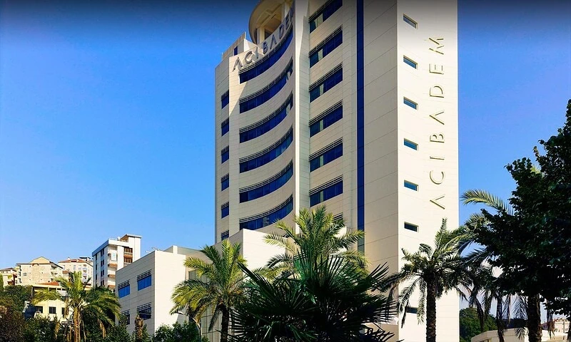


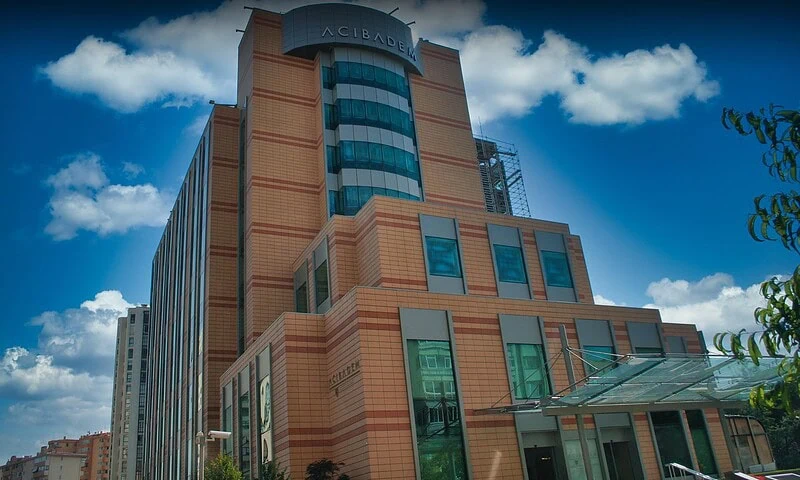

Frequently Asked Questions
What affects the cost of ESWL?
The cost is influenced by stone location and density, required imaging, specialist consultation, pain control or sedation needs, hospital setting, follow-up tests and whether repeat sessions or additional procedures are needed.
How can I get a personalised ESWL quote from Acibadem?
You can request a free consultation and share your medical records, imaging reports and current symptoms. A urology team can review suitability and provide a personalised treatment plan and quotation based on what your care requires.
Is ESWL always the lowest-cost kidney stone treatment?
Not necessarily. ESWL can be cost-effective for suitable stones, but if the stone is unlikely to fragment or pass, another option may be more appropriate. A specialist decides suitability after reviewing imaging and clinical factors.
What should I check before comparing ESWL packages?
Ask whether the quote includes consultation, imaging review, the ESWL session, medications, anaesthesia or sedation support, interpreter services, follow-up imaging and management of possible additional procedures.
Can travel and accommodation change the total cost?
Yes. Flights, hotel stays, transfers, companion travel, interpreter support and the length of stay can affect the total budget. International patient teams can help coordinate these logistics and clarify what is included.