Eye Melanoma Treatment: How It Works, Results and What to Expect

Eye melanoma, also called ocular or uveal melanoma, usually develops in the pigmented middle layer of the eye. Radiation therapy is commonly used for tumors that can be treated while preserving the eye.
Key Takeaways
- Eye melanoma, also called ocular or uveal melanoma, usually develops in the pigmented middle layer of the eye.
- Radiation therapy is commonly used for tumors that can be treated while preserving the eye.
- Surgery may remove the tumor or, in selected situations, the entire eye when it is the safest option.
- Treatment planning includes scans to check whether melanoma has spread, particularly to the liver.
- Regular eye and whole-body follow-up remains important after treatment, even when local tumor control is successful.
Eye melanoma treatment is tailored to the tumor’s size, location and behavior, with the goals of controlling the cancer, preserving vision when possible and protecting overall health. A specialist eye-cancer team helps patients understand each option, expected recovery and the need for long-term follow-up.
Overview: How Eye Melanoma Treatment Works
Eye melanoma treatment aims to destroy or remove melanoma cells while preserving the eye, vision and comfort whenever this can be done safely. The approach depends on where the tumor is located, its thickness and width, its growth pattern, whether it affects important structures such as the optic nerve or macula, and whether there is evidence of spread outside the eye.
Most eye melanomas arise from the uvea, the pigmented middle layer of the eye. This includes the choroid, ciliary body and iris. Uveal melanoma is different from melanoma of the eyelid or conjunctiva, which may be managed more similarly to skin or surface cancers. For patients with a diagnosis of melanoma, specialist assessment is essential because the subtype and site of disease affect treatment decisions.
For many localized tumors, focused radiation is the main eye-preserving treatment. Other options include laser-based treatment, surgery to remove a tumor, removal of the eye in selected cases, and systemic medicines when cancer has spread. Care is typically coordinated by ocular oncologists, radiation oncologists, medical oncologists, retinal specialists, pathologists and imaging specialists.
Treatment Planning and Candidacy
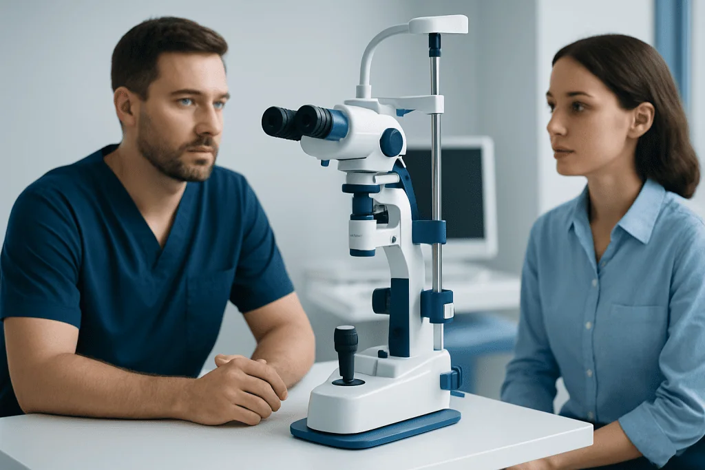
Before recommending treatment, the clinical team confirms the diagnosis and maps the tumor carefully. An ophthalmic examination may include dilated retinal assessment, ocular ultrasound, optical coherence tomography, fluorescein angiography and photographs of the eye. These tests help establish the tumor’s dimensions, location and effects on nearby tissues.
Patients also undergo staging to look for disease outside the eye. This often includes liver blood tests and imaging of the liver, as the liver is a common site of spread for uveal melanoma. Depending on individual findings, chest and abdominal imaging may also be recommended. In some cases, a small sample of tumor tissue is obtained for genetic testing, which can provide information about prognosis and follow-up planning.
Eye-preserving treatment may be suitable when the tumor is limited to the eye and can be targeted without unacceptable harm to critical structures. Removal of the eye may be considered for a very large tumor, an eye that is painful or has lost useful vision, extensive involvement of the optic nerve, or when other treatments are unlikely to provide adequate control. The recommendation should reflect both medical safety and the patient’s priorities.
Radiation Therapy for Eye Melanoma

Radiation therapy treats eye melanoma by delivering energy that damages cancer-cell DNA, preventing the cells from continuing to divide. The radiation is planned precisely to target the tumor while reducing exposure to surrounding tissues. The two commonly used approaches are plaque brachytherapy and carefully planned external-beam proton therapy, although availability and suitability vary by center and country.
In plaque brachytherapy, a small radioactive disc, called a plaque, is surgically placed on the outside wall of the eye directly over the tumor. It remains in place for several days while it delivers radiation to the tumor, then is removed in a second procedure. In proton beam therapy, external beams are shaped to the tumor and delivered over several treatment sessions. Some patients require placement of small markers on the outer eye before planning, helping the team target treatment accurately.
Radiation can often control the tumor without removing the eye. However, preserving the eye does not always mean vision will remain unchanged. The risk to vision depends particularly on whether the tumor or treatment field is close to the macula, optic nerve, lens or front of the eye. Patients should discuss both tumor-control goals and realistic visual expectations before proceeding.
For people considering a radiation-based approach, radiation therapy should be planned by specialists experienced in ocular tumors, as even small differences in anatomy can influence treatment accuracy and side effects.
Surgery, Laser Treatment and Systemic Therapy
Surgery may have different roles in eye melanoma treatment. In selected small or accessible tumors, an experienced ocular surgeon may remove the tumor while preserving the eye. This is technically complex and is not appropriate for every location or tumor type. Surgery may also be used to place and remove a radiation plaque or planning markers for proton therapy.
Enucleation is surgery to remove the entire eye. Although this can sound difficult, it may be the most appropriate and comfortable option when a tumor is large, has caused severe complications, or cannot be treated safely with eye-preserving methods. After healing, many people can be fitted with a custom ocular prosthesis. A prosthetic eye does not restore sight, but it can provide a natural appearance and supports eyelid shape.
Laser therapy may occasionally be used for selected small tumors or as an additional treatment after radiation. Techniques such as transpupillary thermotherapy use heat to damage tumor tissue. Laser treatment is not suitable as the only treatment for many melanomas and is chosen cautiously because complete tumor control must remain the priority.
If melanoma has spread beyond the eye, management is led by medical oncology and may include immunotherapy, targeted therapy in selected circumstances, clinical trials, local treatment for metastases or supportive care. Treatment is individualized according to the sites and extent of disease, overall health and tumor characteristics.
What to Expect: Procedure Steps and Recovery
The exact treatment day experience depends on the chosen method. For plaque brachytherapy, patients first undergo an operation under anesthesia to position the plaque. They may stay in hospital or be reviewed closely while it remains in place. A planned second operation removes the plaque after the prescribed treatment period. The eye can feel sore, red, watery or sensitive to light for a short time after either procedure.
For proton beam therapy, preparation may involve imaging, custom positioning and, in some cases, a short procedure to place markers. During radiation sessions, the patient lies in a carefully arranged position and is asked to keep the eye still, sometimes using a fixation target. The radiation itself is painless, and daily sessions are usually brief, though the planning process is detailed.
Recovery varies. Mild discomfort and surface irritation often improve over days to weeks, while changes within the eye can develop gradually over months or years. Vision may be temporarily blurred after treatment, and the final visual outcome may not be clear for some time. The clinical team provides individualized guidance about eye drops, activity, driving, work and follow-up appointments.
Long-term monitoring includes examinations of the treated eye to confirm tumor regression and manage complications early. Ongoing liver imaging and other surveillance are also important because treatment of the eye controls the local tumor but cannot eliminate the need to check for distant spread.
Benefits, Risks and Long-Term Outlook
The main benefit of local treatment is control of the melanoma in the eye. Modern eye-preserving therapies allow many patients to avoid removal of the eye, particularly when the tumor is detected at a treatable stage. The best option is not necessarily the one that preserves the most vision in the short term; it is the approach that offers appropriate cancer control while considering the person’s eye health and quality of life.
Possible complications vary by treatment and tumor location. Radiation-related effects can include dry eye, cataract, retinal damage, swelling in the macula, optic nerve damage, glaucoma, bleeding inside the eye and gradual vision loss. Surgery can lead to infection, bleeding, retinal detachment, pressure changes or changes in vision. Not everyone develops these complications, and many can be monitored or treated when identified early.
Even after successful treatment, a tumor can occasionally recur locally or spread elsewhere in the body. Prognosis is influenced by tumor size, location, cell type and genetic features, as well as whether spread is present. Patients may find it helpful to ask their clinicians how their individual findings affect follow-up, rather than relying on general prognostic information.
Emotional adjustment is also part of recovery. Changes in vision, uncertainty during surveillance and the possibility of eye removal can be challenging. Counseling, low-vision services, peer support and rehabilitation can help patients and families adapt practically and emotionally.
Follow-Up, Self-Care and When to Seek Medical Care
After treatment, patients should attend every scheduled eye examination and systemic surveillance visit. They should use prescribed drops exactly as instructed, avoid rubbing the treated eye, protect the eye from injury and tell the care team about all medicines and health changes. A balanced diet, regular activity as tolerated, sleep and avoiding tobacco support general health, although they do not replace medical follow-up.
Patients should contact their eye-care team promptly if they develop increasing eye pain, marked redness or swelling, a sudden drop in vision, flashes or a curtain-like shadow, new double vision, pus-like discharge, fever after a procedure, or persistent nausea and vomiting with eye pain. These symptoms do not always indicate a serious problem, but they require timely assessment.
Anyone with a new dark spot on the iris, unexplained blurred vision, visual-field loss, flashing lights or a change noticed during a routine eye examination should arrange an ophthalmology review. Eye melanoma may cause no symptoms, so routine dilated eye examinations remain valuable, especially for people with a known suspicious eye lesion.
Acibadem International’s multidisciplinary specialists and JCI-accredited hospitals assess and treat eye melanoma for international patients, coordinating ophthalmology, oncology, imaging and radiation care when needed.
Frequently asked questions
What is the most common treatment for eye melanoma?
Focused radiation therapy is commonly used for localized uveal melanoma, particularly when the goal is to control the tumor while preserving the eye. Plaque brachytherapy and proton beam therapy are two established approaches. The most appropriate option depends on the tumor’s size, position and effects on vision.
Can eye melanoma be cured without removing the eye?
Many localized eye melanomas can be treated successfully without removing the eye, often with targeted radiation. Whether this is appropriate depends on the tumor and the eye’s condition. Preserving the eye is possible in many cases, but vision may still be affected by the tumor or its treatment.
How long does recovery take after eye melanoma treatment?
Surface discomfort after a procedure may settle over days or weeks, while internal eye healing and vision changes can evolve over months. Some radiation-related effects can appear later, which is why long-term follow-up is essential. The treating team can provide a recovery plan based on the exact treatment used.
Does eye melanoma treatment hurt?
Procedures such as plaque placement and eye removal are performed with anesthesia, so patients should not feel pain during surgery. Radiation delivery itself is painless, although positioning and preparation can be tiring. Mild soreness, redness, dryness or light sensitivity may occur after treatment and can usually be managed with medical guidance.
Can eye melanoma spread after treatment?
Local treatment is designed to control the tumor in the eye, but uveal melanoma can sometimes spread elsewhere in the body. This risk is influenced by the tumor’s characteristics and is not determined solely by the local treatment method. Regular imaging and medical follow-up help detect changes as early as possible.
Will vision return after eye melanoma radiation?
Vision may improve if a tumor-related problem such as fluid buildup settles after treatment, but this is not guaranteed. Vision can also decline when the tumor or radiation affects the macula, optic nerve, lens or retina. An ocular oncology team can explain the likely visual outlook based on the tumor’s precise location.
References
- National Cancer Institute
- American Cancer Society
- American Academy of Ophthalmology
- Cancer Research UK
- National Comprehensive Cancer Network
This article is for general information only and is not a substitute for professional medical advice. Please consult a qualified doctor about your individual situation.
Melanoma in Turkey — costs, top hospitals & a free quote
JCI-accredited · board-certified surgeons · reply within 24h
Add us as a Preferred Source to see more of our trusted health content across Google Search, AI Overviews and Discover.

Medical Oncology Department
Medical treatment of cancer with chemotherapy, immunotherapy and targeted therapies under a multidisciplinary tumor board.
60 specialists in this unit