

Ultrasound Imaging: Abdomen, Pelvis, Thyroid, and Pregnancy Uses

Ultrasound imaging uses high-frequency sound waves, not X-rays, to create images from inside the body. Abdominal ultrasound can assess organs such as the liver, gallbladder, kidneys, pancreas, spleen, and major blood vessels.
Key Takeaways
- Ultrasound imaging uses high-frequency sound waves, not X-rays, to create images from inside the body.
- Abdominal ultrasound can assess organs such as the liver, gallbladder, kidneys, pancreas, spleen, and major blood vessels.
- Pelvic ultrasound helps evaluate the uterus, ovaries, bladder, prostate, and causes of pelvic pain or abnormal bleeding.
- Thyroid ultrasound can examine thyroid size, nodules, cysts, inflammation, and guide needle biopsy when needed.
- Pregnancy ultrasound checks fetal development, gestational age, heartbeat, placenta position, and some structural concerns.
- Preparation varies by scan type; some patients may need to fast or arrive with a full bladder.
Ultrasound imaging is a widely used diagnostic test that creates real-time images of organs, tissues, blood flow, and pregnancy development using sound waves. It is noninvasive, does not use ionizing radiation, and helps doctors evaluate many conditions in the abdomen, pelvis, thyroid, and during pregnancy.
Overview: What Is Ultrasound Imaging?
Ultrasound imaging, also called sonography, is a diagnostic test that uses high-frequency sound waves to produce pictures of structures inside the body. A handheld device called a transducer sends sound waves through the skin and receives echoes as they bounce back from organs and tissues. A computer then converts these echoes into moving images that can be viewed in real time.
Because ultrasound does not use ionizing radiation, it is commonly used for pregnancy imaging and for repeated follow-up when appropriate. It can show the shape, size, texture, and movement of many organs. With Doppler ultrasound, doctors can also assess the direction and speed of blood flow in selected blood vessels.
Ultrasound is often chosen because it is accessible, quick, and generally comfortable. It can help doctors decide whether symptoms need treatment, further imaging, laboratory tests, or a procedure such as image-guided biopsy or fluid drainage.
Abdominal Ultrasound Uses
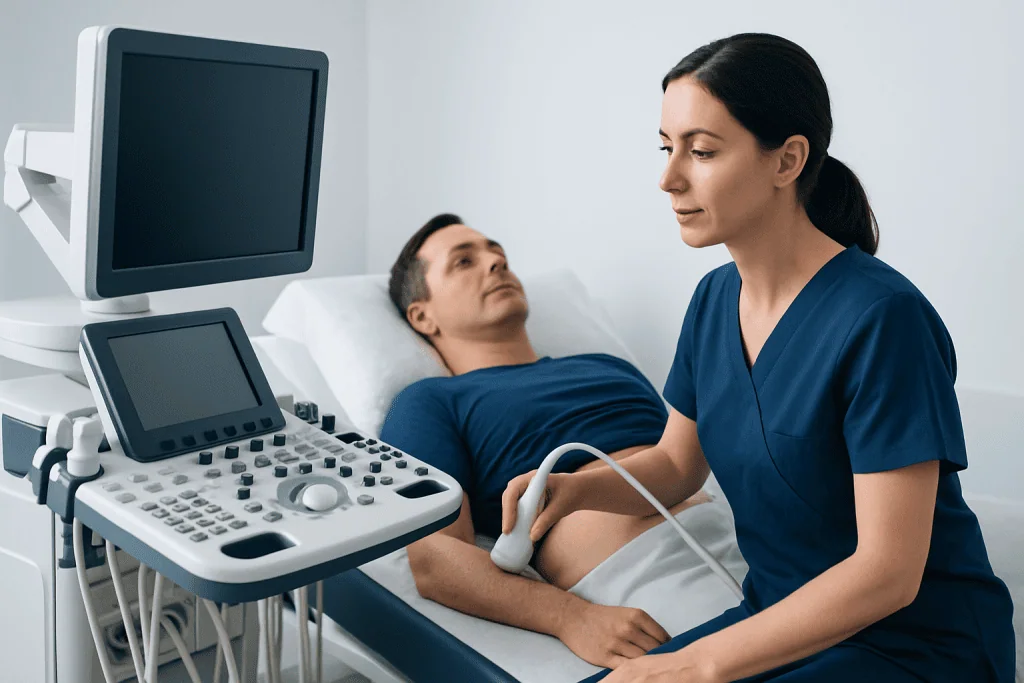
Abdominal ultrasound is used to examine organs and structures in the upper and lower abdomen. It may include the liver, gallbladder, bile ducts, pancreas, spleen, kidneys, bladder, abdominal aorta, and nearby blood vessels. Doctors may request it for abdominal pain, nausea, abnormal liver tests, kidney symptoms, suspected gallstones, swelling, or unexplained changes found during a physical examination.
For the gallbladder and bile ducts, ultrasound can help detect gallstones, inflammation, or blockage. For the liver, it can show changes in size, fatty infiltration, cysts, masses, or signs that may require additional evaluation. For the kidneys, it may detect stones, swelling from urine blockage, cysts, or structural changes.
Doppler ultrasound may be added when blood flow needs evaluation, such as in the portal vein, hepatic vessels, renal arteries, or abdominal aorta. In some patients, ultrasound can also screen or monitor an abdominal aortic aneurysm when clinically indicated. The exact scope of the examination depends on the symptoms and the doctor’s request.
Pelvic Ultrasound Uses
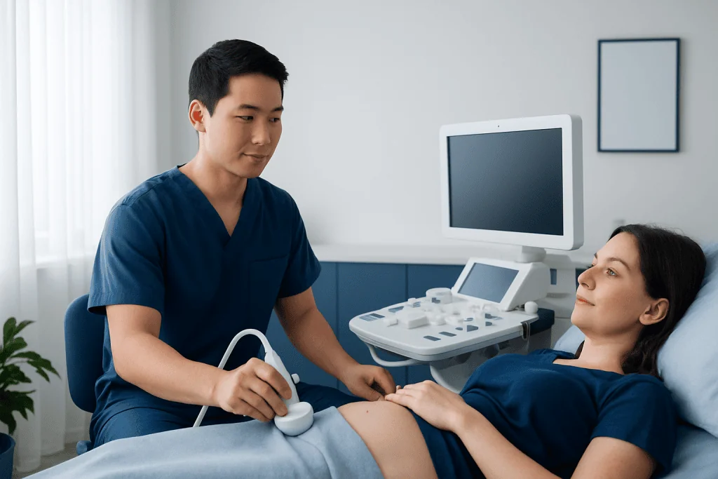
Pelvic ultrasound evaluates organs in the lower abdomen and pelvis. In women, it is commonly used to assess the uterus, endometrium, cervix, fallopian tube region, ovaries, and nearby pelvic structures. It may be recommended for pelvic pain, abnormal menstrual bleeding, suspected ovarian cysts, fibroids, fertility evaluation, or follow-up of known gynecologic conditions.
Pelvic ultrasound may be performed through the abdomen with a full bladder, through the vagina using a slim transvaginal transducer, or both. A transvaginal ultrasound often provides more detailed images of the uterus and ovaries because the transducer is closer to the pelvic organs. The patient’s comfort, age, medical history, pregnancy status, and clinical need are considered when choosing the approach.
In men, pelvic ultrasound can evaluate the bladder, prostate, seminal vesicles, and residual urine after urination. It may be used for urinary symptoms, prostate enlargement assessment, bladder problems, or guidance for selected procedures. As with all ultrasound exams, findings are interpreted together with symptoms, laboratory results, and the physical examination.
Thyroid and Neck Ultrasound Uses
Thyroid ultrasound focuses on the thyroid gland in the front of the neck. It can measure thyroid size, assess the gland’s texture, and identify nodules, cysts, inflammation, or enlargement. It is often requested after a doctor feels a lump, finds abnormal thyroid blood tests, or needs to monitor a known thyroid nodule.
Ultrasound helps describe thyroid nodules by their size, shape, borders, internal appearance, and other features. These details help clinicians decide whether a nodule can be monitored, needs follow-up imaging, or should be sampled with a fine-needle aspiration biopsy. Ultrasound can also guide the biopsy needle precisely into the target area when tissue sampling is needed.
In the neck, ultrasound may also evaluate lymph nodes, salivary glands, or soft-tissue lumps. It cannot determine thyroid hormone levels by itself, so blood tests remain important for assessing thyroid function. A normal or abnormal ultrasound result should always be interpreted in the context of the patient’s medical history and examination.
Pregnancy Ultrasound Uses
Pregnancy ultrasound is used to monitor the health and development of the embryo or fetus and to support safe prenatal care. In early pregnancy, it can help confirm that the pregnancy is in the uterus, estimate gestational age, detect a heartbeat when appropriate for the stage of pregnancy, and identify multiple pregnancies such as twins.
Later in pregnancy, ultrasound may assess fetal growth, anatomy, position, amniotic fluid, placenta location, and cervical length when indicated. A detailed anatomy scan is commonly performed during the second trimester to evaluate many fetal structures. Additional scans may be recommended if there are symptoms, medical conditions, growth concerns, or findings that need follow-up.
Pregnancy ultrasound is considered safe when performed by trained professionals for medical reasons. As a precaution, professional organizations advise using the lowest ultrasound exposure that provides the necessary diagnostic information. Non-medical “keepsake” scanning is generally discouraged because it may involve unnecessary exposure and may not be interpreted by qualified medical staff.
How to Prepare and What to Expect
Preparation depends on the type of ultrasound. For many abdominal scans, patients may be asked to avoid eating or drinking for several hours so the gallbladder is better seen and bowel gas is reduced. For some pelvic or bladder examinations, patients may be asked to drink water and arrive with a comfortably full bladder. Thyroid ultrasound usually requires no special preparation.
During the scan, the patient typically lies on an examination table while a clear gel is applied to the skin. The gel helps transmit sound waves and allows the transducer to move smoothly. The technologist or doctor moves the transducer over the area being examined and may ask the patient to change position, hold their breath briefly, or empty the bladder during certain pelvic exams.
Most ultrasound examinations are painless, although gentle pressure may cause temporary discomfort if the area is tender. A transvaginal ultrasound may feel similar to a pelvic examination and should be explained clearly beforehand. Patients can ask questions and should inform the healthcare team if they are uncomfortable, have allergies, are pregnant, or have difficulty following preparation instructions.
Results, Safety, Limitations, and When to See a Doctor
Ultrasound images are usually reviewed by a radiologist or another qualified physician, who prepares a report for the referring doctor. The report describes relevant findings and may suggest follow-up, additional imaging, blood tests, or specialist evaluation if needed. Some results are available quickly, while others may take longer depending on the healthcare setting and complexity of the exam.
Ultrasound is very useful, but it has limitations. Image quality can be affected by bowel gas, body habitus, wound dressings, patient movement, or the location of the organ being examined. Some conditions are better evaluated with CT, MRI, endoscopy, laboratory tests, or tissue sampling. A “normal” ultrasound does not always rule out every possible cause of symptoms.
Patients should seek medical advice promptly for severe or persistent pain, fever, jaundice, heavy or unusual bleeding, fainting, sudden swelling, a rapidly enlarging neck lump, urinary blockage, or concerning pregnancy symptoms such as bleeding, severe abdominal pain, or reduced fetal movement later in pregnancy. Acibadem International’s multidisciplinary specialists and JCI-accredited hospitals diagnose and treat conditions evaluated by ultrasound for international patients, with care plans guided by each patient’s findings and medical needs.
Frequently asked questions
Is ultrasound imaging safe?
Ultrasound imaging is generally considered safe when performed by trained healthcare professionals for medical reasons. It does not use ionizing radiation, unlike X-rays or CT scans. As with all medical tests, it should be used when clinically appropriate and interpreted by qualified clinicians.
Does an ultrasound hurt?
Most ultrasound exams are painless. Patients may feel mild pressure from the transducer, especially if the area being scanned is already tender. Transvaginal ultrasound can cause mild discomfort for some patients, but it should not be painful and the patient can ask to pause at any time.
Why do some ultrasounds require fasting?
Fasting is often requested for abdominal ultrasound because food can make the gallbladder contract and may increase bowel gas. This can make certain organs harder to see. The healthcare team will provide specific instructions based on the type of scan.
Why is a full bladder needed for pelvic ultrasound?
A full bladder can move bowel loops away and create a better window for viewing pelvic organs during transabdominal ultrasound. It also helps outline the bladder and nearby structures. Some pelvic exams may include a second part after the patient empties the bladder.
Can ultrasound detect cancer?
Ultrasound can identify lumps, nodules, masses, or changes that may raise concern, but it usually cannot diagnose cancer by itself. Further evaluation may include blood tests, CT, MRI, biopsy, or specialist consultation. The next step depends on the organ involved and the ultrasound features.
How soon are ultrasound results available?
In many cases, the images are reviewed after the examination and a written report is sent to the referring doctor. Timing varies by facility and urgency. Patients should ask when and how they will receive results, especially if symptoms are ongoing.
References
- American College of Radiology
- Radiological Society of North America
- American Institute of Ultrasound in Medicine
- World Health Organization
- American College of Obstetricians and Gynecologists
This article is for general information only and is not a substitute for professional medical advice. Please consult a qualified doctor about your individual situation.
Ultrasound in Turkey — costs, top hospitals & a free quote
JCI-accredited · board-certified surgeons · reply within 24h
Add us as a Preferred Source to see more of our trusted health content across Google Search, AI Overviews and Discover.

Check-up & Preventive Medicine
Comprehensive health screenings designed for early detection and prevention.
3 specialists in this unitMore from the Health Library



Related Specialists

Prof. Dr. Ercan Karaarslan
Radiology
Prof. Dr. Bora Peynircioğlu
Interventional Radiology
Prof. Dr. Koray Güven
Interventional Radiology
Assoc. Prof. Dr. Deniz Can Aliş
Radiology