Evoked Potential Tests: How Neurophysiology Evaluates Nerve Pathways

Evoked potential tests record the brain or spinal cord response after a controlled visual, sound, touch, or electrical stimulus. The most common types are visual evoked potentials, brainstem auditory evoked responses, and somatosensory evoked potentials.
Key Takeaways
- Evoked potential tests record the brain or spinal cord response after a controlled visual, sound, touch, or electrical stimulus.
- The most common types are visual evoked potentials, brainstem auditory evoked responses, and somatosensory evoked potentials.
- These tests are usually painless, non-invasive, and performed with small electrodes placed on the scalp or skin.
- Results can show delayed, reduced, or absent nerve signals, but they are interpreted together with symptoms, examination findings, MRI, and other tests.
- Evoked potentials are useful in conditions such as multiple sclerosis, optic nerve disorders, spinal cord problems, hearing pathway disorders, and some pediatric neurological evaluations.
Evoked potential tests are non-invasive neurophysiology studies that measure how quickly and reliably signals travel through selected nerve pathways. They can help neurologists investigate symptoms related to vision, hearing, sensation, the spinal cord, or diseases that affect the protective covering of nerves.
Overview: What Are Evoked Potential Tests?
Evoked potential tests are specialized neurophysiology studies that measure the electrical activity produced by the nervous system in response to a specific stimulus. The stimulus may be a changing visual pattern, a clicking sound, a gentle electrical pulse to a nerve in the arm or leg, or another controlled signal. Sensors placed on the scalp or skin record how the brain, spinal cord, and nerves respond.
The main purpose is to evaluate the function of nerve pathways, especially how fast and consistently information travels from the eyes, ears, limbs, spinal cord, or brainstem to the brain. In many neurological conditions, the signal may still arrive, but it may be slower than expected. This delay can provide useful information about inflammation, demyelination, compression, injury, or other changes affecting the nervous system.
Evoked potentials do not replace a neurological examination or imaging tests such as MRI. Instead, they add functional information: how well a pathway is working. In modern clinical neurophysiology, the results are interpreted alongside the person’s symptoms, medical history, examination findings, blood tests, imaging, and sometimes other electrical studies such as EEG or nerve conduction tests.
Main Types of Evoked Potential Tests
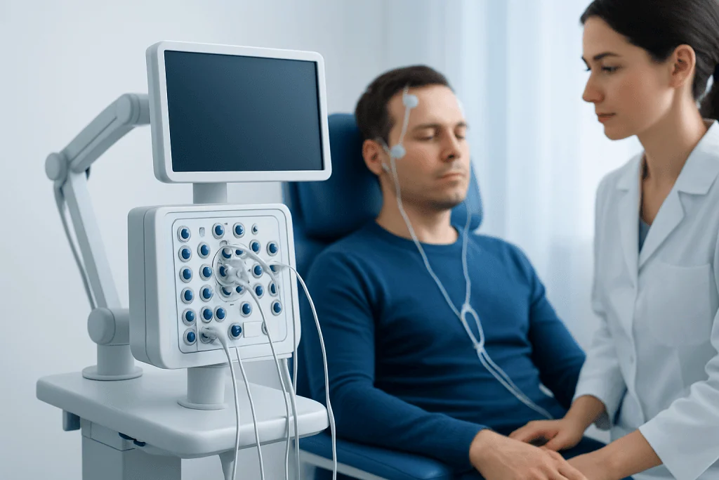
Different evoked potential tests assess different sensory or motor pathways. A neurologist chooses the type of test based on the person’s symptoms and the clinical question. Some people need only one study, while others may have a combination of tests during the same appointment.
The most frequently used tests include:
- Visual evoked potential (VEP): measures the brain’s response to visual stimulation, often a checkerboard pattern on a screen. It is commonly used to evaluate the optic nerves and visual pathways.
- Brainstem auditory evoked response (BAER or BAEP): measures responses to clicking sounds delivered through earphones. It helps assess the auditory nerve and brainstem hearing pathways.
- Somatosensory evoked potential (SSEP): records responses after a mild electrical stimulus is applied to a peripheral nerve, often at the wrist or ankle. It evaluates sensory pathways through the nerves, spinal cord, brainstem, and brain.
- Motor evoked potential (MEP): assesses motor pathways from the brain to muscles, often using magnetic stimulation. It may be used in selected neurological evaluations and during certain surgical procedures.
In eye-related neurological problems, visual evoked potentials may be used together with a detailed eye and neurological assessment. This can be particularly helpful in neuro-ophthalmology, where symptoms such as blurred vision, reduced color perception, or unexplained visual changes may involve the optic nerve or visual pathways rather than the eye alone.
When Doctors May Recommend Evoked Potential Testing

A doctor may recommend evoked potential testing when symptoms suggest that signals may not be traveling normally through a sensory or motor pathway. Examples include unexplained visual loss, double vision, numbness, tingling, weakness, balance problems, hearing-related concerns, or symptoms that suggest spinal cord involvement. The test may also be used when the neurological examination is subtle or when symptoms have improved but a past nerve pathway problem is still suspected.
One important use is in the evaluation of demyelinating diseases, where the immune system affects myelin, the protective covering around nerve fibers. Multiple sclerosis is a well-known example. In this setting, evoked potentials may show delayed conduction in a pathway, even when symptoms are mild or have occurred in the past. The test does not diagnose multiple sclerosis by itself, but it may support the overall clinical picture when interpreted with MRI and other investigations in neuroimmunology.
Evoked potentials can also be helpful in children, especially when communication is limited or when a functional assessment is needed alongside imaging and clinical observation. In selected pediatric cases, they may support evaluation of visual or hearing pathway concerns, developmental neurological conditions, spinal cord problems, or inherited neurological disorders. Pediatric testing is adapted to the child’s age and ability to cooperate, and specialists in neuropediatrics may be involved when appropriate.
How to Prepare and What Happens During the Test
Preparation is usually simple. Patients are often asked to arrive with clean, dry hair and to avoid hair oils, gels, or heavy skin creams that can interfere with electrode contact. They should bring glasses or contact lenses if they use them, because visual evoked potential testing is usually performed with the person’s best corrected vision. Medicines are generally continued unless the doctor gives different instructions.
During the test, a technologist places small recording electrodes on the scalp and sometimes on the neck, back, arms, or legs. The skin may be cleaned gently to improve signal quality. The electrodes record activity; they do not deliver a harmful current to the brain. For a VEP, the patient watches a screen with a shifting pattern or flashing light. For a BAER, the patient hears clicking sounds through earphones. For an SSEP, a mild repeated pulse is applied to a nerve, which may feel like a tapping or tingling sensation.
The test is usually performed while the patient is seated or lying comfortably. It is important to remain as relaxed and still as possible, because muscle tension and movement can create electrical “noise” in the recording. Each response is very small, so the equipment averages many repeated responses to create a clear waveform. Depending on the type and number of studies, the appointment may take from a short session to more than an hour.
Understanding Results: What the Measurements Mean
Evoked potential results are analyzed by looking at the timing, shape, and size of recorded waveforms. Timing is especially important. If a signal takes longer than expected to reach the brain, this is called increased latency or delayed conduction. If the response is very small or absent, it may suggest reduced signal transmission, but the meaning depends on the pathway tested and the clinical context.
For example, a delayed visual evoked potential may suggest previous or current involvement of the optic nerve, even if a person’s vision has partly recovered. An abnormal auditory evoked response may point to a problem along the hearing nerve or brainstem pathway. An abnormal somatosensory evoked potential may help localize dysfunction somewhere along the pathway from the limb to the spinal cord and brain.
Results are not interpreted in isolation. Age, height, body temperature, attention, vision correction, hearing ability, medications, previous surgery, and technical factors may influence the recording. Normal results are reassuring but do not rule out every neurological condition. Abnormal results indicate that a pathway is not conducting as expected, but they do not always identify the exact cause. A neurologist explains how the findings fit with the person’s symptoms and whether further tests are needed.
Benefits, Safety, and Limitations
Evoked potential tests are valued because they provide objective information about nervous system function. They can detect slowed conduction in pathways that may not be fully assessed by a routine examination. They are also non-invasive, do not involve radiation, and can be repeated if needed to monitor changes over time or compare results with future studies.
Most people tolerate the tests well. The electrodes may leave a small amount of paste in the hair, and the skin may feel slightly irritated where it was cleaned. The mild electrical stimulation used in SSEP testing can feel unusual but is adjusted to be tolerable. Flashing lights can be uncomfortable for some people, and patients with a history of photosensitive seizures should inform the team before testing. Motor evoked potential testing has additional screening considerations and is used when clinically appropriate.
The main limitation is that evoked potentials show how a pathway conducts signals, not why the pathway is abnormal. For this reason, they are often combined with MRI, laboratory testing, eye examinations, hearing tests, lumbar puncture in selected cases, or other neurological studies. The most useful interpretation comes from matching the test result to the patient’s story and examination rather than relying on a single number.
After the Test and When to See a Doctor
After evoked potential testing, most people can return to normal daily activities immediately. There is usually no recovery period, sedation is not typically required, and the electrodes are removed before leaving the clinic. If conductive paste was used, it can be washed out with regular shampoo. The recorded data are reviewed by a trained physician, often a neurologist or clinical neurophysiologist, and a report is sent to the referring doctor.
A person should seek medical advice if they have new or persistent neurological symptoms such as vision loss, numbness, weakness, balance difficulty, unexplained hearing changes, or problems with bladder control or walking. Urgent assessment is appropriate for sudden neurological symptoms, especially if they involve one side of the body, speech, vision, severe dizziness, or acute weakness. Evoked potentials may be part of the evaluation, but the first step is timely assessment by a qualified clinician.
For international patients, Acibadem International’s multidisciplinary specialists and JCI-accredited hospitals provide diagnostic evaluation and treatment planning for neurological conditions, including situations where evoked potential testing is clinically indicated. The goal of testing is to clarify nerve pathway function and help the care team choose the most appropriate next steps.
Frequently asked questions
Are evoked potential tests painful?
Most evoked potential tests are not painful. Visual and auditory tests involve watching a pattern or listening to clicks, while somatosensory testing uses a mild electrical pulse that may feel like tapping or tingling. The intensity is adjusted so the test remains tolerable.
How long does an evoked potential test take?
The time depends on which pathways are being tested. A single study may be relatively brief, while a combination of visual, auditory, and somatosensory tests may take longer. The clinic will usually explain the expected appointment length in advance.
Can evoked potential tests diagnose multiple sclerosis?
Evoked potential tests cannot diagnose multiple sclerosis on their own. They may show slowed nerve conduction that supports evidence of demyelination, especially in the optic nerve or other pathways. Doctors interpret the results together with symptoms, neurological examination, MRI findings, and other tests.
What is the difference between an EEG and an evoked potential test?
An EEG records the brain’s ongoing electrical activity, often to evaluate seizures or changes in brain function. An evoked potential test records the nervous system’s response to a specific repeated stimulus, such as a visual pattern, sound, or mild nerve stimulation. Both use electrodes, but they answer different clinical questions.
Do patients need sedation for evoked potential testing?
Adults usually do not need sedation because the test is non-invasive and requires cooperation, such as looking at a screen or staying relaxed. In young children or patients who cannot remain still, the care team may discuss special preparation or alternative approaches. Any decision about sedation is individualized and supervised by medical professionals.
What happens if the result is abnormal?
An abnormal result means that a nerve pathway did not conduct signals in the expected way during the test. It does not automatically identify the cause, so the doctor will compare the finding with symptoms, examination results, and other investigations. Further testing or follow-up may be recommended depending on the overall clinical picture.
References
- American Academy of Neurology
- International Federation of Clinical Neurophysiology
- National Institute of Neurological Disorders and Stroke
- Mayo Clinic
- European Academy of Neurology
This article is for general information only and is not a substitute for professional medical advice. Please consult a qualified doctor about your individual situation.
Neurophysiology in Turkey — costs, top hospitals & a free quote
JCI-accredited · board-certified surgeons · reply within 24h