Diagnostic Mammogram: When It Is Used and What to Expect
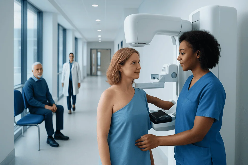
A diagnostic mammogram is used to investigate a breast symptom or clarify an abnormal screening mammogram. It usually includes extra images focused on a specific area of the breast.
Key Takeaways
- A diagnostic mammogram is used to investigate a breast symptom or clarify an abnormal screening mammogram.
- It usually includes extra images focused on a specific area of the breast.
- Most abnormal findings are not cancer, but follow-up imaging is important for an accurate diagnosis.
- The test is brief, and breast compression is temporary but can feel uncomfortable.
- Results may be discussed the same day, and some people may also need ultrasound or biopsy.
A diagnostic mammogram is a more focused type of breast imaging used when there is a symptom, a physical finding, or a screening mammogram that needs a closer look. It provides additional views to help radiologists understand what is causing a change and whether more testing is needed.
Overview
A diagnostic mammogram is a specialized breast X-ray exam performed when a person has a breast symptom or when a screening mammogram shows an area that needs closer evaluation. While a screening mammogram looks for early changes in people without symptoms, a diagnostic mammogram focuses on a specific concern. It is designed to provide more detailed information and help the radiologist decide whether the finding is likely benign, needs short-term follow-up, or requires additional testing.
This type of imaging may be recommended for a new lump, breast pain in a certain area, nipple discharge, skin thickening, or a change seen on routine screening. It can also be used to evaluate changes after breast surgery or to follow a finding that has been monitored over time. In many cases, the test gives reassurance by showing a harmless cause such as overlapping tissue, a cyst, or benign calcifications.
Diagnostic mammography is usually performed in a breast imaging center by trained technologists and interpreted by radiologists with expertise in breast imaging. The exam often includes standard images plus extra views, such as magnified or spot-compression images, to better examine one area. Depending on what is seen, the radiologist may also recommend a breast ultrasound or, less commonly, MRI for further assessment.
When a Diagnostic Mammogram Is Used
A diagnostic mammogram is commonly used after an abnormal screening mammogram. Screening images may show an asymmetry, an area of distortion, calcifications, or a possible mass that cannot be fully characterized on the first exam. Additional diagnostic views help determine whether the finding is simply normal tissue overlap or something that needs closer follow-up.
It is also used when a person notices symptoms in the breast. These can include a new lump, persistent focal pain, nipple inversion that is new, bloody or clear nipple discharge, redness or thickening of the skin, or swelling in one area. Although many breast symptoms are caused by noncancerous conditions, they should still be assessed properly.
Other reasons for diagnostic imaging include checking an area seen on breast ultrasound, monitoring a probably benign finding over time, or evaluating the breast after treatment or surgery. In some cases, a radiologist may compare the current study with prior mammograms to look for changes. If there is a strong clinical concern, further evaluation may include a breast biopsy to obtain a tissue sample.
How It Differs From a Screening Mammogram

The main difference between a screening and diagnostic mammogram is the purpose of the test. A screening mammogram is a routine exam for people without symptoms, aiming to detect breast cancer early before it can be felt or seen. A diagnostic mammogram is more targeted and is ordered because there is a symptom, a physical finding, or an image that needs clarification.
A screening exam usually includes a standard set of images of each breast. A diagnostic exam often takes more images, and the technologist may reposition the breast several times to focus on the area of concern. The radiologist may review images during the appointment and request additional views right away, which is why the visit can take longer than a routine screening exam.
Another difference is how results are delivered. With screening mammography, results may be sent later after review. With a diagnostic mammogram, many centers provide preliminary findings on the same day, sometimes followed by a same-day ultrasound if needed. This more immediate evaluation can help reduce uncertainty and guide the next step efficiently.
What to Expect During the Procedure
Before the exam, the patient will usually be asked not to apply deodorant, powder, lotion, or creams on the underarm or breast area, as some products can appear on X-rays. It is helpful to wear a two-piece outfit, since clothing from the waist up will need to be removed. The imaging team may also ask about breast symptoms, previous surgeries, hormone use, and family or personal history of breast disease.
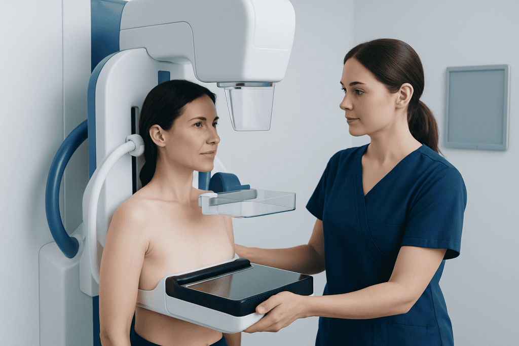
During the exam, each breast is placed on the mammography unit and gently compressed for a few seconds while images are taken. Compression is important because it spreads the breast tissue, reduces motion, and improves image quality while keeping the radiation dose as low as reasonably possible. It can feel uncomfortable, but the pressure is temporary and should ease as soon as each image is completed.
If more detailed views are needed, the technologist may take spot-compression, magnification, or angled images. In some cases, a marker may be placed on the skin to indicate the location of a lump or symptom. A breast ultrasound may follow if the radiologist wants to look more closely at a mass, dense tissue, or an area behind the nipple. When the finding remains unclear, advanced imaging such as breast MRI may occasionally be considered.
The entire visit may last longer than a screening mammogram because of the extra views and radiologist review, but the image-taking portion is usually still fairly short. Patients can usually return to normal activities right away. If the breasts are tender, scheduling the exam for a time when they are less sensitive may make the experience more comfortable.
Understanding the Results
After the images are reviewed, the radiologist describes the findings and assigns an assessment category, often using the BI-RADS system. This helps communicate whether the result is negative, benign, probably benign, suspicious, or highly suggestive of malignancy. It also explains the recommended next step, such as routine screening, short-interval follow-up, more imaging, or tissue sampling.
Many diagnostic mammogram findings turn out to be noncancerous. Benign causes may include cysts, fibroadenomas, fat necrosis, postsurgical changes, benign calcifications, or normal tissue overlap. Even so, follow-up recommendations should be taken seriously, because imaging cannot answer every question with certainty in one visit.
If the radiologist sees something that needs more evaluation, an ultrasound may be added or a biopsy may be advised. This does not mean cancer is certain; it means a closer look is needed to make a clear diagnosis. Some patients may also be evaluated for related breast conditions such as breast cancer or fibrocystic breast changes, depending on the imaging pattern and symptoms.
Benefits, Limitations, and Safety
The main benefit of a diagnostic mammogram is that it provides a more detailed assessment of a breast concern. It can help distinguish harmless findings from changes that require closer follow-up and can detect abnormalities too small to be felt. This allows patients and doctors to make informed decisions about next steps without unnecessary delay.
Like all tests, mammography has limitations. It may be harder to interpret in very dense breasts, and some findings still need ultrasound or biopsy for confirmation. A normal mammogram also does not completely rule out cancer, especially if there is a persistent lump or another concerning symptom. For that reason, imaging results should always be considered alongside a physical examination and medical history.
Mammography uses a low dose of radiation, and the amount is carefully controlled. For most patients, the benefit of diagnosing a significant breast problem outweighs the small radiation exposure from the test. If a patient is pregnant or thinks pregnancy is possible, the imaging team should be told before the exam so the safest approach can be planned.
Preparation, Follow-Up, and When to See a Doctor
Patients can prepare by bringing previous breast imaging if it was done at a different facility, since comparison with earlier studies can be very helpful. It is also useful to note when symptoms started, whether they change with the menstrual cycle, and whether there is any nipple discharge or skin change. Sharing this information can help the radiologist tailor the exam.
After the test, follow the imaging center’s recommendations carefully. Some people will return to routine screening, while others may need short-term follow-up imaging, ultrasound, or biopsy. If symptoms continue even after a reassuring result, it is important to tell a doctor, because persistent or changing symptoms sometimes need re-evaluation.
A person should seek medical advice promptly for a new breast lump, bloody nipple discharge, skin dimpling, persistent one-sided breast pain, breast swelling, or a change in the shape of the breast. Immediate emergency care is not usually necessary for most breast concerns, but timely assessment is important. Near the end of the diagnostic pathway, patients may benefit from coordinated care at specialist centers; Acibadem International’s multidisciplinary specialists in JCI-accredited hospitals diagnose and treat breast conditions for international patients, including imaging follow-up and breast cancer treatment when needed.
Frequently asked questions
Is a diagnostic mammogram the same as a regular mammogram?
No. A screening mammogram is a routine test for people without symptoms, while a diagnostic mammogram is used to examine a specific concern more closely. It usually includes additional images and may involve same-day radiologist review.
Does needing a diagnostic mammogram mean cancer is likely?
Not necessarily. Many people are called back for extra imaging because of overlapping tissue, benign cysts, or other noncancerous changes. The purpose of a diagnostic mammogram is to clarify what was seen and decide whether any further testing is needed.
How painful is a diagnostic mammogram?
Most people describe it as uncomfortable rather than painful. The breast is compressed for a few seconds at a time to get clear images, and the pressure stops quickly after each picture. If breasts are especially tender, scheduling at a more comfortable time in the menstrual cycle may help.
How long does the appointment take?
It is often longer than a screening mammogram because extra views may be needed and the radiologist may review the images during the visit. The exact time varies, but many appointments are completed within about 30 minutes to an hour. If ultrasound is added, the visit may take longer.
Can a diagnostic mammogram detect all breast cancers?
No single test detects every breast cancer. Mammography is very useful, but some cancers can be harder to see, especially in dense breast tissue. If symptoms persist or the physical exam is concerning, a doctor may recommend ultrasound, MRI, or biopsy even if the mammogram is not clearly abnormal.
What happens if the results are abnormal?
An abnormal result means more evaluation is needed, not that cancer is certain. The next step may be short-term follow-up imaging, ultrasound, or a biopsy to examine tissue. The radiologist or doctor will explain what was found and why the recommended follow-up is appropriate.
This article is for general information only and is not a substitute for professional medical advice. Please consult a qualified doctor about your individual situation.
Explore treatments in Turkey — costs, top hospitals & a free quote
JCI-accredited · board-certified surgeons · reply within 24h
Add us as a Preferred Source to see more of our trusted health content across Google Search, AI Overviews and Discover.

More from the Health Library

When Do You Need a Second Opinion on Imaging Results?

Neurophysiology for Seizures and Fainting: Which Tests Help Find the Cause?
PET-CT Scan: How It Helps Detect and Stage Cancer

Neuropsychological Testing: Memory, Attention, and Brain Function Assessment

Diagnostic Criteria for PTSD: How F43.1 Is Diagnosed