Diagnostic Ultrasound: How It Works and What It Can Detect

Diagnostic ultrasound creates images using sound waves, not X-rays. It can help evaluate organs, soft tissues, blood flow, and pregnancy in real time.
Key Takeaways
- Diagnostic ultrasound creates images using sound waves, not X-rays.
- It can help evaluate organs, soft tissues, blood flow, and pregnancy in real time.
- The test is generally painless, widely available, and does not involve ionizing radiation.
- Preparation depends on the body area being examined, such as fasting or having a full bladder.
- Results may lead to further imaging or treatment depending on what the scan shows.
Diagnostic ultrasound is a common imaging test that uses high-frequency sound waves to create real-time pictures inside the body. It helps doctors assess many organs, blood vessels, soft tissues, and pregnancy without using ionizing radiation.
Overview of Diagnostic Ultrasound
Diagnostic ultrasound, also called ultrasonography, is an imaging method that uses high-frequency sound waves to produce pictures of structures inside the body. A small handheld device called a transducer sends sound waves into the body and receives the echoes that bounce back. A computer then converts these echoes into images that can be viewed in real time.
Because it shows movement as it happens, ultrasound is especially useful for examining soft tissues, blood flow, and developing babies during pregnancy. It is commonly used to look at the abdomen, pelvis, thyroid, breasts, heart, muscles, tendons, and blood vessels. Different forms of ultrasound, such as Doppler ultrasound, can also help assess how blood moves through arteries and veins.
One of the main advantages of diagnostic ultrasound is that it does not use ionizing radiation. For many patients, this makes it a practical first-line imaging test. It is also usually quick, noninvasive, and well tolerated, although the exact experience depends on the type of exam being performed.
How Ultrasound Works
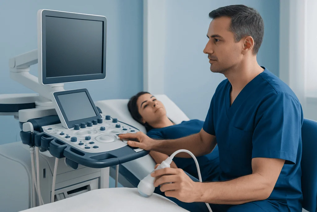
Ultrasound works by sending sound waves that are too high-pitched for human ears to hear into the body. When these waves encounter tissues, fluids, or organs, they reflect back at different speeds and strengths. The ultrasound machine interprets those returning signals and creates shades of gray or color images based on the properties of the tissues being examined.
The transducer is placed on the skin with a clear gel that helps the sound waves travel efficiently. In some situations, the transducer may be inserted into the body, such as in transvaginal or transrectal ultrasound, to obtain clearer images of nearby structures. During the exam, the sonographer or physician moves the transducer to capture the needed views.
Several types of ultrasound may be used depending on the clinical question:
-
Standard ultrasound: creates still or moving images of organs and soft tissues.
-
Doppler ultrasound: measures the direction and speed of blood flow.
-
Echocardiography: a specialized ultrasound of the heart.
-
3D or 4D ultrasound: provides more detailed or dynamic views in selected cases, often in obstetrics.
The quality of an ultrasound image can depend on the body area, the patient’s anatomy, and the amount of gas or bone in the path of the sound waves. For this reason, ultrasound is excellent for many soft-tissue questions but may be less informative for some deeper or air-filled structures.
What Diagnostic Ultrasound Can Detect

Diagnostic ultrasound can help identify a wide range of normal and abnormal findings. In the abdomen, it may be used to evaluate the liver, gallbladder, pancreas, spleen, kidneys, and abdominal aorta. Doctors often request it to look for gallstones, kidney obstruction, fluid collections, enlarged organs, cysts, or masses.
In the pelvis, ultrasound is commonly used to assess the uterus, ovaries, and bladder, and in pregnancy it helps confirm gestational age, check fetal growth, monitor heartbeat and movement, and evaluate the placenta and amniotic fluid. In breast, thyroid, or soft-tissue imaging, it can help distinguish between cystic and solid lumps and guide decisions about follow-up.
Ultrasound also plays an important role in blood vessel imaging. Doppler studies may detect narrowed arteries, reduced blood flow, or blood clots in veins, including deep vein thrombosis. In cardiology, echocardiography helps assess heart chambers, valves, pumping function, and fluid around the heart. Musculoskeletal ultrasound can be used to examine tendons, ligaments, joint fluid, and some muscle injuries.
In addition to diagnosis, ultrasound may guide certain procedures. For example, it can help doctors place a needle accurately during fluid drainage, biopsy, or injections, improving precision while avoiding nearby structures.
When Doctors Recommend an Ultrasound
Doctors may recommend an ultrasound when symptoms suggest a problem in soft tissues or blood vessels, or when they want a safe, real-time first look at an internal structure. Common reasons include abdominal pain, pelvic pain, swelling, a palpable lump, abnormal bleeding, suspected gallbladder disease, urinary symptoms, or concerns during pregnancy.
Ultrasound is also often used when a physical examination or blood test suggests a condition that needs imaging confirmation. For instance, it may help evaluate abnormal liver tests, thyroid enlargement, a suspected ovarian cyst, or a possible hernia. It is frequently chosen because it is accessible and can often answer the clinical question without the need for more complex imaging.
Sometimes ultrasound is the starting point rather than the final test. If the images are limited or the findings need more detail, a doctor may recommend further studies such as MRI scanning or CT imaging. The choice depends on the body area involved and the type of information needed.
What to Expect Before, During, and After the Scan
Preparation for an ultrasound depends on the part of the body being examined. Some abdominal scans require fasting for several hours to reduce gas in the intestines and improve the view of organs such as the gallbladder. Pelvic ultrasound may require a full bladder, as this can help provide clearer images. In many other cases, no special preparation is needed.
During the test, the patient usually lies on an examination table while gel is applied to the skin. The transducer is then moved gently over the area. The exam is usually painless, though slight pressure may be felt, especially if the area is tender. Internal ultrasound exams, such as transvaginal ultrasound, may cause mild discomfort but are generally brief.
Most ultrasound scans take about 15 to 45 minutes, depending on the area studied and how many images are needed. After the exam, patients can typically return to normal activities right away. The images are reviewed by a radiologist or another qualified physician, who sends a report to the referring doctor.
If an ultrasound identifies a concerning finding, the next steps may include repeat imaging, lab tests, referral to a specialist, or image-guided sampling such as a biopsy. The scan itself does not treat disease, but it often provides important information that helps guide care.
Benefits, Limits, and Safety
Ultrasound offers several important benefits. It does not use ionizing radiation, provides real-time images, and can often be performed quickly in outpatient clinics, hospitals, or emergency settings. It is especially useful in pregnancy, pediatric care, and vascular imaging, where repeated evaluation may be needed.
The test does have limitations. Ultrasound waves do not pass well through bone or air, so structures hidden behind the skull, lungs, or gas-filled bowel may be difficult to see clearly. Body size, scar tissue, and the exact location of an abnormality can also affect image quality. In some situations, a normal ultrasound does not completely rule out disease.
When used appropriately by trained professionals, diagnostic ultrasound is considered very safe. Patients often ask whether ultrasound is harmful; in standard medical use, it has an excellent safety record. Even so, it should be performed only when clinically indicated and interpreted in the context of symptoms, examination findings, and other tests.
For some conditions, ultrasound findings may overlap with other diagnoses. For example, a scan may suggest inflammation, a benign cyst, or a suspicious mass, but further evaluation may still be needed to confirm the cause. This is why ultrasound results are best discussed with the doctor who ordered the test.
How Results Are Interpreted and What Happens Next
An ultrasound report describes the appearance, size, shape, and texture of the organs or tissues examined. It may mention whether structures look normal, whether fluid is present, or whether a cyst, stone, blockage, or mass is seen. In Doppler studies, the report may also include information about blood flow patterns.
Some results are straightforward, such as confirming gallstones or showing a normal pregnancy heartbeat. Other findings are less specific and may require comparison with symptoms, blood tests, or prior imaging. A doctor may decide that no further action is needed, recommend monitoring over time, or arrange a specialist consultation.
If additional evaluation is necessary, options can include imaging such as PET-CT in selected oncology settings, MRI, or CT, depending on the suspected condition. In other situations, follow-up ultrasound may be enough to see whether a finding changes over time.
For international patients who need coordinated evaluation, Acibadem International’s multidisciplinary specialists and JCI-accredited hospitals diagnose and treat a wide range of conditions using advanced imaging and follow-up care plans tailored to the patient’s needs.
When to Speak With a Doctor
Patients should speak with a doctor if they have persistent symptoms that may need imaging, such as unexplained pain, swelling, a new lump, abnormal bleeding, urinary changes, or symptoms during pregnancy that raise concern. An ultrasound can be helpful, but the decision to order it should be based on a full medical assessment.
It is also important to discuss the results with a qualified clinician rather than trying to interpret the report alone. Many ultrasound findings are benign, and even abnormal results often need clinical context before they can be fully understood. A doctor can explain what was seen, whether follow-up is necessary, and what the next steps should be.
Urgent medical attention may be needed if symptoms are severe, sudden, or associated with fainting, heavy bleeding, chest pain, shortness of breath, high fever, or signs of stroke. In these situations, imaging may be only one part of immediate medical care.
Self-care and Practical Tips for Patients
Although ultrasound itself does not require recovery, patients can make the process smoother by following preparation instructions carefully. Drinking water when asked, fasting if advised, and bringing previous imaging reports can all help improve the usefulness of the exam. Wearing comfortable clothing may also make the visit easier.
Patients may want to prepare a few questions before the appointment, such as why the ultrasound is being ordered, what body area will be examined, and when results are expected. Asking whether the scan is likely to answer the main concern can also be helpful, especially if symptoms are ongoing.
After the scan, the most important step is follow-up. Even if the ultrasound is normal, persistent or worsening symptoms should be reviewed again. Imaging is one tool in diagnosis, and good care often depends on combining the scan findings with medical history, examination, and, when needed, further testing.
Frequently asked questions
Is diagnostic ultrasound the same as an X-ray?
No. Diagnostic ultrasound uses sound waves to create images, while X-rays use ionizing radiation. Ultrasound is often preferred for viewing soft tissues, blood flow, and pregnancy because it provides real-time images without radiation exposure.
Can ultrasound detect cancer?
Ultrasound can identify abnormal masses or changes in tissue that may need further evaluation, but it usually cannot confirm cancer by itself. A doctor may recommend additional imaging or a biopsy to determine exactly what a finding means.
Is an ultrasound painful?
Most ultrasound exams are painless. Patients may feel cool gel on the skin and mild pressure from the transducer, and some internal exams can cause brief discomfort, but the test is generally well tolerated.
How should someone prepare for an ultrasound scan?
Preparation depends on the body part being examined. Some scans require fasting, while others need a full bladder, and many need no preparation at all. The imaging center or doctor’s office usually gives specific instructions before the appointment.
How long does it take to get ultrasound results?
This varies by facility and by the urgency of the test. In many cases, a radiologist reviews the images and sends a report to the referring doctor within a short time, after which the doctor explains the results and next steps.
Are there any risks from diagnostic ultrasound?
When used appropriately in medical care, diagnostic ultrasound is considered very safe and has a strong safety record. It does not use ionizing radiation, but like any medical test, it should be performed for a clear clinical reason and interpreted by trained professionals.
This article is for general information only and is not a substitute for professional medical advice. Please consult a qualified doctor about your individual situation.
Ultrasound in Turkey — costs, top hospitals & a free quote
JCI-accredited · board-certified surgeons · reply within 24h
Add us as a Preferred Source to see more of our trusted health content across Google Search, AI Overviews and Discover.

Check-up & Preventive Medicine
Comprehensive health screenings designed for early detection and prevention.
1 specialists in this unitMore from the Health Library

Pediatric EEG: How Brain Wave Testing Is Done and How to Prepare

Can Diagnostic Tests Miss a Problem? Understanding False Negative and False Positive Results
MRI Scan: How It Works, Safety, and What Results Can Show

EMG and Nerve Conduction Studies: Diagnosing Neuromuscular Disease
Ultrasound-Guided vs CT-Guided Procedures: What Is the Difference?