What Happens During a Diagnostic Imaging Exam: Step-by-Step Guide

Most diagnostic imaging exams are quick, structured, and designed to be as comfortable as possible. Preparation depends on the imaging type and may include fasting, avoiding metal, or drinking contrast.
Key Takeaways
- Most diagnostic imaging exams are quick, structured, and designed to be as comfortable as possible.
- Preparation depends on the imaging type and may include fasting, avoiding metal, or drinking contrast.
- Common imaging tests include X-ray, ultrasound, CT, MRI, and nuclear medicine scans.
- A radiologist reviews the images and sends a report to the referring doctor.
- Patients should tell the care team about pregnancy, allergies, implants, kidney problems, or claustrophobia before the exam.
A diagnostic imaging exam helps doctors look inside the body to diagnose, monitor, or guide treatment. While the exact steps vary by test, most imaging exams follow a similar process from preparation and positioning to image capture and follow-up.
Overview: What a Diagnostic Imaging Exam Is
A diagnostic imaging exam is a medical test that creates pictures of the inside of the body. These pictures help doctors investigate symptoms, confirm a diagnosis, monitor a known condition, or plan treatment. Imaging can look at bones, organs, blood vessels, joints, and soft tissues in ways that a physical exam alone cannot.
Several technologies are used in medical imaging. Common examples include X-rays, ultrasound, computed tomography (CT), magnetic resonance imaging (MRI), and nuclear medicine scans such as PET. Each one works differently, so the exact experience can vary from one test to another.
Even so, most imaging exams follow a similar path. A patient typically checks in, may change clothes, receives instructions, is positioned for the scan, and then has images taken by a trained radiology technologist. Afterward, a radiologist interprets the images and sends the results to the doctor who ordered the test.
Why Doctors Order Imaging Tests
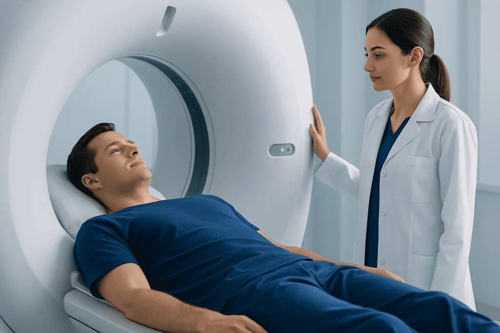
Doctors order a diagnostic imaging exam when they need more information than symptoms, blood tests, or a physical examination can provide. Imaging can help locate an injury, detect inflammation or infection, assess organ function, and check how a condition is changing over time.
For example, an X-ray may be used to look for a fracture, while an ultrasound can assess the gallbladder, kidneys, or pregnancy. CT and MRI provide more detailed images of internal structures, and some scans can help evaluate blood flow, tumors, or nerve-related problems. In many cases, imaging also helps guide procedures such as biopsies or drainage.
Not every scan is used for diagnosis alone. Some exams are done to screen people who have no symptoms but may have certain risk factors, while others are used to monitor recovery after surgery or treatment. The choice of scan depends on the body area being examined, the question the doctor needs answered, and the person’s medical history.
How to Prepare Before the Exam
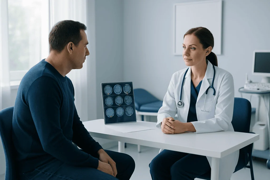
Preparation depends on the type of diagnostic imaging exam. Some tests require no special preparation at all, while others may involve fasting for several hours, drinking water to fill the bladder, or avoiding food, caffeine, or certain medicines for a short time. The imaging center usually gives instructions in advance, and it is important to follow them closely.
Patients may be asked to wear loose clothing or change into a hospital gown. Metal objects such as jewelry, belts, watches, hearing aids, hairpins, and removable dental work may need to be removed, especially before MRI or some X-ray-based exams. Bringing a list of current medicines and any previous scan reports can also be helpful.
Before the exam, the care team should be told about pregnancy or a chance of pregnancy, allergies, kidney disease, asthma, diabetes, implanted devices, and any past reaction to contrast material. People who feel anxious in enclosed spaces should mention this too, particularly before an MRI, because extra support or planning may help make the exam more comfortable.
- Fasting may be needed for some abdominal ultrasound, CT, or nuclear medicine scans.
- A full bladder may be requested for pelvic ultrasound.
- Metal must usually be removed before MRI.
- Contrast material may require questions about allergies and kidney function.
Step by Step: What Happens During the Exam
On arrival, the patient checks in and confirms personal details, the body area being scanned, and any important medical information. A technologist may ask a few safety questions and explain what will happen. If contrast material is needed, it may be given by mouth, through an intravenous line, or less commonly by another route depending on the test.
Next, the patient is positioned on a table, chair, or standing platform. Good positioning is important because it helps produce clear images. The technologist may use cushions, straps, or supports to help the patient stay still and comfortable. For some exams, the patient may be asked to hold their breath for a few seconds or change position during image capture.
The imaging itself is usually painless, although lying still can sometimes feel uncomfortable. X-rays are often completed in minutes. Ultrasound uses a handheld probe with gel on the skin. CT scans involve a moving table that passes through a ring-shaped scanner, while MRI uses a tunnel-like machine and can be noisier and longer. Some people notice warmth, a metallic taste, or a brief flushing feeling if contrast is used, but the team monitors for any concerns.
When the images are complete, the technologist checks that they are clear and complete. Sometimes extra images are needed, which is common and does not necessarily mean something is wrong. After the exam, most patients can return to normal activities right away unless they were given sedation or special instructions.
What to Expect From Different Imaging Types
Each kind of diagnostic imaging exam has its own purpose and feel. X-rays are quick and commonly used for bones and chest imaging. Ultrasound does not use radiation and is often chosen for soft tissues, pregnancy, blood flow, and abdominal organs. CT uses X-rays to create cross-sectional images and is useful when rapid, detailed pictures are needed.
MRI does not use ionizing radiation. Instead, it uses a strong magnetic field and radio waves to create detailed images, especially of the brain, spine, joints, and soft tissues. Because the machine can be narrow and loud, ear protection is usually provided, and some people may benefit from relaxation techniques or discussing sedation options with their doctor beforehand. MRI scanning is often selected when high soft-tissue detail is important.
Nuclear medicine scans use a small amount of radioactive tracer to show how tissues or organs function. These exams can take longer because the tracer may need time to circulate before images are taken. In some situations, doctors may combine tests for a fuller picture, such as CT scanning for anatomy or ultrasound imaging for real-time views of organs and blood flow.
After the Exam and How Results Are Given
After most imaging tests, the patient can leave shortly after the scan is finished. If contrast was used, the team may recommend drinking fluids unless another medical condition limits this. If sedation was given, the patient may need observation for a short time and should usually arrange for someone to accompany them home.
The images are then reviewed by a radiologist, a doctor trained to interpret medical imaging. The radiologist prepares a report that is sent to the referring doctor, who explains the findings in the context of symptoms, examination, and other test results. This is important because an image alone does not always provide the full diagnosis.
Timing for results varies. Some urgent scans are reviewed quickly, while routine studies may take longer. Patients should ask when and how they can expect results and whom to contact if they have questions afterward. If follow-up imaging is recommended, it may be to clarify a finding, monitor healing, or evaluate treatment progress.
Safety, Risks, and Common Concerns
Medical imaging is generally very safe, and the type of risk depends on the exam. X-ray and CT use ionizing radiation, so doctors balance the benefit of the information gained against the small exposure involved. Ultrasound and MRI do not use ionizing radiation, but MRI has special safety rules because of its strong magnet.
Contrast materials are helpful in some scans because they make certain structures easier to see. Most people tolerate contrast well, but side effects and allergic-type reactions can happen. For this reason, patients should always tell the team about previous contrast reactions, kidney disease, asthma, or other relevant health conditions.
Claustrophobia, difficulty lying flat, pain, or trouble staying still are also common concerns and can often be managed with simple adjustments. Children, older adults, and people with complex medical needs may require tailored planning. In experienced centers, the radiology team explains each step clearly and works to make the process as calm and comfortable as possible. Near the end of the care journey, some international patients choose centers such as Acibadem International, where multidisciplinary specialists and JCI-accredited hospitals provide diagnostic imaging and treatment planning.
When to Ask Questions or Contact a Doctor
It is always appropriate for patients to ask questions before a diagnostic imaging exam. They may want to know why the test is needed, whether contrast will be used, how to prepare, how long it will take, and when results will be available. Clear communication often reduces anxiety and helps the exam run smoothly.
Patients should contact the imaging center or their doctor before the appointment if they become unwell, think they may be pregnant, cannot follow the preparation instructions, or have new information such as an implant or allergy. They should also seek advice if they are unable to lie still, have severe claustrophobia, or need language or mobility support.
After the exam, medical advice should be sought promptly if there is unexpected swelling, rash, breathing difficulty, severe dizziness, or another concerning symptom after contrast or sedation. Most people do not experience complications, but it is sensible to know whom to call if something does not feel right.
Frequently asked questions
Is a diagnostic imaging exam painful?
Most imaging exams are not painful. Some people may feel mild discomfort from lying still, holding a position, or having an IV placed for contrast, but the imaging itself is usually painless.
How long does an imaging exam take?
The length depends on the type of scan and whether contrast is needed. Simple X-rays may take only a few minutes, while MRI or nuclear medicine studies can take longer, sometimes an hour or more.
Do all imaging tests use radiation?
No. X-rays and CT scans use ionizing radiation, but ultrasound and MRI do not. The doctor chooses the most suitable test based on the medical question and the person's situation.
What is contrast material and why might it be used?
Contrast material is a substance used to make certain tissues, blood vessels, or organs show more clearly on images. It may be swallowed, injected, or given in another way depending on the exam.
Can someone eat and drink before a diagnostic imaging exam?
Sometimes yes, sometimes no. Some exams need no preparation, while others require fasting or drinking water beforehand, so it is important to follow the instructions given by the imaging center.
Who explains the imaging results?
A radiologist reviews the images and writes a report. The referring doctor usually explains what the findings mean in relation to symptoms, medical history, and any next steps.
This article is for general information only and is not a substitute for professional medical advice. Please consult a qualified doctor about your individual situation.
Explore treatments in Turkey — costs, top hospitals & a free quote
JCI-accredited · board-certified surgeons · reply within 24h
Add us as a Preferred Source to see more of our trusted health content across Google Search, AI Overviews and Discover.

More from the Health Library

Urine Test Results Explained: Infection, Protein, Blood, and Other Common Findings
Open MRI vs Standard MRI: Which Patients May Benefit?

What Is a Diagnostic Test? Types, Purpose, and How Results Help Doctors

Doppler Ultrasound: Checking Blood Flow in Veins and Arteries

Neurophysiology for Seizures and Fainting: Which Tests Help Find the Cause?