Skin Cancer vs Benign Moles: How Doctors Tell the Difference
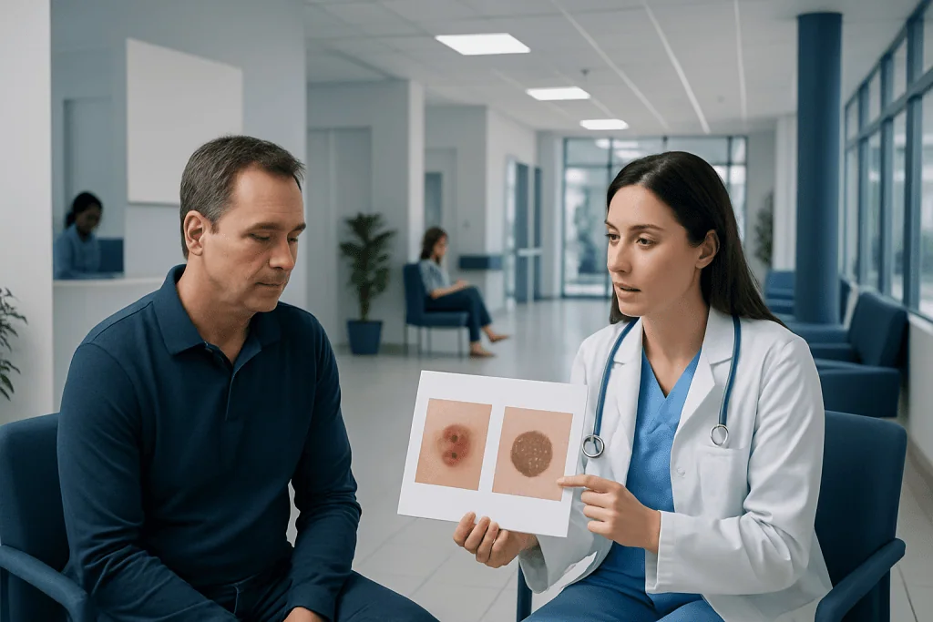
Most moles are benign, but new or changing spots should be checked by a doctor. Doctors often use the ABCDE rule to look for possible melanoma warning signs.
Key Takeaways
- Most moles are benign, but new or changing spots should be checked by a doctor.
- Doctors often use the ABCDE rule to look for possible melanoma warning signs.
- A skin examination and dermoscopy can help distinguish suspicious lesions from harmless moles.
- A biopsy is the most reliable way to confirm whether a spot is benign or cancerous.
- Sun protection and regular skin checks help with early detection.
Many moles are harmless, but some skin changes can suggest skin cancer. Doctors look at a mole’s appearance, any recent changes, and sometimes a biopsy to tell the difference accurately.
Overview: Skin Cancer and Benign Moles
Moles are common skin growths made up of pigment-producing cells called melanocytes. Most people have at least a few moles, and many remain stable for years without causing any health problems. Benign moles are usually round or oval, evenly colored, and similar in appearance over time.
Skin cancer develops when skin cells grow abnormally. Some skin cancers arise in or near existing moles, while others appear as entirely new spots. The main types include basal cell carcinoma, squamous cell carcinoma, and melanoma, which is the form most often associated with unusual moles and pigment changes.
The challenge is that not every concerning lesion looks dramatic, and not every odd-looking mole is cancer. Doctors combine visual clues, medical history, risk factors, and specialized tools to decide whether a spot is likely harmless or needs further testing.
Early evaluation matters because skin cancer is generally easier to treat when found sooner. For that reason, any mole or skin lesion that looks different from the others or begins to change deserves attention from a qualified clinician.
What Benign Moles Usually Look Like
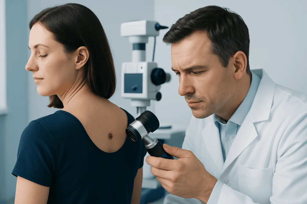
Benign moles often appear in childhood, adolescence, or early adulthood. They may be flat or slightly raised and are commonly tan, brown, or flesh-colored. Many are smaller than 6 millimeters, with smooth borders and a uniform color throughout.
A harmless mole usually stays relatively stable. It may darken slightly with sun exposure or hormonal changes, but it generally does not rapidly enlarge, bleed, crust, or become noticeably uneven. Some benign moles can have hair growing from them, which by itself is not a sign of cancer.
There are also special types of noncancerous moles, such as congenital moles present from birth or atypical moles that look somewhat unusual but are not necessarily malignant. Because some benign lesions can resemble cancer, appearance alone does not always provide certainty.
Other harmless skin spots can also be mistaken for moles, including freckles, seborrheic keratoses, skin tags, and sun spots. A dermatologist can help distinguish among these common findings and explain whether simple observation is appropriate.
Warning Signs That May Suggest Skin Cancer

Doctors often teach the ABCDE rule to help identify suspicious pigmented lesions. This guide is especially useful for spotting possible melanoma, though it does not detect every type of skin cancer.
- A – Asymmetry: one half does not match the other.
- B – Border: edges are irregular, blurred, scalloped, or poorly defined.
- C – Color: more than one shade is present, such as brown, black, red, white, or blue.
- D – Diameter: the spot is larger than about 6 millimeters, although smaller lesions can also be cancerous.
- E – Evolving: the mole changes in size, shape, color, or symptoms over time.
Another useful clue is the “ugly duckling” sign. This means one spot looks noticeably different from a person’s other moles. Even if it does not meet every ABCDE feature, a lesion that stands out should be examined.
Symptoms can also matter. Itching, tenderness, bleeding, crusting, oozing, or a sore that does not heal may suggest cancer, especially for non-melanoma skin cancers. A pearly bump, a scaly patch, or a persistent ulcer may point more toward skin cancer beyond the typical mole pattern.
Not every skin cancer is dark or pigmented. Some melanomas and many basal or squamous cell carcinomas can be pink, red, skin-colored, or translucent. Because of this, any persistent or changing lesion deserves medical assessment, even if it does not look like a classic mole.
Risk Factors Doctors Consider
When evaluating a suspicious spot, doctors do not rely only on appearance. They also consider personal and family risk factors. A history of intense sun exposure, frequent sunburns, indoor tanning, fair skin, light eyes, and easy freckling can all increase skin cancer risk.
Having many moles or atypical moles can make monitoring more important. A personal history of skin cancer or a close family history of melanoma also raises concern. People with weakened immune systems may be at higher risk for certain skin cancers as well.
Age can influence the type of lesion seen, although skin cancer can occur at any age. For example, basal and squamous cell carcinomas are more common later in life, while melanoma can affect younger adults too. Congenital large moles and certain inherited syndromes may also require closer follow-up.
Doctors also ask about change over time. A mole that has looked the same for many years is generally less concerning than one that has recently enlarged, darkened, become irregular, or started to bleed. Patient observations are often an important part of diagnosis.
How Doctors Tell the Difference
The first step is a careful skin examination. A doctor looks at the size, shape, color, borders, and location of the lesion, and may examine the rest of the skin for comparison. This full-body check can reveal whether a spot is unusual for that individual or part of a common mole pattern.
Dermatologists often use dermoscopy, a handheld magnifying device with light that allows them to see structures beneath the skin surface. Dermoscopy can help identify patterns linked with benign moles and patterns that are more suspicious for melanoma or other cancers. This tool improves accuracy, but it still does not replace tissue testing when doubt remains.
Photographs or digital mole mapping may be used for people with many moles or atypical lesions. Comparing images over time helps detect subtle changes that might be hard to notice day to day. In some cases, the doctor may recommend follow-up after a few months if a lesion is not clearly dangerous but not entirely typical.
If a lesion appears suspicious, a skin biopsy is usually the next step. During a biopsy, all or part of the spot is removed and examined under a microscope by a pathologist. This is the most reliable way to confirm whether the lesion is benign, precancerous, or cancerous.
Diagnosis and Treatment Options
If biopsy results show a benign mole, no additional treatment may be needed unless the lesion is irritating, repeatedly traumatized, or cosmetically bothersome. Some benign moles are removed for comfort or because they are in areas that are difficult to monitor. The doctor will explain whether routine observation is enough.
If cancer is diagnosed, treatment depends on the type, depth, and location of the lesion. Many skin cancers are treated with skin cancer treatment that includes surgical removal. In some cases, the doctor may recommend a Mohs surgery approach, especially for selected non-melanoma skin cancers in cosmetically sensitive areas or where preserving healthy tissue is important.
For melanoma, management may include a wider excision and, in some situations, further staging tests or review by oncology specialists. The exact plan depends on pathology findings. Prompt treatment improves the chance of complete removal and helps guide any necessary follow-up care.
Near the end of the care pathway, some patients seek multidisciplinary assessment, especially if the diagnosis is uncertain or treatment planning is complex. Acibadem International’s multidisciplinary specialists and JCI-accredited hospitals diagnose and treat this condition for international patients.
Prevention, Self-Checks, and When to See a Doctor
Although not all skin cancers can be prevented, sun protection lowers risk and helps reduce cumulative skin damage. Helpful steps include seeking shade during strong midday sun, wearing protective clothing, using broad-spectrum sunscreen, and avoiding indoor tanning. These habits are especially important for people with fair skin or a history of sunburns.
Regular self-checks can help people notice changes early. It is useful to examine the entire skin surface once a month, including the scalp, back, soles, and between the toes, using mirrors or a partner when needed. Looking for a new spot, a changing mole, or an “ugly duckling” lesion can make it easier to decide when to book an appointment.
A doctor should evaluate any mole or lesion that changes in size, shape, or color; begins to itch, bleed, or crust; or does not heal. People with many moles, atypical moles, or a personal or family history of skin cancer may benefit from scheduled professional skin checks even when no obvious problem is present.
It is best not to try to diagnose a suspicious spot at home. Many harmless lesions look unusual, and some dangerous ones appear subtle. A timely medical opinion offers reassurance when a mole is benign and supports early treatment when something more serious is found.
Frequently asked questions
How can someone tell if a mole is benign or cancerous?
A benign mole is often symmetrical, evenly colored, and stable over time. A cancerous lesion may change in size, shape, or color, develop irregular borders, or start bleeding or itching. Because appearance alone is not always enough, a doctor’s examination is the safest way to know.
Are all changing moles cancer?
No, not every changing mole is cancerous. Moles can change for noncancerous reasons, including irritation, sun exposure, age, or hormonal shifts. Still, any noticeable change should be checked by a qualified doctor.
What is the ABCDE rule for skin cancer?
The ABCDE rule stands for asymmetry, border irregularity, color variation, diameter, and evolving. It is a simple guide doctors use to recognize features that may suggest melanoma. It is helpful, but it does not detect every skin cancer.
Do doctors always biopsy suspicious moles?
Not always, but a biopsy is recommended when a lesion looks suspicious or cannot be confidently identified as benign. Some spots can be safely monitored if the doctor thinks they are low risk and follow-up is appropriate. If there is significant concern, biopsy is the most reliable test.
Can skin cancer look like a normal mole?
Yes, some skin cancers can resemble ordinary moles, especially in early stages. Others may look pink, red, pearly, or scaly rather than dark brown or black. That is why new or unusual spots should not be ignored.
When should someone see a dermatologist for a mole?
A dermatologist should evaluate a mole that is new in adulthood, looks different from surrounding moles, or shows changes in color, border, size, or texture. An appointment is also important if a lesion bleeds, crusts, itches, or does not heal. People with many atypical moles or a history of skin cancer may need regular skin checks.
References
- World Health Organization
- American Academy of Dermatology
- National Cancer Institute
- American Cancer Society
- National Health Service
This article is for general information only and is not a substitute for professional medical advice. Please consult a qualified doctor about your individual situation.
Skin Cancer in Turkey — costs, top hospitals & a free quote
JCI-accredited · board-certified surgeons · reply within 24h
Add us as a Preferred Source to see more of our trusted health content across Google Search, AI Overviews and Discover.