How the Heart Works: Chambers, Valves, and Blood Flow Explained Simply
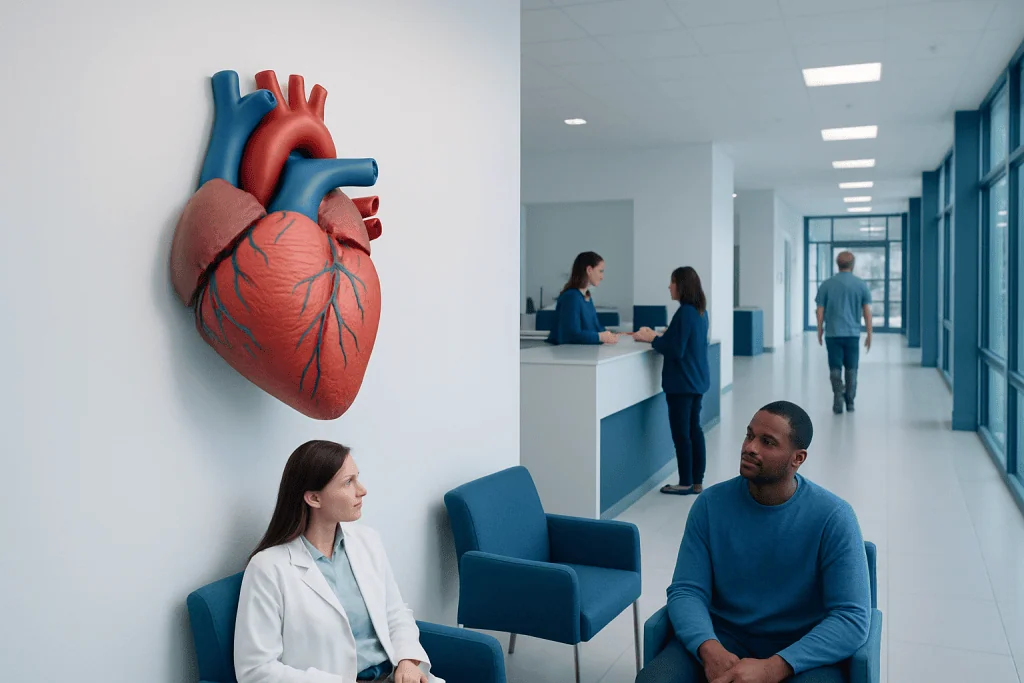
The heart has four chambers: two atria and two ventricles. Four valves keep blood moving in one direction through the heart.
Key Takeaways
- The heart has four chambers: two atria and two ventricles.
- Four valves keep blood moving in one direction through the heart.
- The right side sends blood to the lungs, and the left side sends oxygen-rich blood to the body.
- Electrical signals coordinate each heartbeat so the chambers pump in the correct order.
- Common heart problems can affect the muscle, valves, rhythm, or blood vessels.
The heart is a muscular pump that moves blood through the lungs and the rest of the body. Understanding its chambers, valves, and blood flow can make heart health easier to follow and discuss with a doctor.
Overview: what the heart does
The heart is a strong, fist-sized muscle located slightly to the left of the center of the chest. Its main job is to pump blood continuously so that oxygen, nutrients, hormones, and immune cells can reach the body’s tissues. At the same time, blood carries carbon dioxide and other waste products away to be removed.
The heart works as part of the cardiovascular system, which includes the blood vessels and blood itself. Arteries carry blood away from the heart, veins bring blood back, and tiny capillaries allow exchange with the body’s cells. Even while a person is resting, the heart is active every moment.
Although the heart may seem complicated, its basic function is simple: receive blood, pump it to the lungs for oxygen, and then pump oxygen-rich blood to the body. This process repeats with each heartbeat, usually without any conscious effort.
The four chambers of the heart
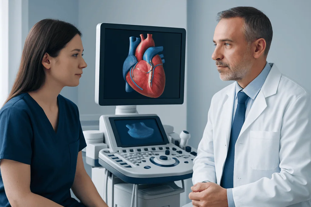
The heart has four chambers arranged in two sides. The upper chambers are called the right atrium and left atrium. The lower chambers are called the right ventricle and left ventricle. Each chamber has a specific role in moving blood forward.
The atria are smaller receiving chambers. The right atrium collects oxygen-poor blood returning from the body. The left atrium receives oxygen-rich blood coming back from the lungs. The ventricles are the stronger pumping chambers. The right ventricle sends blood to the lungs, while the left ventricle pumps blood out to the entire body.
The left ventricle has the thickest muscle wall because it must generate enough pressure to move blood through the body’s large network of vessels. The right ventricle does not need to pump as forcefully because the lungs are nearby and require lower pressure.
- Right atrium: receives blood from the body
- Right ventricle: pumps blood to the lungs
- Left atrium: receives blood from the lungs
- Left ventricle: pumps blood to the body
How the heart valves keep blood moving

Between the chambers and at the exits of the heart are four valves. These valves act like one-way doors. They open to let blood move forward and close to help prevent blood from flowing backward.
The tricuspid valve sits between the right atrium and right ventricle. The pulmonary valve is located between the right ventricle and the pulmonary artery, which carries blood to the lungs. On the left side, the mitral valve lies between the left atrium and left ventricle, and the aortic valve sits between the left ventricle and the aorta, the body’s main artery.
Healthy valves open and close in a carefully timed sequence with each heartbeat. If a valve becomes narrow, stiff, or leaky, blood flow can be disrupted. This may happen in heart valve disease, where the heart may need to work harder to maintain normal circulation.
Blood flow through the heart and lungs
Blood travels through the heart in a continuous loop. Oxygen-poor blood from the body returns through large veins into the right atrium. It then passes through the tricuspid valve into the right ventricle. When the right ventricle contracts, blood moves through the pulmonary valve and into the pulmonary arteries, which carry it to the lungs.
In the lungs, blood releases carbon dioxide and picks up fresh oxygen. This oxygen-rich blood returns to the heart through the pulmonary veins and enters the left atrium. It then flows through the mitral valve into the left ventricle.
When the left ventricle contracts, blood is pumped through the aortic valve into the aorta and then distributed to the body. After delivering oxygen and nutrients, blood returns through the veins to the right side of the heart, and the cycle begins again. This is the basic answer to the question of how the heart works.
How the heartbeat is controlled
The heart has its own electrical system that controls the timing of each beat. A natural pacemaker called the sinoatrial node, located in the right atrium, starts the electrical signal. This signal spreads across the atria, causing them to contract and push blood into the ventricles.
The signal then reaches the atrioventricular node, where there is a very brief pause. This delay gives the ventricles time to fill properly before they contract. The signal then travels through specialized pathways in the ventricles, triggering a coordinated squeeze that sends blood to the lungs and body.
If the electrical system becomes irregular, the heart may beat too fast, too slowly, or unevenly. These rhythm problems are known as arrhythmias. Some are harmless, while others may need evaluation or treatment. In selected cases, tests or procedures such as electrophysiology study and ablation may help diagnose or treat abnormal rhythms.
What can affect normal heart function
Several types of conditions can interfere with how the heart pumps blood. Problems may affect the heart muscle, the valves, the electrical system, or the blood vessels that supply the heart itself. For example, narrowed coronary arteries can reduce blood flow to the heart muscle and may lead to coronary artery disease.
High blood pressure can make the heart work harder over time. Valve disorders may cause obstruction or leakage. Damage to the heart muscle may weaken pumping ability, a problem often seen in heart failure. Rhythm disorders can also reduce the heart’s efficiency and sometimes cause palpitations, dizziness, or fainting.
Risk factors for heart disease include smoking, diabetes, high cholesterol, obesity, physical inactivity, chronic stress, poor sleep, and a family history of cardiovascular problems. Age also increases risk. Understanding how the heart normally works can make it easier to see why these factors matter and why prevention is so important.
How doctors assess heart health
Doctors assess heart health by combining symptoms, physical examination, and testing when needed. A person may be evaluated for chest discomfort, shortness of breath, swelling, fatigue, palpitations, or reduced exercise tolerance. Listening to the heart can sometimes reveal a murmur, extra sounds, or an irregular rhythm.
Common tests include an electrocardiogram to check the heart’s electrical activity and an echocardiogram to show the chambers, valves, and pumping function. Blood pressure measurement, blood tests, stress testing, and imaging may also be used depending on the person’s symptoms and medical history.
When blood vessel disease is suspected, more specialized imaging or procedures may be recommended. In some situations, treatment may include coronary angiography to assess coronary arteries or cardiac bypass surgery if circulation to the heart muscle needs to be restored. The right approach depends on the underlying problem rather than on symptoms alone.
Protecting heart health and when to seek medical care
Everyday habits play a major role in keeping the heart working well. A heart-healthy lifestyle usually includes regular physical activity, balanced nutrition, avoiding tobacco, limiting alcohol, managing stress, and getting adequate sleep. Controlling conditions such as high blood pressure, diabetes, and high cholesterol is also important.
People should seek medical advice if they have ongoing chest discomfort, unexplained shortness of breath, swelling in the legs, frequent palpitations, fainting, or unusual fatigue. Sudden severe chest pain, severe breathlessness, or signs of stroke need urgent emergency care. Early evaluation often helps identify problems before they become more serious.
Learning the basics of how the heart works can help patients ask informed questions and recognize when symptoms should not be ignored. For international patients who need evaluation or treatment, Acibadem International’s multidisciplinary specialists and JCI-accredited hospitals provide care for a wide range of heart conditions.
Frequently asked questions
What is the main function of the heart?
The heart’s main function is to pump blood throughout the body. This delivers oxygen and nutrients to tissues and helps remove carbon dioxide and other waste products.
How many chambers does the heart have?
The heart has four chambers. These are the right atrium, right ventricle, left atrium, and left ventricle, and each one helps move blood in a specific part of the circulation cycle.
What do heart valves do?
Heart valves help blood move in one direction through the heart. They open and close with each heartbeat to reduce backward flow and support efficient pumping.
Why does blood go to the lungs before the body?
Blood goes to the lungs to pick up oxygen and release carbon dioxide. Once it is oxygen-rich, it returns to the left side of the heart and is pumped out to the rest of the body.
What controls the heartbeat?
The heartbeat is controlled by the heart’s electrical system. A natural pacemaker in the right atrium starts the signal, and this signal travels through the heart to coordinate the pumping sequence.
Can a person live normally with a heart valve or rhythm problem?
Many people with heart valve or rhythm problems live active, normal lives, especially when the condition is monitored and treated appropriately. The outlook depends on the type of problem, its severity, and how early it is addressed.
References
- American Heart Association
- National Heart, Lung, and Blood Institute
- British Heart Foundation
- European Society of Cardiology
- World Health Organization
This article is for general information only and is not a substitute for professional medical advice. Please consult a qualified doctor about your individual situation.
Heart care in Turkey — expert evaluation and treatment
JCI-accredited · board-certified surgeons · reply within 24h
Add us as a Preferred Source to see more of our trusted health content across Google Search, AI Overviews and Discover.