Cerebral Angiography vs MR Angiography: Which Brain Vessel Test Is Right?

MR angiography is noninvasive and is often used first for many brain vessel questions. Cerebral angiography is more invasive but provides highly detailed images and can guide treatment during the same procedure.
Key Takeaways
- MR angiography is noninvasive and is often used first for many brain vessel questions.
- Cerebral angiography is more invasive but provides highly detailed images and can guide treatment during the same procedure.
- Each test has different benefits, limits, and safety considerations, including contrast use and device compatibility.
- Doctors choose the test based on symptoms, urgency, suspected condition, and whether an intervention may be needed.
- A neurologist, neuroradiologist, or interventional specialist can explain which option fits the clinical situation best.
Cerebral angiography and MR angiography both examine blood vessels in the brain, but they do so in different ways. The best choice depends on how much detail is needed, whether treatment may be required, and the person’s overall health and medical history.
Overview: How These Two Brain Vessel Tests Differ
When doctors need to look closely at blood vessels in the brain, they may recommend either MR angiography or cerebral angiography. Both tests are designed to show how blood flows through arteries and veins, helping identify problems such as narrowing, blockages, aneurysms, or abnormal vessel connections. Although they sound similar, they are not the same test.
MR angiography, often called MRA, uses magnetic resonance imaging to create pictures of blood vessels. It does not usually require a catheter placed into an artery, so it is considered noninvasive. In many cases, it can be done with or without contrast dye, depending on the question being asked and the type of scanner or protocol used.
Cerebral angiography, often called catheter angiography or digital subtraction angiography, involves inserting a thin catheter into a blood vessel, usually through the wrist or groin, and guiding it toward the arteries that supply the brain. Contrast dye is then injected while X-ray images are taken. This method is more invasive, but it remains the most detailed way to study brain blood vessels in certain situations.
In practice, the choice is not about one test being universally better than the other. It is about selecting the right test for the right purpose. For many people, imaging begins with noninvasive neuroradiology evaluation and then moves to catheter-based testing only if more precision or treatment planning is needed.
When Doctors Use MR Angiography
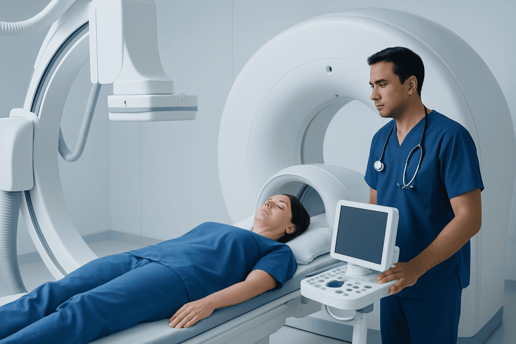
MR angiography is often chosen when doctors want a detailed look at brain vessels without using an invasive procedure. It may be helpful in evaluating headaches, dizziness, suspected aneurysms, blood vessel narrowing, or changes seen on other scans. It can also be useful for follow-up monitoring when a condition has already been identified and repeated imaging may be needed over time.
Because MRA does not usually require an arterial puncture, recovery is simple. Most people can return to normal activities soon after the exam. This makes it especially appealing when the clinical situation is stable and there is enough time to gather information without moving directly to an invasive study.
MRA is commonly used when doctors are evaluating possible reduced blood flow to the brain, including conditions related to carotid artery stenosis. It may also be considered after symptoms suggestive of a transient ischemic attack, depending on the person’s symptoms and the rest of the imaging plan.
Even so, MRA has limitations. Very small vessels or subtle abnormalities may not always be shown as clearly as they are with catheter angiography. Motion during the scan can also reduce image quality, and some people cannot undergo MRI because of certain implanted devices, severe claustrophobia, or other specific safety concerns.
When Cerebral Angiography Is Preferred

Cerebral angiography is usually recommended when doctors need the most precise view of brain vessels or when there is a strong possibility that treatment may be needed. It is especially useful for evaluating aneurysms, arteriovenous malformations, vasculitis patterns, vessel spasm, or complex narrowing that may not be fully defined on noninvasive imaging.
This test can be essential in urgent situations, such as suspected bleeding around the brain or a serious blood vessel blockage. For example, after a subarachnoid hemorrhage, catheter angiography may help find the source of bleeding if other imaging is inconclusive. It can also provide detailed guidance in some cases of suspected stroke when endovascular treatment is being considered.
An important advantage is that diagnosis and treatment may sometimes happen in the same session. If a blocked or abnormal vessel is found, the specialist may be able to proceed with selected endovascular care through interventional neuroradiology or interventional neurology, depending on the condition and the treatment plan.
Because cerebral angiography is invasive, it is not automatically the first choice for every person. Instead, it is reserved for situations where its extra detail, accuracy, or treatment capability is likely to change care in a meaningful way.
What Each Test Can Show
Both tests can help doctors assess the shape, size, and course of blood vessels in the brain. They can identify narrowing, complete blockage, ballooning of the vessel wall such as an aneurysm, and some abnormal connections between arteries and veins. In many routine cases, MRA provides enough information to support diagnosis and treatment planning.
However, cerebral angiography gives a dynamic, highly detailed view of blood flow in real time. It can show how quickly contrast moves through the vessels and how different vascular territories connect. This level of detail can matter when small abnormalities are suspected or when doctors need to understand the exact anatomy before a procedure.
MRA may sometimes overestimate or underestimate the degree of narrowing, especially in small or tortuous vessels. It is also less suited for direct therapeutic action. Cerebral angiography, by contrast, is often considered the reference standard when exact vessel anatomy must be confirmed.
In simple terms, MRA is often excellent for screening and follow-up, while cerebral angiography is often best when the stakes are higher, the anatomy is complex, or treatment decisions depend on the most exact possible map of the vessels.
Preparation, Procedure, and Recovery
For MR angiography, preparation is usually straightforward. The person may be asked about kidney function, allergies, pregnancy, and any metal implants or devices. During the test, they lie still on a table that moves into the MRI scanner. The exam is painless, though the machine can feel noisy and enclosed. If contrast is used, it is usually given through a vein in the arm.
Recovery after MRA is minimal. Most people leave soon after the scan unless they received sedation. Drinking fluids may be advised if contrast was used, especially if there are any concerns about kidney function.
Cerebral angiography requires more preparation. Blood tests may be needed, and the care team reviews medicines, including blood thinners, diabetes medications, and any prior contrast reactions. During the procedure, a catheter is inserted into an artery and carefully advanced under imaging guidance. Contrast dye is injected while a series of images is obtained.
After catheter angiography, the puncture site must be monitored for bleeding, swelling, or bruising. The person may need to lie flat for a period, depending on the access site and closure method. Mild soreness can occur, but most people recover without difficulty when the procedure is performed by experienced teams.
Risks, Safety, and Limitations
MR angiography is generally very safe, but it is not risk-free. If contrast is used, there is a small chance of an allergic reaction or concerns related to kidney function in susceptible individuals. MRI itself may not be suitable for some people with certain pacemakers, implanted devices, or metal fragments, although modern device compatibility is broader than in the past and must be checked carefully.
Cerebral angiography carries additional risks because it involves arterial access and contrast injection directly into blood vessels. Possible complications include bleeding or bruising at the catheter site, contrast reactions, kidney strain, blood vessel injury, or, rarely, stroke. These risks are uncommon but are part of the reason the test is used selectively.
Doctors weigh these risks against the expected benefit. If a person’s symptoms suggest a serious vascular problem that requires exact imaging or urgent treatment, the greater precision of cerebral angiography may outweigh its added invasiveness. If the goal is screening or routine follow-up, MRA may be sufficient and more convenient.
One practical limitation is that image quality in MRA can be affected by movement, irregular heart rhythm, or technical factors. In cerebral angiography, the limitation is not usually image clarity but rather the need for a procedure room, a trained interventional team, and short-term observation after the test.
How Doctors Decide Which Test Is Right
The decision is based on the clinical question. If doctors need a broad, noninvasive assessment of brain circulation, MR angiography is often the first step. If they need to confirm a small abnormality, clarify uncertain results, or prepare for a procedure, cerebral angiography may be recommended instead.
Symptoms matter as well. Sudden severe headache, signs of stroke, new neurological deficits, or suspected vascular bleeding may call for a more urgent and precise imaging pathway. In contrast, longstanding stable symptoms or incidental findings may be evaluated with less invasive imaging first.
Personal factors also influence the choice. Kidney health, previous contrast reactions, claustrophobia, implanted devices, ability to lie still, and current medications all matter. Doctors also consider whether the person is likely to need treatment immediately after diagnosis, because catheter angiography can sometimes transition directly into therapy.
Patients benefit from asking a few clear questions: What is this test looking for? Could a noninvasive test answer the same question? Would the result change treatment? Understanding the purpose of the exam often makes the decision feel less overwhelming. Near the end of the care pathway, Acibadem International’s multidisciplinary specialists in JCI-accredited hospitals diagnose and treat brain vessel conditions for international patients when advanced evaluation is needed.
When to Seek Medical Advice Promptly
Medical advice should be sought promptly if a person develops sudden neurological symptoms. These may include weakness on one side, trouble speaking, facial drooping, sudden loss of vision, severe dizziness, confusion, or a sudden intense headache unlike usual headaches. These symptoms can signal a brain blood vessel emergency and should not be ignored.
People should also contact a doctor if they have been told they may have an aneurysm, vessel narrowing, or another vascular abnormality and are unsure which test is appropriate. Questions are especially important before imaging if there is kidney disease, pregnancy, an implanted device, or a previous reaction to contrast dye.
After cerebral angiography, urgent medical attention is needed if there is increasing swelling, persistent bleeding, numbness in the limb used for access, chest pain, shortness of breath, or new neurological symptoms. After MR angiography, concerns are less common, but any unexpected symptoms after contrast or sedation should still be reported.
Timely evaluation can help doctors choose the safest and most useful test. The goal is not simply to obtain pictures, but to answer the right question in a way that supports effective, individualized care.
Frequently asked questions
Is MR angiography safer than cerebral angiography?
MR angiography is generally considered safer because it is noninvasive and usually does not require a catheter placed in an artery. Cerebral angiography is also commonly performed safely, but it has additional procedural risks such as bleeding at the access site and, rarely, stroke.
Which test is more accurate for brain blood vessels?
Cerebral angiography is typically the most detailed and accurate test for defining complex brain vessel anatomy. MR angiography is very useful and often accurate enough for screening, diagnosis, and follow-up, but it may not show very small or subtle abnormalities as clearly.
Does MR angiography always need contrast dye?
No. Some MR angiography techniques can be performed without contrast, while others use intravenous contrast to improve image quality. The decision depends on what the doctor is looking for and the person’s medical history.
Can treatment be done during cerebral angiography?
In some cases, yes. If the problem found is suitable for endovascular treatment, doctors may be able to continue with a catheter-based procedure during the same session or a planned later session. This is one of the major advantages of cerebral angiography.
How long do these tests take?
MR angiography usually takes less time overall and does not require a recovery period unless sedation is used. Cerebral angiography often takes longer because it includes preparation, the procedure itself, and observation afterward.
Who should not have MR angiography?
Some people with certain implanted devices, metal fragments, or severe claustrophobia may not be suitable candidates for MRI-based tests. The imaging team reviews device compatibility and other safety factors before scheduling the exam.
References
- American Heart Association
- American Stroke Association
- Radiological Society of North America
- National Institute of Neurological Disorders and Stroke
- American College of Radiology
This article is for general information only and is not a substitute for professional medical advice. Please consult a qualified doctor about your individual situation.
Explore treatments in Turkey — costs, top hospitals & a free quote
JCI-accredited · board-certified surgeons · reply within 24h