Diagnostic Medical Ultrasound: Who May Need It and What It Can Detect

Diagnostic medical ultrasound uses sound waves, not radiation, to create images inside the body. It can help assess pregnancy, abdominal organs, pelvic organs, blood vessels, the heart, thyroid, breasts, muscles, and soft tissues.
Key Takeaways
- Diagnostic medical ultrasound uses sound waves, not radiation, to create images inside the body.
- It can help assess pregnancy, abdominal organs, pelvic organs, blood vessels, the heart, thyroid, breasts, muscles, and soft tissues.
- Many people may need an ultrasound when they have pain, swelling, lumps, abnormal test results, or signs of circulation problems.
- Most ultrasound exams are quick, noninvasive, and require little or no recovery time.
- Preparation depends on the area being examined and may include fasting or having a full bladder.
- Ultrasound results are interpreted together with symptoms, physical examination, and sometimes other imaging tests.
Diagnostic medical ultrasound is a safe, widely used imaging test that helps doctors look at organs, soft tissues, blood flow, and pregnancy in real time. It may be recommended for symptoms such as pain, swelling, abnormal bleeding, or to monitor a developing baby and certain medical conditions.
Overview of diagnostic medical ultrasound
Diagnostic medical ultrasound is an imaging test that uses high-frequency sound waves to create pictures of structures inside the body. A handheld device called a transducer sends sound waves into the body and receives the echoes that bounce back. A computer then turns these echoes into real-time images that doctors can study.
Unlike X-rays or CT scans, ultrasound does not use ionizing radiation. For this reason, it is often chosen when a radiation-free imaging method is preferred, including during pregnancy and for repeated follow-up exams. It is especially useful for viewing soft tissues, fluid-filled structures, moving organs, and blood flow.
Ultrasound can be performed in different ways depending on the body area. Common types include abdominal, pelvic, obstetric, breast, thyroid, vascular, musculoskeletal, and echocardiographic ultrasound. Some exams are done through the skin, while others use internal probes, such as transvaginal or transrectal ultrasound, when a closer view is needed.
Who may need an ultrasound
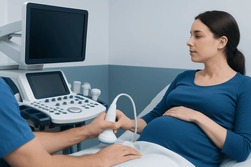
A doctor may recommend an ultrasound when a person has symptoms that suggest a problem in the abdomen, pelvis, blood vessels, heart, or soft tissues. Common reasons include abdominal pain, pelvic pain, swelling, a palpable lump, abnormal vaginal bleeding, urinary symptoms, or abnormal blood test results. Ultrasound is also often used to investigate suspected gallstones, kidney stones, liver problems, thyroid nodules, and fluid collections.
Pregnant patients commonly have ultrasound to confirm pregnancy, estimate gestational age, check fetal growth, assess the placenta, and look for structural concerns. In gynecology, ultrasound can help evaluate the uterus and ovaries in people with pain, cysts, fibroids, or fertility concerns. In vascular medicine, it may be used to look for poor circulation, narrowed arteries, or blood clots such as deep vein thrombosis.
Ultrasound may also be needed to guide medical procedures. Examples include helping place a needle into a fluid collection, cyst, joint, thyroid nodule, or breast lesion. Real-time imaging can improve accuracy and help doctors avoid nearby structures during biopsies, aspirations, and some pain-management or drainage procedures.
What diagnostic medical ultrasound can detect

Ultrasound can detect many different findings, but what it shows depends on the body part being examined. In the abdomen, it may reveal gallstones, liver enlargement, fatty liver changes, bile duct widening, kidney swelling, kidney cysts, and some masses. In the pelvis, it may show ovarian cysts, uterine fibroids, endometrial thickening, or signs related to ovarian cysts.
In pregnancy, ultrasound can confirm that a pregnancy is in the uterus, estimate dates, assess fetal heartbeat and movement, and evaluate growth and anatomy. In the blood vessels, Doppler ultrasound can show how blood is flowing and help detect reduced circulation, varicose vein problems, or clots. In the neck, ultrasound can assess thyroid size, inflammation, and nodules, including features that may need follow-up or biopsy.
Ultrasound is also useful for breast imaging, especially in younger patients or for evaluating a lump found on exam or mammography. In muscles, tendons, and joints, it can identify fluid, tears, inflammation, and some soft tissue masses. The heart can be examined with echocardiography, a specialized ultrasound that helps assess heart chambers, valves, pumping function, and fluid around the heart.
Although ultrasound is very helpful, it does have limits. Air in the intestines, obesity, deep structures, and some bone-protected areas can make images harder to interpret. In some situations, doctors may recommend additional imaging such as MRI or CT scan to answer a question more clearly.
How the test is performed and how to prepare
Most ultrasound exams are simple and are done in an outpatient setting. During a standard ultrasound, gel is placed on the skin and the transducer is moved over the area being examined. The gel helps sound waves travel efficiently and improves image quality. The test is usually painless, though mild pressure may be felt if the area is tender.
Preparation depends on the type of ultrasound. For an abdominal ultrasound, the patient may be asked not to eat or drink for several hours so the gallbladder and upper abdominal organs can be seen more clearly. For a pelvic ultrasound, a full bladder may be needed because it can improve the view of the uterus and ovaries. For many other exams, no special preparation is required.
Some ultrasounds use internal probes for a more detailed view. A transvaginal ultrasound may be recommended to evaluate the uterus, ovaries, early pregnancy, or pelvic pain. A transrectal ultrasound may be used for the prostate or nearby structures. These tests can feel unfamiliar or slightly uncomfortable, but they are usually well tolerated and are performed with attention to privacy and patient comfort.
How doctors interpret ultrasound results
Ultrasound images are typically reviewed by a radiologist or another specially trained physician, then shared with the doctor who ordered the test. The report describes what was seen, whether the appearance is normal or abnormal, and whether any follow-up is recommended. The findings are interpreted together with symptoms, examination results, and laboratory tests.
Sometimes an ultrasound gives a clear answer right away, such as identifying gallstones, a simple cyst, or a blood clot. In other cases, the result may be indeterminate, meaning the imaging shows a change but cannot fully explain its cause. This does not necessarily mean something serious is present; it simply means more information may be needed.
If the images raise further questions, doctors may suggest repeat ultrasound after a period of monitoring, additional laboratory tests, or another imaging study such as PET-CT in selected oncology settings. When a tissue diagnosis is necessary, ultrasound can also help guide a biopsy safely and precisely.
Benefits, limitations, and safety
One of the main advantages of diagnostic medical ultrasound is that it provides real-time images without radiation exposure. It is widely available, relatively quick, and often less costly than more complex imaging methods. Because it can show motion, it is particularly useful for evaluating blood flow, fetal movement, heart function, and the movement of tendons or joints.
Ultrasound is generally considered very safe when used appropriately by trained healthcare professionals. It has been used in obstetrics and many other areas of medicine for decades. The exam itself does not usually require sedation, and most people can return to their usual activities immediately afterward.
Its limitations are important to understand. Ultrasound does not show all organs or conditions equally well, and image quality depends on the body area, the patient, and the examiner’s technique. It is best seen as one part of diagnosis rather than a stand-alone answer for every medical question.
When to speak with a doctor
A person should speak with a doctor if they have ongoing pain, swelling, a new lump, abnormal bleeding, unexplained digestive symptoms, urinary changes, or signs of poor circulation such as leg swelling or calf pain. A doctor can decide whether ultrasound is the right first test or whether another examination would be more useful. Prompt assessment can help clarify the cause and guide the next steps.
People should also seek medical advice if an ultrasound report recommends follow-up imaging, a specialist visit, or biopsy. Many ultrasound findings are benign, but follow-up matters because it helps confirm whether a finding is stable, harmless, or in need of treatment. Questions about the report should be discussed with the treating clinician, who can explain the result in context.
For international patients who need imaging and coordinated specialist care, Acibadem International offers diagnostic assessment and treatment through multidisciplinary teams in JCI-accredited hospitals. This can be helpful when ultrasound findings need further evaluation by radiology, gynecology, cardiology, vascular, or surgical specialists.
Frequently asked questions
Is diagnostic medical ultrasound safe?
Yes. Diagnostic medical ultrasound is generally considered safe because it uses sound waves rather than ionizing radiation. It has been used for many years in pregnancy and many other medical settings when performed by trained professionals.
Does an ultrasound show cancer?
Ultrasound can show lumps, nodules, cysts, and other abnormal changes, but it cannot always determine whether a finding is cancer. If something looks suspicious, the doctor may recommend follow-up imaging or a biopsy to make a diagnosis.
What is the difference between ultrasound and Doppler ultrasound?
Standard ultrasound creates pictures of organs and soft tissues. Doppler ultrasound is a special technique that shows how blood is moving through vessels, which helps doctors assess circulation and look for narrowing or clots.
How long does an ultrasound take?
Many ultrasound exams take about 15 to 45 minutes, depending on the body area and the reason for the test. More detailed studies, such as some pregnancy or heart ultrasounds, may take longer.
Do people need to fast before an ultrasound?
Sometimes. Fasting may be needed before an abdominal ultrasound, while a full bladder may be requested for some pelvic scans. The imaging center or doctor usually gives specific instructions based on the type of exam.
Can ultrasound detect pregnancy problems early?
Ultrasound can help detect some pregnancy-related concerns early, such as the location of the pregnancy, fetal heartbeat, growth, and certain structural findings. The timing and detail depend on how far along the pregnancy is and the type of ultrasound performed.
This article is for general information only and is not a substitute for professional medical advice. Please consult a qualified doctor about your individual situation.
Ultrasound in Turkey — costs, top hospitals & a free quote
JCI-accredited · board-certified surgeons · reply within 24h
Add us as a Preferred Source to see more of our trusted health content across Google Search, AI Overviews and Discover.

Check-up & Preventive Medicine
Comprehensive health screenings designed for early detection and prevention.
1 specialists in this unitMore from the Health Library

What Is a Lab Test Panel and Why Are Tests Grouped Together?
PET-CT Scan: How It Helps Detect and Stage Cancer

Quest Diagnostics Hours: When Labs Open and How to Plan a Visit

Quest Diagnostics Login and MyQuest: What the Patient Portal Lets You Do

Diagnostic Tests for Breast Symptoms: Mammogram, Ultrasound, or MRI?