Peripheral Vision Loss: Causes and When Neuro-Ophthalmology Is Needed

Peripheral vision loss may develop suddenly or gradually and can affect one or both eyes. Common causes include glaucoma, retinal disease, optic nerve disorders, stroke, and other neurological conditions.
Key Takeaways
- Peripheral vision loss may develop suddenly or gradually and can affect one or both eyes.
- Common causes include glaucoma, retinal disease, optic nerve disorders, stroke, and other neurological conditions.
- Formal visual field testing and a detailed eye and neurological examination help find the cause.
- Sudden vision changes, headache, weakness, double vision, or eye pain need urgent medical attention.
- Neuro-ophthalmology is especially useful when visual field loss may be linked to the optic nerve, brain, or eye movement pathways.
Peripheral vision loss means reduced side vision, sometimes described as tunnel vision or missing areas in the visual field. It can happen for several reasons, ranging from eye conditions to problems affecting the optic nerve or brain, so proper evaluation is important.
Overview
Peripheral vision loss is a reduction in the ability to see objects to the side while looking straight ahead. Many people call it tunnel vision, although the pattern can vary. Some notice bumping into objects, difficulty driving, trouble navigating crowds, or missing people approaching from the side.
This symptom is not a diagnosis by itself. Instead, it is a sign that can be caused by several different conditions affecting the eyes, optic nerves, or brain. The change may happen in one eye or both, and it may begin suddenly or progress gradually over time.
Because side vision depends on healthy structures along the entire visual pathway, the cause is not always limited to the eye itself. In some cases, a person may need assessment by a neuro-ophthalmology team when the pattern of vision loss suggests involvement of the optic nerve or the brain’s visual pathways.
Symptoms and How It May Feel
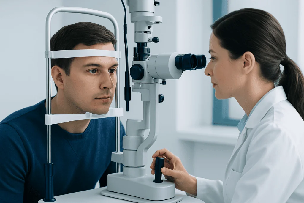
People experience peripheral vision loss in different ways. Some describe darkened or blurred edges of vision, while others notice missing patches, difficulty seeing in dim light, or a narrowed field of view. The change may be subtle at first and only become obvious during driving, walking in unfamiliar places, or sports.
Symptoms may affect daily life in practical ways. Reading central text can remain normal in some conditions, which can delay recognition of the problem. Others may notice poor depth awareness, increased tripping, trouble with stairs, or difficulty locating objects unless they turn their head more than usual.
Additional symptoms can offer clues about the underlying cause. These may include:
- Blurred vision or reduced sharpness
- Eye pain, especially with eye movement
- Flashing lights or new floaters
- Headache
- Double vision
- Numbness, weakness, imbalance, or speech difficulty
- Night vision problems
Any sudden visual field change should be taken seriously, especially if it occurs with neurological symptoms. While not every case is an emergency, urgent assessment may be needed to protect vision and address time-sensitive causes.
Causes and Risk Factors

Peripheral vision loss can result from disorders of the eye, optic nerve, or brain. One of the best-known eye causes is glaucoma, which can gradually damage the optic nerve and reduce side vision before a person notices symptoms. Retinal disorders, including retinal detachment, may also cause part of the peripheral field to disappear, sometimes suddenly.
Conditions affecting the optic nerve can produce characteristic visual field defects. These include optic neuritis, ischemic optic neuropathy, compression from masses near the optic pathways, and inherited or inflammatory optic nerve disease. Neuro-ophthalmology becomes especially important when the pattern of loss suggests a problem beyond the eyeball itself.
Brain-related causes include stroke, head injury, tumors, and disorders involving the visual pathways in the occipital lobe or deeper brain structures. For example, a stroke can cause loss of the same side of vision in both eyes, even if the eyes themselves are healthy. Migraine with visual symptoms may temporarily affect the visual field, but persistent or repeated episodes still need medical review.
Risk factors depend on the cause but can include older age, diabetes, high blood pressure, smoking, autoimmune disease, severe nearsightedness, family history of glaucoma, past eye injury, and known neurological disease. Some medications and toxins can also affect vision, though this is less common.
When Neuro-Ophthalmology Is Needed
A neuro-ophthalmologist is a specialist who evaluates vision problems related to the optic nerve, eye movement system, and brain. Referral is often helpful when the cause of peripheral vision loss is not clearly limited to the front structures of the eye, or when standard eye examination findings do not fully explain the symptoms.
Neuro-ophthalmology may be needed if visual field loss is accompanied by double vision, unequal pupils, drooping eyelid, unexplained optic nerve swelling, color vision loss, eye movement problems, or signs of neurological disease. It is also appropriate when imaging suggests a problem near the optic chiasm or visual pathways, or when one eye specialist suspects a neurological cause.
Examples include unexplained visual field defects, suspected optic neuritis, compressive lesions, pituitary-related vision changes, and stroke-related visual pathway injury. In these situations, coordinated assessment may involve ophthalmology, neurology, neuroradiology, and when appropriate brain tumor surgery or stroke treatment teams.
The goal is not simply to confirm vision loss but to identify where along the visual pathway the problem is occurring. That location often determines the next tests, the urgency of care, and the most suitable treatment plan.
How Doctors Diagnose Peripheral Vision Loss
Diagnosis begins with a careful history. Doctors ask when the problem started, whether it is sudden or gradual, whether one or both eyes are involved, and whether there are associated symptoms such as headache, pain, flashes, weakness, or double vision. Past eye disease, medications, vascular risk factors, and family history are also important.
The examination usually includes visual acuity, pupil testing, eye pressure measurement, assessment of color vision, and a dilated eye examination to look at the retina and optic nerve. Formal visual field testing is especially important because it maps the pattern of missing vision and can provide strong clues about whether the cause is retinal, optic nerve-related, or neurological.
Additional testing depends on the suspected cause. Doctors may use optical coherence tomography to examine retinal and optic nerve layers, blood tests for inflammatory or autoimmune conditions, and imaging such as MRI or CT when a brain or optic pathway problem is suspected. If retinal disease is the concern, retinal imaging and specialist assessment may be needed.
In some cases, urgent imaging is recommended right away, particularly when the onset is sudden or accompanied by neurological signs. If a structural cause is identified, further evaluation by specialists may include MRI imaging and, when surgery is considered, consultation for neurosurgery.
Treatment Options
Treatment for peripheral vision loss depends entirely on the underlying cause. There is no single remedy that fits all cases. Some causes can be treated promptly to preserve remaining vision, while others focus on slowing progression, managing symptoms, or improving safety and adaptation.
If glaucoma is responsible, treatment may involve eye drops, laser procedures, or surgery to reduce eye pressure and protect the optic nerve. Retinal tears or detachments often require urgent retinal care. Inflammatory optic nerve conditions may be treated with medications directed by a specialist, while compression of the visual pathway may require medical or surgical treatment depending on the source.
For brain-related causes, management may include stroke care, treatment of a tumor, control of inflammation, or rehabilitation after neurological injury. When vision loss cannot be fully reversed, visual rehabilitation strategies can still help. These may include scanning training, mobility advice, driving guidance based on local regulations, and low-vision support.
People should avoid starting or stopping treatment on their own without medical advice. Because causes vary widely, the safest approach is a targeted plan based on an accurate diagnosis and regular follow-up when recommended.
Prevention and Self-Care
Not every cause of peripheral vision loss can be prevented, but some risks can be reduced. Regular eye examinations are important, especially for people over a certain age, those with diabetes or high blood pressure, individuals with a family history of glaucoma, and anyone who has already been told they are at risk for eye disease.
General vascular health also matters because blood vessel problems can affect both the eyes and the brain. Managing blood pressure, cholesterol, and blood sugar, staying physically active, not smoking, and following treatment for chronic conditions all support long-term eye and neurological health.
Practical self-care is useful while evaluation is in progress or after diagnosis. Good lighting at home, reducing trip hazards, moving slowly in crowded areas, and turning the head more deliberately can improve safety. Driving should be discussed with a doctor if vision loss affects field requirements or awareness of surroundings.
For people receiving specialist care, follow-up appointments are important even when symptoms seem stable. Near the end of the care pathway, a center such as Acibadem International may coordinate evaluation through multidisciplinary specialists and JCI-accredited hospitals for international patients with eye, optic nerve, or brain-related causes of visual field loss.
When to See a Doctor
A doctor should evaluate any new peripheral vision loss, even if it seems mild. Gradual changes can still reflect important conditions, including glaucoma or neurological disease, and early detection may help preserve vision and function.
Urgent medical attention is needed if the loss is sudden, painful, or associated with flashes, many new floaters, a curtain-like shadow, severe headache, weakness, numbness, confusion, trouble speaking, or double vision. These features can point to retinal detachment, stroke, optic nerve inflammation, or other conditions that may need prompt treatment.
People should also seek reassessment if symptoms worsen, affect daily activities, or continue despite previous treatment. A second opinion may be appropriate when the cause remains unclear or when neuro-ophthalmology expertise is recommended.
Frequently asked questions
What does peripheral vision loss mean?
Peripheral vision loss means reduced ability to see objects at the edges of the visual field while looking straight ahead. Some people describe it as tunnel vision, but the exact pattern can differ depending on the cause.
Can peripheral vision loss be temporary?
Sometimes it can be temporary, such as with certain migraine-related visual symptoms. However, any new or repeated visual field change should still be assessed because temporary symptoms can also mimic more serious eye or neurological problems.
Is peripheral vision loss always caused by an eye disease?
No. It may be caused by eye conditions such as glaucoma or retinal disease, but it can also result from optic nerve disorders or problems in the brain's visual pathways. That is why some people need neuro-ophthalmology evaluation.
How is peripheral vision loss tested?
Doctors usually perform a complete eye examination and formal visual field testing. Depending on findings, they may also order retinal scans, blood tests, or brain imaging such as MRI or CT.
Can lost peripheral vision come back?
Recovery depends on the underlying cause and how quickly treatment begins. Some causes are reversible or partly reversible, while others mainly require treatment to prevent further loss and to support adaptation.
When is peripheral vision loss an emergency?
It should be treated as urgent if it starts suddenly or occurs with eye pain, flashes, many new floaters, a curtain-like shadow, severe headache, weakness, or speech trouble. These symptoms can signal retinal detachment, stroke, or other conditions needing prompt care.
References
- American Academy of Ophthalmology
- National Eye Institute
- American Academy of Neurology
- National Institute of Neurological Disorders and Stroke
- Royal College of Ophthalmologists
This article is for general information only and is not a substitute for professional medical advice. Please consult a qualified doctor about your individual situation.
Explore treatments in Turkey — costs, top hospitals & a free quote
JCI-accredited · board-certified surgeons · reply within 24h
Add us as a Preferred Source to see more of our trusted health content across Google Search, AI Overviews and Discover.
Check your numbers in seconds
BMI, calories, due date, blood pressure and 30+ more clinical calculators — free, instant, doctor-reviewed ranges.