

Brain PET-CT in Dementia Evaluation: When Is It Used?
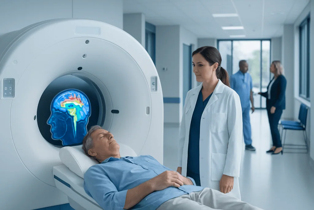
Brain PET-CT shows how different parts of the brain are functioning, not just what they look like structurally. It may help distinguish between some causes of dementia when the diagnosis remains uncertain after routine evaluation.
Key Takeaways
- Brain PET-CT shows how different parts of the brain are functioning, not just what they look like structurally.
- It may help distinguish between some causes of dementia when the diagnosis remains uncertain after routine evaluation.
- PET-CT is one part of a broader assessment that often includes history, cognitive testing, blood tests, and MRI or CT.
- The scan requires a small amount of radioactive tracer and is generally well tolerated.
- Results must be interpreted by specialists because PET findings can overlap and do not diagnose dementia by themselves.
Brain PET-CT can help doctors understand patterns of brain function that may support the evaluation of dementia when symptoms, examination, and standard tests do not give a clear answer. It is usually used selectively, alongside clinical assessment and other imaging, rather than as a first test for every person with memory problems.
Overview: What Brain PET-CT Means in Dementia Assessment
Brain PET-CT is an imaging test that combines positron emission tomography (PET) with computed tomography (CT). In dementia evaluation, the PET part is usually the most important because it shows patterns of brain metabolism or other disease-related changes. The CT part helps with anatomical localization and image correction.
Unlike a standard CT or MRI, which mainly show brain structure, PET can provide information about how active different brain regions are. This can be helpful because some neurodegenerative disorders affect brain function before clear structural changes are seen. In everyday practice, the most commonly used form is FDG PET, which shows how the brain uses glucose, its main energy source.
Brain PET-CT is not usually the first test ordered for memory concerns. Doctors typically begin with a medical history, neurological examination, cognitive testing, blood work, and structural imaging such as MRI or CT. PET-CT becomes more useful when these steps do not fully explain the symptoms or when there is a need to distinguish between conditions with similar presentations.
When Is Brain PET-CT Used?
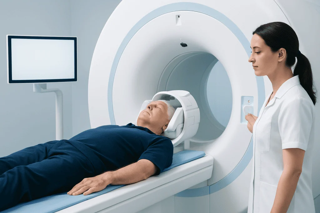
Brain PET-CT is generally used when a person has cognitive decline, behavioral change, language problems, or other symptoms suggesting dementia, but the exact cause remains uncertain. It can be especially helpful when the clinical picture is atypical, when symptoms begin at a younger age than expected, or when more than one diagnosis is possible.
For example, PET-CT may help support the distinction between Alzheimer’s disease and some other neurodegenerative conditions such as frontotemporal dementia or dementia with Lewy bodies. It may also be considered when symptoms involve unusual patterns, such as prominent visual-spatial difficulties, language loss, or marked changes in personality and behavior.
Doctors do not rely on PET-CT alone. The scan is interpreted in the context of the person’s symptoms, test results, medicines, mood, sleep, and general medical health. This matters because memory and thinking problems can also be affected by depression, thyroid disease, vitamin deficiencies, sleep disorders, stroke, or other neurological illnesses.
In some centers, more specialized PET studies may be used in selected cases. Depending on availability and the clinical question, these may include imaging related to amyloid or other disease markers, though such tests are not needed for everyone and are usually reserved for carefully chosen situations.
What the Scan Can Show
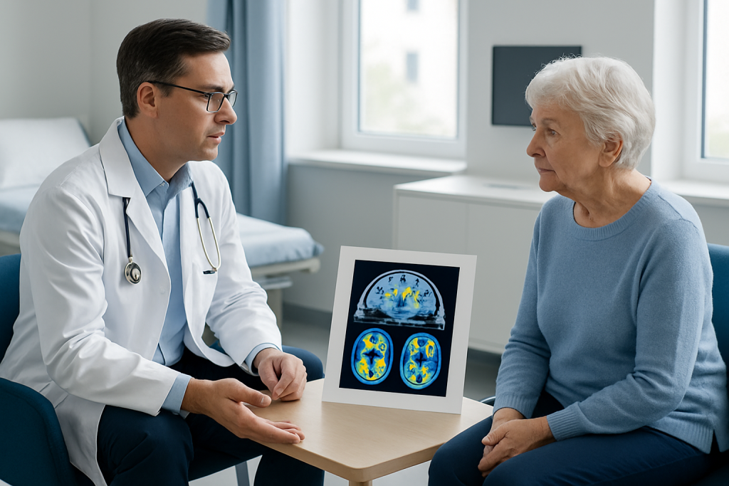
In FDG PET, areas of the brain that are less active use less glucose and appear as regions of reduced metabolism. Different dementia syndromes can produce different patterns. For instance, Alzheimer’s disease often affects the parietal and temporal regions and parts of the posterior cingulate cortex, while frontotemporal dementia more often involves the frontal and anterior temporal regions.
These patterns do not act like a simple yes-or-no test, but they can provide strong supporting evidence. PET findings may help explain why a person has memory loss, language difficulty, poor judgment, or changes in behavior. In some cases, the scan can also suggest that another condition should be considered instead of a primary neurodegenerative dementia.
The scan may be particularly helpful when MRI findings are mild or nonspecific. Structural imaging can show shrinkage, prior strokes, tumors, <a href="https://acibademinternational.com/diseases/normal-pressure-hydrocephalus/”>normal pressure hydrocephalus, or other causes of symptoms, while PET adds functional information. This is one reason PET is often considered an adjunct rather than a replacement for MRI or CT.
People who are being assessed for cognitive decline may also undergo other imaging tests when appropriate, such as brain MRI or PET-CT imaging, depending on the doctor’s question and the center’s protocols.
How the Test Is Performed and What to Expect
Before a brain PET-CT, the patient is usually asked to follow simple preparation instructions, which may include fasting for several hours. A small amount of radioactive tracer is injected into a vein. After the injection, there is a waiting period so the tracer can circulate and be taken up by the brain.
During the scan, the person lies still on a table while the PET-CT machine takes images. The test is painless, although staying still can feel tiring for some people. The scanner is open enough for many patients to tolerate well, but anyone with severe claustrophobia or difficulty lying flat should mention this in advance.
The radiation exposure is generally low and carefully controlled. PET-CT is avoided or used with special caution in pregnancy unless clearly necessary. Patients should also tell the medical team about diabetes, recent illness, medications, or movement problems, because these factors can affect image quality or interpretation.
After the scan, most people can return to normal activities unless their care team advises otherwise. A specialist in nuclear medicine or neuroradiology reviews the images and prepares a report for the doctor who is coordinating the dementia evaluation.
Benefits, Limits, and Accuracy
The main benefit of brain PET-CT is that it can reveal functional changes that are not obvious on standard structural scans. This can improve diagnostic confidence in selected patients and sometimes help families understand the likely nature of a person’s symptoms. A clearer diagnosis may also guide treatment planning, safety decisions, follow-up, and supportive care.
At the same time, PET-CT has limitations. It cannot by itself confirm how severe dementia is, predict exactly how fast symptoms will progress, or replace a careful neurological assessment. Some patterns overlap between conditions, and reduced metabolism may be influenced by previous stroke, epilepsy, head injury, severe depression, infection, or other brain disorders.
Accuracy is usually best when the scan is ordered for a specific clinical reason and interpreted by experienced specialists together with other test results. A normal or near-normal PET scan may be reassuring in some settings, but it does not always exclude disease, particularly in very early stages or in less typical presentations.
Because dementia is a broad term rather than a single disease, the imaging result is often discussed alongside possible conditions such as Alzheimer’s disease or frontotemporal dementia when doctors are narrowing the diagnosis.
How Brain PET-CT Fits Into the Full Diagnostic Workup
Evaluation of dementia usually begins long before any PET-CT is considered. Doctors first look closely at the person’s symptoms, how they developed over time, and whether daily activities are affected. Family observations are often very valuable because changes in memory, behavior, language, or planning may be easier for relatives to notice than for the patient.
Common parts of the workup include cognitive screening or formal neuropsychological testing, blood tests to look for reversible causes, and structural brain imaging. Depending on the situation, doctors may also assess sleep, mood, hearing, vision, gait, or signs of other neurological disorders. This broader view helps avoid mislabeling symptoms that might have a treatable cause.
PET-CT is best understood as a problem-solving tool. It can be considered when there is a significant question that standard assessment has not answered. In selected patients, further evaluation may also involve neurology consultation or other specialist input, especially if symptoms are complex or rapidly changing.
In experienced centers, multidisciplinary review may include neurologists, geriatric specialists, neuroradiologists, nuclear medicine physicians, psychiatrists, and neuropsychologists. Near the end of this process, Acibadem International’s multidisciplinary specialists and JCI-accredited hospitals may also support international patients who need coordinated diagnosis and treatment planning for cognitive disorders.
When to See a Doctor and Questions to Ask
A person should see a doctor if memory problems, confusion, disorientation, language difficulty, personality change, or reduced ability to manage daily tasks is becoming noticeable or progressive. Earlier evaluation can be helpful because not all causes of cognitive decline are dementia, and some may be treatable or reversible if identified promptly.
Medical attention is especially important if symptoms appear suddenly, worsen quickly, or are accompanied by weakness, severe headache, falls, hallucinations, seizures, fever, or a major change in alertness. These features may point to another urgent medical or neurological problem rather than a slowly progressive dementia syndrome.
Patients and families may want to ask why PET-CT is being recommended, what type of PET study is planned, what the scan can and cannot show, and how the result might change care. It is also reasonable to ask whether an MRI, blood tests, or cognitive testing should be done first, and whether any medications or medical conditions could affect the findings.
Even when a PET-CT result supports a diagnosis, follow-up remains important. Care plans often include symptom management, lifestyle guidance, medication review, safety planning, and support for caregivers over time.
Frequently asked questions
Is brain PET-CT used for everyone with memory loss?
No. Brain PET-CT is usually reserved for situations where the cause of cognitive symptoms is still unclear after a standard evaluation. Many people are first assessed with history, examination, cognitive tests, blood work, and MRI or CT.
Can a PET-CT scan diagnose Alzheimer’s disease by itself?
Not by itself. A PET-CT scan can show a pattern that supports Alzheimer’s disease or another dementia type, but the diagnosis is made by combining imaging with symptoms, examination findings, and other tests. This is why specialist interpretation is important.
What is the difference between MRI and brain PET-CT?
MRI mainly shows brain structure, such as shrinkage, stroke, bleeding, or tumors. Brain PET-CT shows aspects of brain function, such as glucose metabolism, which can change before structural abnormalities become obvious. The two tests often complement each other.
Is brain PET-CT safe?
For most people, yes. The test uses a small amount of radioactive tracer, and the radiation exposure is generally limited and carefully managed. Patients should tell their doctor if they are pregnant, breastfeeding, diabetic, or unable to lie still comfortably.
How should a patient prepare for a brain PET-CT?
Preparation varies by center, but patients are often asked not to eat for several hours before the scan. The imaging team may also give instructions about medications, blood sugar control, and avoiding strenuous activity beforehand. It is best to follow the specific advice from the hospital or imaging department.
What happens if the PET-CT result is normal?
A normal result may make some forms of neurodegenerative disease less likely, but it does not always rule them out, especially in very early stages. The doctor will interpret the result together with the patient’s symptoms and other findings. Sometimes follow-up or additional testing is still needed.
References
- World Health Organization
- National Institute on Aging
- Alzheimer's Association
- Radiological Society of North America
- Society of Nuclear Medicine and Molecular Imaging
This article is for general information only and is not a substitute for professional medical advice. Please consult a qualified doctor about your individual situation.
Dementia in Turkey — costs, top hospitals & a free quote
JCI-accredited · board-certified surgeons · reply within 24h
Add us as a Preferred Source to see more of our trusted health content across Google Search, AI Overviews and Discover.

Neurology
Diagnosis and treatment of disorders of the brain, spinal cord, nerves and muscles.
51 specialists in this unitMore from the Health Library



Related Specialists

Prof. Dr. Cihan Aksoy
Physical Medicine & Rehabilitation
Prof. Dr. Bora Peynircioğlu
Interventional Radiology
Dr. Ulduz Jafarova
Gynecology & Obstetrics
Gülşah Kalaycı
Physical Medicine & Rehabilitation