Neuroimmunology Testing: MRI, Spinal Fluid, and Antibody Tests Explained

Neuroimmunology testing is used to evaluate suspected autoimmune or inflammatory disorders affecting the nervous system. MRI can show inflammation, lesions, or other changes in the brain and spinal cord, but it is only one part of the diagnosis.
Key Takeaways
- Neuroimmunology testing is used to evaluate suspected autoimmune or inflammatory disorders affecting the nervous system.
- MRI can show inflammation, lesions, or other changes in the brain and spinal cord, but it is only one part of the diagnosis.
- Spinal fluid testing may detect signs of inflammation, infection, or abnormal immune activity within the nervous system.
- Antibody tests can support a diagnosis, but results must be interpreted carefully because not every positive or negative result gives a complete answer.
- Doctors often combine imaging, blood tests, spinal fluid analysis, and neurological examination to reach the most accurate diagnosis.
Neuroimmunology testing helps doctors investigate conditions in which the immune system affects the brain, spinal cord, optic nerves, or peripheral nerves. Common tools include MRI scans, spinal fluid analysis, and antibody tests, which are interpreted together with symptoms, examination findings, and other laboratory results.
Overview of Neuroimmunology Testing
Neuroimmunology testing refers to the group of tests doctors use when they suspect that the immune system is affecting the nervous system. This can involve the brain, spinal cord, optic nerves, or peripheral nerves. These tests are often considered when a person has symptoms such as vision changes, numbness, weakness, balance problems, seizures, confusion, unexplained pain, or episodes of neurological symptoms that come and go.
The goal is not simply to find “one positive test,” but to build a clear clinical picture. A neurologist or neuroimmunology specialist usually combines a person’s medical history, neurological examination, MRI findings, spinal fluid analysis, blood work, and sometimes nerve studies or other imaging. This step-by-step approach helps distinguish autoimmune disease from infection, vascular problems, degenerative conditions, or other causes of similar symptoms.
Testing may be used to evaluate conditions such as multiple sclerosis, neuromyelitis optica spectrum disorder, autoimmune encephalitis, myelin oligodendrocyte glycoprotein antibody-associated disease, and some inflammatory neuropathies. In many cases, the same symptoms can occur in more than one disorder, so careful testing helps guide treatment and avoid unnecessary therapies.
When These Tests May Be Recommended

Doctors may suggest neuroimmunology testing when symptoms point to inflammation or immune activity in the nervous system. This may include sudden or subacute weakness, tingling, sensory loss, double vision, pain with vision loss, difficulty walking, bladder problems, tremor, memory changes, mood or behavior changes, or episodes of unexplained neurological decline. Symptoms may appear once, return in attacks, or slowly progress over time.
Sometimes testing is recommended after an abnormal MRI, an episode of optic neuritis, transverse myelitis, unexplained seizures, or encephalitis-like symptoms. It can also be useful when routine evaluations have not found a cause for ongoing neurological complaints. In children and adults alike, the timing and pattern of symptoms often provide important clues.
These tests are also used to rule out other explanations. Infections, vitamin deficiencies, tumors, metabolic disorders, stroke, and inherited conditions can sometimes mimic autoimmune neurological disease. Because treatment may involve immune-suppressing medicines, doctors usually aim to confirm the diagnosis as carefully as possible before starting long-term therapy.
MRI in Neuroimmunology: What It Shows

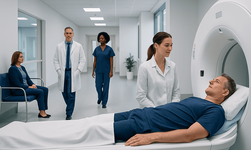
MRI is one of the most important tools in neuroimmunology testing because it creates detailed images of the brain, spinal cord, and sometimes the optic nerves. It can show areas of inflammation, demyelination, swelling, scar-like changes, or damage from previous immune attacks. MRI is often used both at diagnosis and during follow-up to see whether a condition is active, stable, or changing over time.
Doctors may request MRI of the brain, cervical spine, thoracic spine, or orbits depending on the symptoms. Contrast dye may be used in some cases to highlight active inflammation. For example, active lesions may enhance with contrast, while older lesions may remain visible without showing current activity. The pattern, location, and shape of abnormalities can help doctors distinguish between disorders such as multiple sclerosis and neuromyelitis optica.
Still, MRI does not provide the full answer on its own. Some people can have symptoms before changes become visible, and some MRI findings are not specific to autoimmune disease. Small vessel changes, migraine-related findings, prior infections, or age-related changes can sometimes look similar. That is why MRI is interpreted together with the examination, blood tests, and sometimes spinal fluid results.
In practice, MRI is especially helpful for identifying the distribution of disease, monitoring response to treatment, and detecting complications. When necessary, specialists may use advanced MRI imaging protocols to better evaluate suspected inflammatory or demyelinating disease.
Spinal Fluid Testing: Why a Lumbar Puncture May Be Needed
Spinal fluid testing, also called cerebrospinal fluid analysis, is performed through a lumbar puncture. During this procedure, a doctor collects a small amount of fluid from the lower back using a needle. Although many people feel anxious about the idea, the test is commonly performed and can provide valuable information that cannot always be seen in a blood test or MRI.
Spinal fluid can be checked for white blood cells, protein, glucose, signs of infection, and markers of immune activity. In some autoimmune neurological conditions, the fluid may show inflammation or the presence of oligoclonal bands, which suggest abnormal immune activity within the central nervous system. These findings can support a diagnosis, especially when paired with symptoms and MRI changes.
This test is also important for ruling out other causes. Some infections, cancers, and inflammatory disorders can present with symptoms similar to autoimmune disease. In suspected autoimmune encephalitis or unexplained inflammation of the brain or spinal cord, spinal fluid may help guide further antibody testing or treatment decisions.
After the procedure, some people experience temporary headache or back soreness. These effects are usually manageable and tend to improve with rest, hydration, and medical advice. If a lumbar puncture is recommended, the care team usually explains the purpose, preparation, and possible side effects in advance.
Antibody Tests and Blood Work Explained
Antibody tests look for immune proteins that may target parts of the nervous system. These tests are especially useful in disorders where a specific antibody is strongly associated with a condition. Examples include aquaporin-4 antibodies in neuromyelitis optica spectrum disorder, MOG antibodies in MOG-associated disease, and certain neuronal antibodies in autoimmune encephalitis. Depending on the situation, doctors may test blood, spinal fluid, or both.
Blood work may also include general inflammatory markers, infection screening, vitamin levels, thyroid studies, autoimmune panels, and tests that check for systemic diseases such as lupus or sarcoidosis. These tests help doctors understand whether the problem is isolated to the nervous system or part of a broader immune condition affecting other organs.
Antibody testing is helpful, but results must be interpreted carefully. A positive result does not always mean that the antibody is causing the symptoms, and a negative result does not always exclude disease. The quality of the laboratory method, the type of specimen tested, the timing of the illness, and the overall clinical context all matter. For this reason, specialists focus on the full pattern rather than one result in isolation.
When autoimmune encephalitis is suspected, doctors may also consider EEG testing alongside antibody studies to evaluate brain activity, especially if seizures, confusion, or altered behavior are present. In selected cases, broader immune or cancer-related evaluations may be needed because some antibodies can be associated with tumors elsewhere in the body.
How Doctors Combine Test Results to Make a Diagnosis
Diagnosing an autoimmune neurological disorder often requires a structured process rather than a single definitive test. Doctors look at the timing of symptoms, neurological examination findings, MRI patterns, spinal fluid results, blood tests, and sometimes additional studies such as visual testing, EEG, or nerve conduction studies. Each piece contributes part of the answer.
For example, a person with vision loss and spinal cord inflammation may need evaluation for autoimmune encephalitis if brain symptoms are also present, or for demyelinating disorders if the MRI and antibody profile suggest a different cause. The diagnosis depends on how the symptoms fit together, not only on one abnormal result. This is especially important because different disorders may require very different treatments and follow-up plans.
In some cases, the first round of tests may not provide a complete answer. A repeat MRI, repeat antibody testing, or follow-up neurological examination may be needed over time. Doctors may monitor changes before confirming a diagnosis, especially when symptoms are mild, early, or atypical. This careful approach helps reduce the risk of misdiagnosis.
Treatment Planning, Follow-Up, and Self-Care
Once a diagnosis is made, treatment is tailored to the specific disorder, symptom severity, and whether the condition is causing an acute attack or needs long-term control. Some people may need corticosteroids, plasma exchange, intravenous immunoglobulin, or other immune-targeting medicines. Others may need rehabilitation, pain management, bladder care, vision support, or mental health support alongside medical treatment.
Testing also helps with monitoring. Follow-up MRI, repeat laboratory work, and ongoing neurological assessment can show whether treatment is working or whether disease activity continues. In some conditions, early recognition and regular follow-up may help reduce future damage, preserve function, and improve quality of life.
Self-care remains important even when specialist treatment is needed. People are usually encouraged to keep a symptom diary, attend follow-up appointments, take medicines as prescribed, ask about vaccines and infection prevention, and discuss new symptoms promptly. Rest, balanced nutrition, physical activity within a doctor’s guidance, and emotional support can all be valuable parts of living with a chronic neurological condition.
Near the end of the diagnostic journey, some patients seek care in specialized centers with coordinated imaging, laboratory, and neurology expertise. Acibadem International’s multidisciplinary specialists and JCI-accredited hospitals diagnose and treat neuroimmunological conditions for international patients, including access to services such as neurology care when a comprehensive assessment is needed.
When to See a Doctor
A person should seek medical attention if they develop new neurological symptoms such as vision loss, weakness, numbness, difficulty walking, severe dizziness, unexplained bladder changes, confusion, memory problems, or seizures. Symptoms that appear over hours to days, or that worsen quickly, should be assessed urgently because prompt evaluation can be important.
Emergency care is especially important for sudden inability to move a limb, severe or rapidly progressive weakness, new seizures, loss of consciousness, serious breathing difficulty, or marked confusion. These symptoms can have several possible causes, including stroke, infection, or inflammation, and they need immediate medical assessment.
Even when symptoms are milder or intermittent, ongoing evaluation matters if they keep returning or remain unexplained. Early referral to a neurologist may help clarify the cause, guide testing, and reduce uncertainty. People should not interpret MRI or antibody results on their own, since proper diagnosis depends on expert clinical correlation.
Frequently asked questions
What is neuroimmunology testing used for?
Neuroimmunology testing is used to investigate conditions in which the immune system may be attacking or inflaming the nervous system. It helps doctors evaluate symptoms affecting the brain, spinal cord, optic nerves, or peripheral nerves and distinguish autoimmune disease from other causes.
Can an MRI alone diagnose an autoimmune neurological disease?
No. MRI is very helpful, but it usually cannot confirm the diagnosis by itself. Doctors interpret MRI findings together with symptoms, examination results, spinal fluid analysis, antibody tests, and other laboratory studies.
Why might a lumbar puncture be necessary?
A lumbar puncture allows doctors to analyze cerebrospinal fluid for signs of inflammation, infection, or abnormal immune activity. It can provide important information when MRI or blood tests do not give a complete answer.
Are antibody tests always accurate?
Antibody tests can be very useful, but they are not perfect. A positive result may need confirmation in the right clinical setting, and a negative result does not always rule out disease, especially early in the illness or in less typical cases.
How long does it take to get a diagnosis?
The timeline varies depending on the symptoms, urgency, and which tests are needed. Some people receive answers quickly, while others need repeat imaging or follow-up evaluations over time before the diagnosis becomes clear.
Do all people with suspected neuroimmunological disease need the same tests?
No. Testing is individualized based on symptoms, examination findings, age, medical history, and the suspected condition. One person may need brain and spine MRI plus spinal fluid testing, while another may need targeted antibody tests or additional neurological studies.
This article is for general information only and is not a substitute for professional medical advice. Please consult a qualified doctor about your individual situation.
Neuroimmunology in Turkey — costs, top hospitals & a free quote
JCI-accredited · board-certified surgeons · reply within 24h
Add us as a Preferred Source to see more of our trusted health content across Google Search, AI Overviews and Discover.

More from the Health Library

Quest Diagnostics Hours: When Labs Open and How to Plan a Visit
Diagnostic Mammogram: Who Needs It and What Results Mean

What Is a Lab Test Panel and Why Are Tests Grouped Together?

Ultrasound vs Mammogram: How Doctors Choose the Right Breast Imaging Test
Ultrasound-Guided vs CT-Guided Procedures: What Is the Difference?