Brain MRI With Contrast: Why It Is Ordered and What Patients Should Expect
Brain MRI with contrast can show more detail than a non-contrast MRI in many situations. The contrast material most often used is gadolinium-based and is given through a vein.
Key Takeaways
- Brain MRI with contrast can show more detail than a non-contrast MRI in many situations.
- The contrast material most often used is gadolinium-based and is given through a vein.
- The test is painless, but patients need to lie still in the MRI scanner for clear images.
- Doctors may order it for headaches with warning signs, seizures, tumors, infection, inflammation, or stroke-related concerns.
- Most people tolerate contrast well, but kidney function, allergies, implants, and pregnancy should be discussed beforehand.
- Results are interpreted by a radiologist and reviewed with the treating doctor in the context of symptoms and medical history.
A brain MRI with contrast is an imaging test that uses a contrast agent to make certain brain tissues, blood vessels, and abnormal areas easier to see. It is commonly ordered to help diagnose symptoms, evaluate known conditions, or monitor treatment over time.
Overview: What a Brain MRI With Contrast Is
A brain MRI with contrast is a magnetic resonance imaging test that creates detailed pictures of the brain and nearby structures. MRI uses a strong magnet and radio waves rather than ionizing radiation. When contrast is added, doctors can see certain tissues, blood vessels, and abnormal changes more clearly than on a standard scan alone.
The contrast material is usually a gadolinium-based agent given through a vein, often in the arm or hand. After it circulates through the bloodstream, it can highlight areas where the blood-brain barrier is disrupted, where there is inflammation, or where blood flow differs from normal. This added information can help distinguish between different possible causes of symptoms.
Not every brain MRI requires contrast. In some cases, non-contrast images provide enough information. In others, the radiologist or referring physician may request both non-contrast and contrast-enhanced images to answer a specific clinical question more accurately.
Why Doctors Order a Brain MRI With Contrast
Doctors order brain MRI with contrast when they need a closer look at brain tissue or blood vessels and want to detect abnormalities that may not be fully visible on non-contrast images. It is often used when a person has neurological symptoms that need explanation, such as seizures, weakness, changes in vision, or persistent headaches with concerning features.
This test can help evaluate a wide range of conditions. These include brain tumors, infection, inflammation, multiple sclerosis, abscesses, bleeding in selected settings, and certain vascular problems. It may also be used to assess the pituitary gland, the lining around the brain, or changes after surgery or treatment.
Brain MRI with contrast is also valuable for follow-up care. If someone already has a diagnosed condition, the scan may help show whether it is stable, improving, or progressing. For example, it can help monitor a known brain tumor or assess changes related to multiple sclerosis. In some situations, the findings may guide additional testing or procedures such as MRI imaging or specialist treatment planning.
Symptoms and Situations That May Lead to This Test

A doctor may recommend this scan when symptoms suggest a problem involving the brain, nerves, or surrounding tissues. The purpose is not simply to image a symptom, but to look for a cause that may need treatment or closer observation. The exact decision depends on the person’s age, health history, exam findings, and how symptoms started.
Common reasons for a brain MRI with contrast include:
- New seizures or unexplained episodes of loss of awareness
- Persistent or worsening headaches with neurological warning signs
- Weakness, numbness, balance problems, or difficulty speaking
- Vision changes, double vision, or hearing-related neurological concerns
- Suspected brain infection, inflammation, or demyelinating disease
- Follow-up of a known tumor, prior surgery, or radiation treatment
- Evaluation of the pituitary region or cranial nerves
Sometimes the test is ordered urgently, while in other cases it is part of planned outpatient care. If symptoms suggest stroke, the imaging approach may vary depending on timing and urgency, and doctors may choose MRI, CT, or vascular imaging based on the situation. The scan is one part of evaluation and is interpreted together with the clinical picture.
How to Prepare and What Happens During the Scan
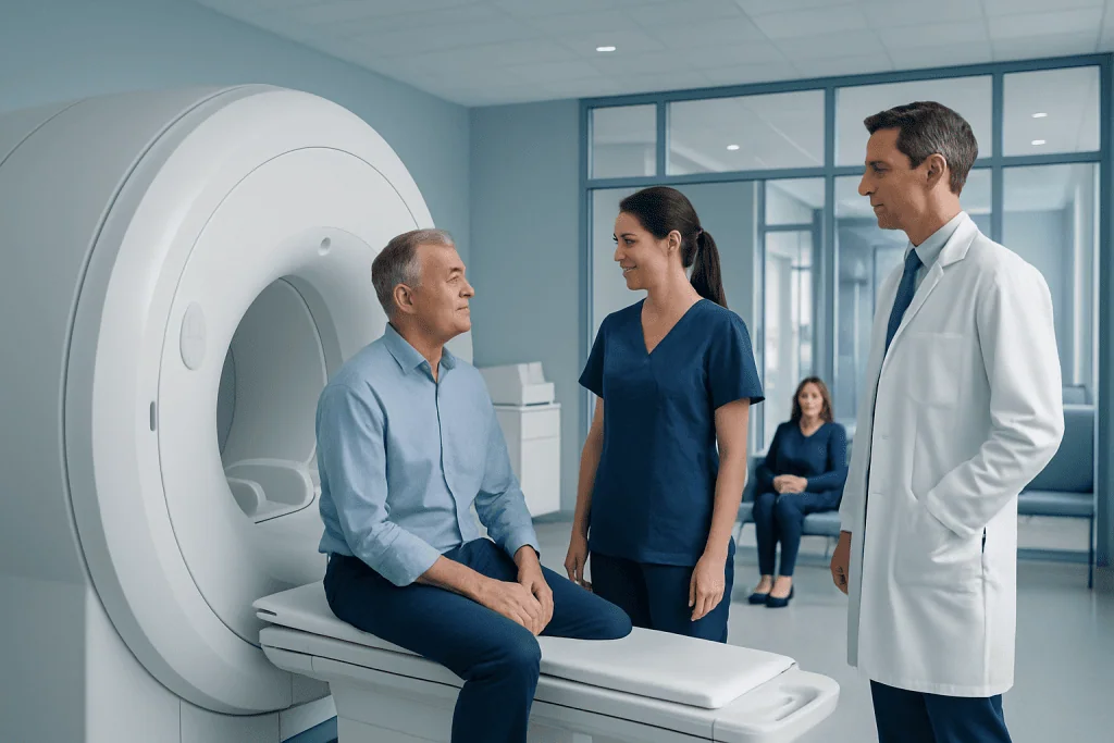
Before the scan, the imaging team usually asks about medical history, allergies, kidney disease, pregnancy, and any implanted devices. MRI safety is especially important because the machine uses a strong magnet. Patients should mention pacemakers, aneurysm clips, cochlear implants, metal fragments, infusion ports, joint hardware, or any prior surgeries, since some devices are MRI-compatible and others need special review.
Patients are usually asked to remove jewelry, watches, hairpins, hearing aids, and clothing with metal fasteners. In many centers, eating and drinking are allowed unless sedation or a special protocol is planned. If a person is very anxious in enclosed spaces, it is helpful to mention this in advance so the team can discuss comfort measures.
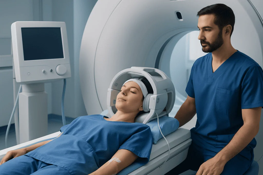
During the test, the patient lies on a narrow table that moves into the MRI scanner. A technologist places a small IV line for the contrast injection. The machine makes loud tapping or thumping sounds, so ear protection is provided. The most important part is staying as still as possible so the images remain sharp. The scan is painless, though some people notice temporary coolness in the arm from the contrast injection or mild discomfort from lying still.
The study may take about 30 to 60 minutes, depending on the protocol. Some images are taken before contrast, then the gadolinium-based contrast is injected and additional images are acquired. In selected cases, advanced sequences may be added to evaluate blood flow, tissue characteristics, or surgical planning, sometimes alongside tests such as MR angiography.
Benefits, Risks, and Safety Considerations
The main benefit of brain MRI with contrast is improved visibility of certain abnormalities. Contrast can help doctors define the size, location, and pattern of a lesion more clearly. This can make diagnosis more accurate and may help distinguish among infection, inflammation, tumor, scar tissue, or treatment-related changes.
For most people, gadolinium-based contrast agents are well tolerated. Side effects are uncommon and are often mild when they occur, such as nausea, headache, or temporary discomfort at the injection site. Allergic-like reactions can happen but are rare. The imaging team is trained to respond if a reaction occurs.
Kidney health is an important part of safety screening. In people with severe kidney impairment, certain contrast agents may carry added risk, so doctors may check kidney function before the scan or choose a different approach. Patients should also tell their doctor if they are pregnant or breastfeeding, as the need for contrast is considered carefully in these situations.
MRI itself does not use radiation, which is reassuring for many patients. However, the scanner can feel confined, and the loud noise may be uncomfortable without ear protection. People with claustrophobia, chronic pain, or difficulty lying flat may need additional planning to complete the study comfortably. When needed, related imaging or computed tomography may be considered for specific clinical questions.
How Results Are Read and What They Can Show
After the scan, a radiologist reviews the images in detail and prepares a report for the referring doctor. The report describes normal findings as well as any areas of abnormal signal, swelling, bleeding, enhancement, mass effect, or vascular change. The meaning of the results depends on where the abnormality is located, how it appears, and how those findings match the patient’s symptoms and examination.
Contrast enhancement does not automatically mean a serious condition. It simply means an area takes up contrast in a way that stands out on MRI. This can happen with many different processes, including inflammation, infection, active demyelination, postoperative change, and tumors. The pattern of enhancement helps narrow the possibilities, but it is usually interpreted together with the full scan and medical history.
Sometimes the results are normal, which can also be useful. A normal brain MRI with contrast may help rule out many structural causes of symptoms and guide doctors toward other explanations. In other cases, the report may recommend follow-up imaging, additional blood tests, referral to a neurologist or neurosurgeon, or further procedures depending on what is seen.
After the Test, Follow-Up, and When to Seek Medical Advice
Most people can return to normal activities soon after the scan. If contrast was used, drinking fluids afterward is commonly advised unless a doctor has given a fluid restriction for another medical reason. If sedation was used, the patient may need someone to accompany them home and should follow the facility’s instructions about rest and daily activities.
Patients should contact a healthcare professional if they develop unexpected symptoms after the test, such as a rash, breathing difficulty, or significant swelling at the IV site. These problems are uncommon, but they should be assessed promptly. Ongoing or worsening neurological symptoms should also be reported, even if the scan has already been completed.
It is helpful for patients to ask when and how they will receive results. Some findings are straightforward, while others require discussion with a neurologist, neurosurgeon, or another specialist. If the scan identifies a condition that needs treatment, care may involve medication, monitoring, surgery, or other targeted therapies such as Gamma Knife radiosurgery in selected cases.
For international patients who need advanced evaluation, Acibadem International offers diagnosis and treatment through multidisciplinary specialists in JCI-accredited hospitals. A qualified doctor can explain whether contrast MRI is appropriate, what the findings mean, and what next steps are most suitable for the individual patient.
Frequently asked questions
What is the difference between a brain MRI and a brain MRI with contrast?
A standard brain MRI creates detailed images without an injected contrast agent. A brain MRI with contrast adds a gadolinium-based material through a vein to make certain tissues and abnormal areas easier to see. Doctors choose contrast when they need more detail to answer a specific question.
Is a brain MRI with contrast safe?
For most people, it is considered safe and well tolerated. The MRI itself does not use radiation, and serious reactions to gadolinium contrast are uncommon. Safety screening is still important, especially for people with kidney disease, pregnancy, or certain implanted devices.
How long does a brain MRI with contrast take?
Many exams take about 30 to 60 minutes, although timing can vary by protocol and the reason for the scan. Images are often taken before and after the contrast injection. Extra time may be needed if advanced sequences or additional positioning are required.
Does a brain MRI with contrast hurt?
The scan itself is painless. The only discomfort for many patients is the placement of the IV line and the need to lie still in the scanner. Some people also find the noise or enclosed space uncomfortable, but ear protection and support from the imaging team usually help.
Do patients need to fast before a brain MRI with contrast?
Often, no fasting is required for a routine brain MRI with contrast. However, instructions can vary depending on the facility, whether sedation is planned, or whether another test is being done at the same time. It is best to follow the specific preparation guidance provided before the appointment.
Can a brain MRI with contrast detect a tumor?
It can help detect and characterize many brain tumors by showing their size, location, and enhancement pattern more clearly. However, the scan does not by itself provide every answer about the exact tumor type. Sometimes follow-up imaging, specialist review, or biopsy is needed for a definitive diagnosis.
References
- Radiological Society of North America
- American College of Radiology
- National Institute of Neurological Disorders and Stroke
- National Institute of Biomedical Imaging and Bioengineering
- Mayo Clinic
This article is for general information only and is not a substitute for professional medical advice. Please consult a qualified doctor about your individual situation.
Explore treatments in Turkey — costs, top hospitals & a free quote
JCI-accredited · board-certified surgeons · reply within 24h
Add us as a Preferred Source to see more of our trusted health content across Google Search, AI Overviews and Discover.