Dermatofibroma
Dermatofibroma is a common benign skin lump. Learn symptoms, causes, diagnosis, treatment options and when to see a dermatologist.
Quick answer
Dermatofibroma is a common, benign skin nodule that usually appears as a small, firm bump, often on the legs, and is typically diagnosed with a skin examination and, when needed, a biopsy to confirm it is not another lesion. At Acibadem in Turkey, dermatologists assess the spot and, if treatment is needed for symptoms, appearance, or diagnostic certainty, management may…
What is dermatofibroma?
A dermatofibroma is a common, benign (non-cancerous) growth of the skin. It forms a small, firm bump, most often on the arms or legs, and is made up of fibrous tissue: the same type of supportive tissue that gives skin its structure. Doctors sometimes call it a fibrous histiocytoma, which simply refers to the mix of cells found inside the lesion when it is examined under a microscope. In medical coding systems, a dermatofibroma falls under benign skin neoplasms (ICD-10 code D23.9).
If you are wondering what is dermatofibroma in everyday terms, it helps to think of it as a small, harmless knot of scar-like tissue in the skin. It is not a mole, not a wart, and not skin cancer. Dermatofibromas do not spread to other parts of the body, and in most cases they cause no health problems at all beyond their appearance or occasional tenderness.
Dermatofibromas can occur in anyone, but they are most often seen in adults between roughly 20 and 50 years of age, and they appear somewhat more frequently in women than in men. Many people have just one, though some people develop several over time. Having multiple dermatofibromas that appear rapidly is uncommon and may prompt your doctor to look for an underlying condition affecting the immune system, but for the vast majority of people a dermatofibroma is an isolated, harmless finding.
Symptoms of dermatofibroma
Dermatofibroma symptoms are usually mild, and many people notice the bump by chance rather than because it causes discomfort. Typical features include:
- A small, firm bump: usually between a few millimeters and about one centimeter across, though occasionally larger.
- Location: most often on the lower legs, but also on the arms, shoulders, or trunk.
- Color: pink, red, brown, gray, or a mix of shades; in people with darker skin, the lesion often appears darker than the surrounding skin.
- Texture: firm or hard to the touch, like a small button or pea just under the skin surface.
- The “dimple sign”: when the skin on either side of the bump is gently pinched, the lesion often dips inward or puckers rather than rising up. This is a well-known clue that doctors use during examination.
- Itching or tenderness: some dermatofibromas itch or feel mildly sore, especially when rubbed, shaved over, or knocked.
- Slow or no change over time: most dermatofibromas stay roughly the same size for years.
How the lesion looks can shift slightly over its “life.” A newer dermatofibroma may look pinker or redder because of increased blood supply, while an older one often turns brown or gray and may fade somewhat at the center while keeping a darker rim. These changes are gradual. A bump that grows quickly, bleeds without an obvious cause, breaks down into an open sore, or changes color suddenly does not fit the usual pattern of a dermatofibroma and should be checked by a doctor.
There are also less common types. A so-called cellular dermatofibroma tends to be somewhat larger and, after removal, has a slightly higher chance of coming back at the same spot, although it remains benign. An aneurysmal dermatofibroma contains small blood-filled spaces and may look bruised or change size more noticeably. These variants are identified under the microscope rather than by appearance alone, which is one reason doctors sometimes recommend a biopsy (removal of a small tissue sample for laboratory examination) when a lesion looks unusual.
Causes and risk factors
The exact dermatofibroma causes are not fully understood. The most widely accepted explanation is that many dermatofibromas represent an exaggerated local reaction of the skin to minor injury. In many cases, patients recall a small trauma at the site, for example an insect bite, a thorn prick, a splinter, an ingrown hair, or a nick from shaving, weeks or months before the bump appeared. The skin’s repair cells, called fibroblasts, appear to keep producing fibrous tissue after the injury has healed, creating the firm nodule. However, plenty of dermatofibromas arise with no remembered injury at all, so this cannot be the whole story. Some researchers consider certain dermatofibromas to be true benign growths rather than purely reactive scars, and the two ideas are not mutually exclusive.
Factors that appear to make dermatofibromas more likely include:
- Age: they are most common in young and middle-aged adults, though they can occur at any age, including in children.
- Sex: women are affected somewhat more often than men, possibly in part because leg shaving increases minor skin trauma on a common site.
- Minor skin injury: bites, pricks, scratches, and folliculitis (inflammation of hair follicles) at the site.
- Immune system conditions: the sudden appearance of many dermatofibromas at once, known as multiple eruptive dermatofibromas, has been reported in people with weakened or altered immune function, such as those with certain autoimmune diseases or those taking medicines that suppress the immune system. This is uncommon.
It is important to stress what dermatofibromas are not caused by. They are not infections, they are not contagious, and they are not caused by poor hygiene. They are also not caused by sun exposure in the way many skin cancers are. There is nothing you did wrong to develop one, and there is no reliable way to prevent them.
Diagnosis
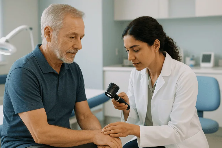
Dermatofibroma diagnosis usually begins, and often ends, with a careful physical examination by a doctor, ideally a dermatologist (a physician specializing in skin conditions). Experienced clinicians can recognize a typical dermatofibroma by its firm feel, characteristic color, common location on the limbs, and the dimple sign described above. In many cases no further testing is needed.
When the doctor wants additional certainty, several tools may be used:
- Dermoscopy: a handheld magnifying instrument with a light, called a dermatoscope, lets the doctor see structures in the skin that are invisible to the naked eye. A typical dermatofibroma often shows a pale, scar-like center surrounded by a fine brown network at the edges. This pattern helps distinguish it from moles and from melanoma, a serious form of skin cancer.
- Skin biopsy: if the appearance is unusual, if the lesion has changed, or if there is any doubt about the diagnosis, the doctor may remove part or all of the lesion and send it to a pathology laboratory. Under the microscope, a dermatofibroma shows a characteristic pattern of fibrous cells in the dermis (the deeper layer of the skin). A biopsy is the only way to confirm the diagnosis with certainty and to identify variants such as cellular or aneurysmal dermatofibroma.
- Imaging: imaging tests such as ultrasound are not usually needed for a typical dermatofibroma. They may occasionally be used for deeper or larger lumps to help decide whether the growth is in the skin itself or in the tissue beneath it.
A key part of diagnosis is ruling out look-alike conditions. Lesions that can resemble a dermatofibroma include moles, cysts, keloid scars, and, rarely but importantly, dermatofibrosarcoma protuberans (DFSP), an uncommon skin cancer that can superficially mimic a large dermatofibroma, as well as melanoma when the lesion is darkly pigmented. This is why any skin bump that is growing, changing color, bleeding, or behaving unusually deserves professional evaluation rather than self-diagnosis. At Acibadem hospitals, this evaluation is carried out within the dermatology department, which manages both the assessment and, when needed, the removal of skin lesions.
Treatment options
Dermatofibroma treatment is often not needed at all. Because the lesion is benign and usually stable, the most common and entirely reasonable approach is watchful waitingsimply leaving it alone and keeping an eye on it. Treatment is generally considered when the lesion is painful, itchy, repeatedly irritated (for example by shaving or clothing), cosmetically bothersome, or when the diagnosis is uncertain.
The main options your doctor may discuss include:
- Observation (watchful waiting): for a typical, symptom-free dermatofibroma, no treatment is required. Your doctor may suggest monitoring it and returning if it changes.
- Surgical excision: the most definitive treatment is cutting out the entire lesion under local anesthesia (numbing medicine injected into the skin). Because a dermatofibroma extends into the deeper layer of the skin, complete removal requires excising down through the dermis, which always leaves a scar. Removal also allows the tissue to be examined in the laboratory, confirming the diagnosis. In many cases the resulting scar is a line similar in length to, or slightly longer than, the original bump, so patients weighing removal for cosmetic reasons should discuss the likely trade-off with their doctor.
- Shave removal: shaving off the raised part of the lesion flattens it but leaves the deeper portion in place, so the dermatofibroma may partially regrow or remain palpable. It can be an option when the main concern is the raised surface.
- Cryotherapy: freezing the lesion with liquid nitrogen can flatten it and reduce symptoms in some cases, but because the deeper tissue often survives, the lesion may persist or recur, and freezing can leave lighter or darker patches of skin, especially in darker skin tones.
- Corticosteroid injection: injecting a steroid (an anti-inflammatory medicine) into the lesion may soften it or reduce tenderness in some patients, though results vary and the lesion usually does not disappear.
- Laser treatment: certain lasers have been used to improve the color or surface of dermatofibromas. Results are variable, and laser treatment does not remove the deeper fibrous core.
There is no cream, tablet, or home remedy that reliably makes a dermatofibroma go away, and attempting to cut, burn, or tie off the lesion at home is unsafe and strongly discouraged: it risks infection, worse scarring, and, most importantly, destroying tissue that might have been needed to rule out a more serious condition. Even after complete surgical removal, a small proportion of dermatofibromas, particularly the cellular variant, can recur at the same site, in which case re-excision may be discussed.
Choosing between these options is a personal decision made with your doctor, balancing symptoms, cosmetic concerns, scar expectations, and diagnostic certainty. For most people, reassurance after a professional examination is the only “treatment” required.
Living with dermatofibroma and outlook
The outlook for people with a dermatofibroma is excellent. The lesion is benign, does not turn into cancer in any established way, and does not spread. Most dermatofibromas remain stable for many years; some fade partially and flatten over time, though complete spontaneous disappearance is uncommon. For most people, living with a dermatofibroma means very little day to day.
Practical points that many patients find helpful:
- Protect it from repeated irritation: if the bump sits where you shave, consider shaving around it or using an electric razor to reduce nicks and inflammation.
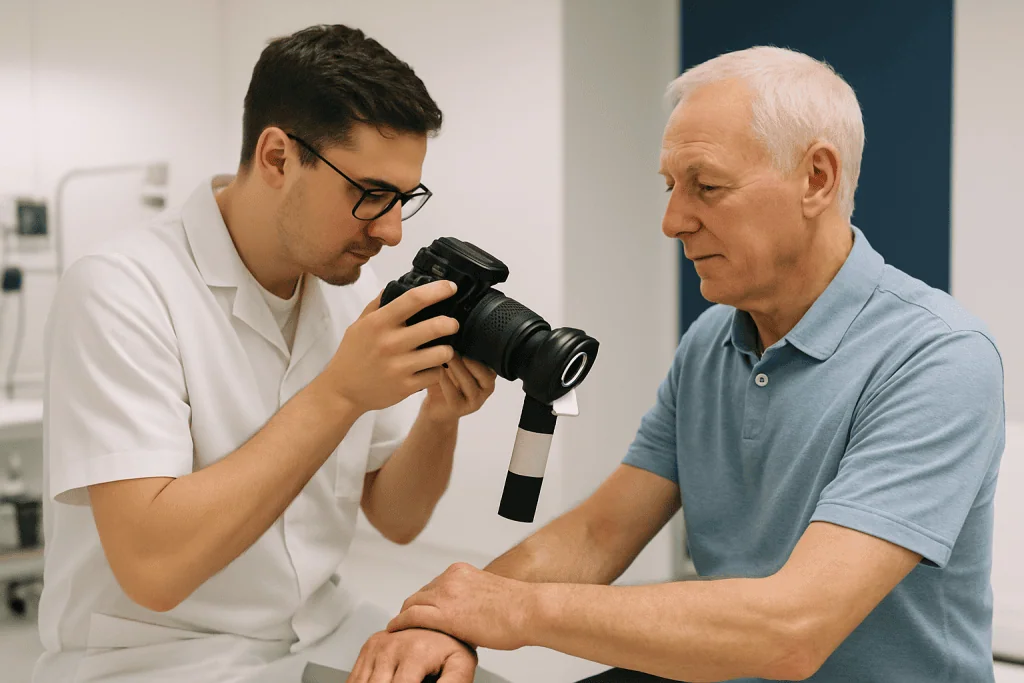
- Know your lesion: take note of its size and color, or photograph it occasionally, so that any genuine change is easy to spot and describe to your doctor.
- Keep routine skin awareness: having a dermatofibroma does not increase your risk of skin cancer, but everyone benefits from being familiar with their skin and reporting new or changing lesions.
- After removal: if you choose excision, follow your doctor’s wound-care instructions; scars on the lower leg can take longer to heal than scars elsewhere, and healing quality varies from person to person.
No doctor can promise that a dermatofibroma will never change, recur after removal, or that a new one will never appear elsewhere. What can be said honestly is that, once the diagnosis is confirmed, a dermatofibroma poses no threat to your general health in the overwhelming majority of cases.
Frequently asked questions
What is dermatofibroma and is it dangerous?
A dermatofibroma is a benign, firm bump in the skin made of fibrous, scar-like tissue, most often found on the arms or legs. It is not cancer and does not spread to other parts of the body. Once a doctor has confirmed the diagnosis, it is generally considered harmless, although it can sometimes itch or feel tender. Any bump that is growing quickly or behaving unusually should still be examined, because other conditions can look similar.
Can a dermatofibroma go away on its own?
Most dermatofibromas persist for many years without treatment. Some flatten or fade partially over time, but complete spontaneous disappearance is uncommon. If the lesion does not bother you, leaving it alone is a perfectly acceptable option; if it is painful, irritated, or cosmetically troubling, your doctor can discuss removal and other options with you.
What are the typical dermatofibroma symptoms I should look for?
The classic picture is a small, firm, pink-to-brown bump, usually under a centimeter across, that dimples inward when the surrounding skin is pinched. It may itch or feel slightly tender when rubbed or knocked, and it typically changes very little from year to year. Rapid growth, bleeding, ulceration, or sudden color change are not typical dermatofibroma symptoms and warrant a medical review.
How is dermatofibroma diagnosis confirmed?
In many cases, a dermatologist can recognize a dermatofibroma by examining and feeling it, often with the help of a dermatoscope, a magnifying instrument that reveals characteristic patterns in the skin. When there is any doubt, for example, if the lesion is large, changing, or unusually colored, a skin biopsy is performed. Laboratory examination of the tissue is the only way to confirm the diagnosis with certainty.
What is the best dermatofibroma treatment if it bothers me?
There is no single “best” treatment for everyone. Complete surgical excision under local anesthesia is the most definitive option because it removes the entire lesion and allows laboratory confirmation, but it always leaves a scar. Less invasive approaches such as cryotherapy, steroid injection, or shave removal may improve symptoms or appearance, though the lesion can persist or return. Your doctor can help you weigh symptom relief, cosmetic outcome, and scarring for your particular situation.
Can a dermatofibroma turn into skin cancer?
A dermatofibroma is benign, and there is no established pathway by which a typical dermatofibroma transforms into cancer. The genuine concern is different: rare skin cancers, such as dermatofibrosarcoma protuberans or a pigmented melanoma, can occasionally be mistaken for a dermatofibroma. This is why a bump that grows steadily, ulcerates, bleeds, or otherwise changes should be evaluated by a doctor rather than assumed to be harmless.
What is recovery like after dermatofibroma removal?
Removal is usually a short outpatient procedure performed under local anesthesia, after which you can typically go home the same day. The wound is closed with stitches and generally heals over one to a few weeks, depending on its size and location; wounds on the lower leg often heal more slowly. Some soreness, and later a scar, is expected. Your doctor will advise on wound care, stitch removal, and any activity restrictions during healing.
When to see a doctor
Any new or changing skin lump deserves a professional look, even when it seems minor, because look-alike conditions range from harmless to serious. Arrange a medical review promptly if you notice any of the following red flags:
- Rapid growth: a bump that is clearly enlarging over weeks to months.
- Bleeding or ulceration: a lesion that bleeds without injury or breaks down into an open sore that does not heal.
- Sudden color change: darkening, new black areas, or uneven, spreading pigment.
- Increasing pain: a previously quiet lesion that becomes persistently painful.
- Irregular or changing borders: edges that look ragged, notched, or are shifting over time.
- Many new bumps appearing quickly: multiple lesions erupting over a short period, especially if you have an immune condition or take medicines that suppress the immune system.
- Signs of infection after a biopsy or removal: spreading redness, warmth, swelling, pus, or fever.
If none of these apply and your bump matches the typical description of a dermatofibroma, the situation is rarely urgent, but a one-time examination by a dermatologist is still the safest way to confirm what it is. A confirmed diagnosis gives you peace of mind and a clear basis for deciding whether to leave the lesion alone or have it treated.
Medically reviewed by the Acıbadem International Medical Board September 2, 2026
See our medical review board →
Update history
- PublishedJune 8, 2026
- Medical review approvedSeptember 2, 2026
- Last content updateSeptember 2, 2026