

14 week ultrasound: A Complete Medical Guide for Patients

A 14 week ultrasound can confirm fetal heartbeat, growth, and gestational age. It is not always a routine scan in every pregnancy, but it may be recommended for specific medical reasons.
Key Takeaways
- A 14 week ultrasound can confirm fetal heartbeat, growth, and gestational age.
- It is not always a routine scan in every pregnancy, but it may be recommended for specific medical reasons.
- Some basic anatomy can be seen at 14 weeks, but the detailed anatomy scan is usually done later.
- The scan is commonly performed through the abdomen, though a transvaginal ultrasound may sometimes be needed.
- Results should be interpreted by a qualified obstetric clinician in the context of symptoms, history, and other prenatal tests.
A 14 week ultrasound is an early second-trimester pregnancy scan used to check fetal growth, heartbeat, dating, and the general progress of the pregnancy. It may also help guide next steps if there are symptoms such as pain, bleeding, uncertainty about dates, or questions after an earlier scan.
Overview: what a 14 week ultrasound is
A 14 week ultrasound is a pregnancy scan performed at about 14 weeks of gestation, which is the beginning of the second trimester. In many cases, it is used to confirm that the pregnancy is progressing as expected by checking the fetal heartbeat, growth, movement, and overall position in the uterus. It can also help estimate gestational age if earlier dating is uncertain.
This scan is not always the main routine ultrasound in every pregnancy. Some people have their first-trimester scan earlier, and then the next standard anatomy scan later, often around 18 to 22 weeks. However, a 14 week ultrasound may be advised when there are symptoms, when dates are unclear, after earlier inconclusive imaging, or when a doctor wants an early look at the pregnancy.
At this stage, the fetus is larger and easier to see than in very early pregnancy, so the scan can often provide reassuring information. Some parts of early anatomy may be visible, but this is not usually considered a complete structural assessment. For a full review of fetal development and organs, the later detailed scan remains important.
Why a doctor may recommend it
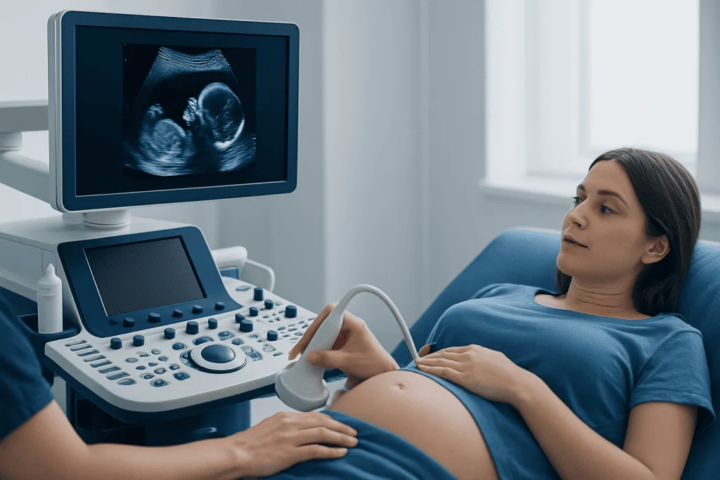
A doctor may recommend a 14 week ultrasound for several reasons. One common reason is to confirm the expected due date if earlier menstrual dates are uncertain or if there was no earlier dating scan. Accurate dating helps guide the timing of prenatal tests and follow-up care throughout pregnancy.
The scan may also be used to evaluate symptoms such as light bleeding, pelvic pain, or cramping. In these situations, ultrasound can help check fetal viability, look at the uterus and cervix, and identify whether there is a cause that needs further medical attention. If a patient has had previous pregnancy complications, the doctor may also suggest an earlier second-trimester review.
In some pregnancies, this scan is part of follow-up after findings from an earlier visit. For example, a clinician may want to review fetal growth, recheck a suspected twin pregnancy, or assess the placenta’s early location. It may also be used alongside other prenatal screening decisions, especially if there are questions about family history or previous results.
When ultrasound is being used to look more closely at pregnancy health, it may be part of a broader imaging plan such as obstetric ultrasound evaluation. If symptoms suggest a nonviable pregnancy or earlier concern, doctors may also discuss related conditions such as miscarriage in a careful and supportive way.
What the scan can show at 14 weeks
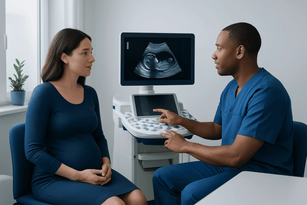
At 14 weeks, ultrasound can usually show the fetal heartbeat, head, body, arms, and legs clearly enough to assess general growth and movement. The sonographer or doctor will often measure the fetus and compare these measurements with the expected gestational age. If growth appears appropriate, this can support accurate dating and provide reassurance.
The scan can also help confirm whether there is one fetus or more than one, and it may give information about the placenta’s early position and the amount of surrounding fluid. The uterus and nearby pelvic structures may also be reviewed if there are symptoms such as pain or bleeding.
Although some anatomy may be visible at this stage, the scan has limitations. It is too early to exclude every structural condition with certainty, and some findings may only become clear later in pregnancy. For that reason, a normal 14 week ultrasound does not replace the detailed mid-pregnancy anatomy scan.
In selected cases, an early review of the cervix or placenta may be helpful, especially if there is concern about bleeding or prior pregnancy history. If clinicians are evaluating broader pregnancy complications, they may recommend follow-up with more advanced prenatal imaging or specialist care rather than relying on one early second-trimester scan alone.
What to expect during the appointment
Most 14 week ultrasounds are performed through the abdomen. A gel is placed on the skin, and a handheld probe is moved over the lower belly to create images. Because the uterus has risen higher out of the pelvis by this stage, abdominal scanning is often enough to get useful views.
Sometimes, however, a transvaginal ultrasound is still recommended. This may happen if the views through the abdomen are limited, if the uterus tilts in a way that makes imaging more difficult, or if the doctor needs a closer look at the cervix or early placenta. A transvaginal scan uses a slim covered probe inserted into the vagina and is generally brief.
The appointment usually includes image recording, fetal measurements, and a check of the heartbeat. The clinician may also ask about symptoms, last menstrual period, previous pregnancies, and any earlier scans. Depending on the purpose of the visit, the results may be explained immediately or reviewed later by an obstetrician or radiologist.
Patients are often advised to wear comfortable clothing and to follow any preparation instructions given by the clinic. Some centers may ask for a moderately full bladder, though this is less commonly needed than in very early pregnancy. If the scan is being done because of symptoms, it is helpful to mention when the symptoms started and whether they are changing.
Understanding the results
A reassuring result usually means the scan shows a live intrauterine pregnancy with measurements that match the expected gestational age, along with no obvious early concerns. The report may mention fetal heart activity, estimated age by ultrasound, number of fetuses, and general observations about the uterus, placenta, and fluid.
Sometimes the results lead to a change in the expected due date. This is not unusual when menstrual cycles are irregular or the date of conception is uncertain. In many pregnancies, ultrasound dating is more reliable than menstrual dating alone, especially if earlier information is limited.
Not every uncertain result means there is a serious problem. For example, the fetus may be in a position that makes certain views incomplete, or the placenta may appear low early on and then shift upward as the uterus grows. In these cases, the doctor may recommend a repeat scan rather than drawing conclusions from one examination.
If an abnormality is suspected, follow-up may include targeted imaging, blood tests, or referral to a maternal-fetal medicine specialist. When there are concerns related to the uterus, ovaries, or pelvic pain, doctors may also consider a broader gynecologic evaluation to rule out other causes of symptoms.
Limits of the scan and how it differs from other prenatal ultrasounds
A 14 week ultrasound is useful, but it does not answer every question about pregnancy health. At this stage, some organs and structures are still developing, and even with good-quality imaging, certain conditions may not be visible yet. That is why a later anatomy scan remains a key part of routine prenatal care.
This scan also differs from a very early first-trimester ultrasound. Earlier scans often focus mainly on confirming the pregnancy location, viability, and number of embryos. By 14 weeks, the fetus is larger, and there is usually more information about growth and movement, but the scan is still not as comprehensive as the mid-pregnancy anatomy assessment.
Another important point is that ultrasound findings should be interpreted alongside symptoms, examination, and any prenatal blood tests. A scan can provide strong clues, but it is one part of a larger medical picture. Patients should feel comfortable asking what the scan can show clearly and what still requires later follow-up.
When there is concern about fetal development, structural differences, or placental issues, a specialist may recommend further assessment through perinatology care. This can help ensure that imaging findings are reviewed in the context of the whole pregnancy rather than in isolation.
Self-care after the scan and preparing for next steps
Most people can return to normal daily activities immediately after a 14 week ultrasound. The scan itself does not usually require recovery time, and it is considered a standard, noninvasive imaging tool in pregnancy. If the scan was done because of symptoms, the next steps depend more on the underlying concern than on the ultrasound procedure itself.
It can help to keep a copy of the report and note any follow-up recommendations, such as repeat imaging, blood tests, or the timing of the anatomy scan. Patients may also want to prepare questions for their next prenatal visit, especially if the report contains terms they do not recognize. Asking for clear explanations is appropriate and often very helpful.
Good pregnancy self-care continues to matter after the scan. This generally includes attending regular prenatal appointments, taking prescribed prenatal vitamins, staying hydrated, eating a balanced diet, and avoiding smoking, alcohol, and nonprescribed substances. Any medication use during pregnancy should be discussed with a qualified clinician.
For international patients who need coordinated pregnancy assessment, Acibadem International’s multidisciplinary specialists and JCI-accredited hospitals provide diagnostic and treatment services tailored to obstetric care. If there are related concerns such as ongoing pelvic symptoms, clinicians may also consider conditions like ectopic pregnancy when clinically appropriate, although this is usually identified earlier in pregnancy.
When to seek medical care
Patients should seek prompt medical advice if they have vaginal bleeding, significant pelvic or abdominal pain, fever, leakage of fluid, fainting, or symptoms that are worsening rather than settling. These symptoms do not always mean there is a serious problem, but they should be assessed by a doctor or maternity care team without delay.
Medical care is also important if the ultrasound report is unclear, if the dates do not match expected growth, or if the clinician recommends repeat imaging or specialist follow-up. Early evaluation can clarify what is happening and help guide safe next steps for both the patient and the pregnancy.
Urgent evaluation is especially important for severe pain, heavy bleeding, shoulder pain, dizziness, or collapse. While many causes are benign, these symptoms can sometimes signal complications that need immediate assessment. If a patient feels unwell or worried, contacting a qualified healthcare professional is the safest course.
Frequently asked questions
Is a 14 week ultrasound routine in pregnancy?
It may be routine in some care settings, but not in all pregnancies. Many people have an earlier dating scan and then a later anatomy scan, while a 14 week ultrasound is often used when there are symptoms, uncertain dates, or a need for follow-up.
Can a 14 week ultrasound show if the baby is developing normally?
It can show important early signs such as heartbeat, growth, movement, and general progress of the pregnancy. However, it cannot rule out all structural or genetic conditions, so later prenatal screening and the detailed anatomy scan are still important.
Is the scan abdominal or transvaginal?
At 14 weeks, the scan is usually done through the abdomen. A transvaginal scan may still be used if better views are needed, especially to assess the cervix, placenta, or a pregnancy that is difficult to see clearly on abdominal imaging.
Can the baby’s sex be seen at 14 weeks?
Sometimes sex may be suggested at 14 weeks, but it is not always accurate at this stage. Fetal position, image quality, and individual development can affect what is visible, so confirmation often comes later.
What if the ultrasound dates are different from my last menstrual period?
A difference in dates is common, especially if cycles are irregular or ovulation occurred later than expected. Your doctor may adjust the estimated due date based on ultrasound measurements if they believe the scan gives a more accurate estimate.
Does a normal 14 week ultrasound mean everything is definitely fine?
A normal result is reassuring, but no single scan can guarantee that every aspect of pregnancy is normal. Ultrasound is one part of prenatal care, and later scans and routine checkups are still needed.
References
- World Health Organization
- American College of Obstetricians and Gynecologists
- International Society of Ultrasound in Obstetrics and Gynecology
- National Institute for Health and Care Excellence
- Centers for Disease Control and Prevention
This article is for general information only and is not a substitute for professional medical advice. Please consult a qualified doctor about your individual situation.
Ultrasound in Turkey — costs, top hospitals & a free quote
JCI-accredited · board-certified surgeons · reply within 24h
Add us as a Preferred Source to see more of our trusted health content across Google Search, AI Overviews and Discover.
Check your numbers in seconds
BMI, calories, due date, blood pressure and 30+ more clinical calculators — free, instant, doctor-reviewed ranges.

Check-up & Preventive Medicine
Comprehensive health screenings designed for early detection and prevention.
3 specialists in this unitMore from the Health Library



Related Specialists

Prof. Dr. Muzaffer Çetingüç
Psychiatry
Assoc. Prof. Dr. Burak Özturan
Orthopedic Surgery & Traumatology
Dr. Aysel Özkaynak
Gynecology & Obstetrics
Prof. Dr. Evrim Kadriye Tezcanlı
Radiation Oncology