Abnormal ECG: What Patients Need to Know

An abnormal ECG is a test finding, not a diagnosis by itself. Some ECG changes are harmless, while others suggest rhythm problems, reduced blood flow, or structural heart disease.
Key Takeaways
- An abnormal ECG is a test finding, not a diagnosis by itself.
- Some ECG changes are harmless, while others suggest rhythm problems, reduced blood flow, or structural heart disease.
- Symptoms such as chest pain, fainting, shortness of breath, or palpitations need prompt medical attention.
- Doctors often confirm the meaning of an abnormal ECG with repeat testing, blood tests, monitoring, or heart imaging.
- Treatment depends on the cause and may range from observation and lifestyle changes to medicines or procedures.
An abnormal ECG means the heart’s electrical activity looked different from the expected pattern on an electrocardiogram. This finding does not always mean serious heart disease, but it does need to be interpreted alongside symptoms, medical history, and sometimes further testing.
Overview: what an abnormal ECG means
An abnormal ECG means the tracing of the heart’s electrical activity looks different from what is typically expected. ECG stands for electrocardiogram, a quick and painless test that records how electrical impulses travel through the heart. The result may show a fast, slow, irregular, or differently shaped rhythm pattern.
Importantly, an abnormal ECG is not a final diagnosis on its own. Some people have ECG changes that are normal for them, related to age, fitness level, body shape, medicines, or minor technical issues during the test. Others may have a change that points to a heart rhythm problem, signs of strain on the heart, or changes that need more investigation.
Doctors interpret ECG findings together with symptoms, physical examination, risk factors, and medical history. For example, the same ECG result may mean something different in a young athlete than in an older adult with chest pain, diabetes, or a history of heart disease.
Because the ECG captures only a short moment in time, it may miss intermittent problems or show changes that need confirmation. That is why follow-up testing is sometimes recommended, even when a person feels well.
What an ECG can show
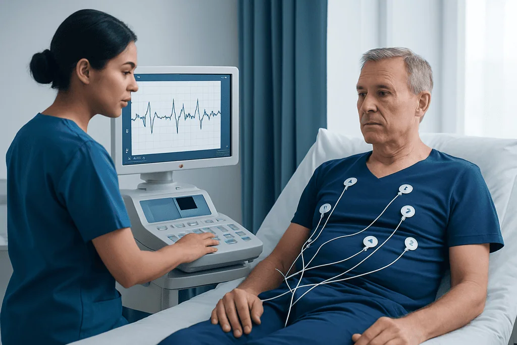
An ECG records the timing and pattern of electrical signals in the heart. From this, a doctor can look for clues about the heart rate, rhythm, and whether electrical conduction is moving normally through the chambers of the heart.
Common abnormalities may include rhythms that are too fast or too slow, extra beats, delays in conduction, or patterns that suggest the heart muscle is under stress. In some cases, the tracing may suggest previous damage to heart muscle or changes linked to reduced blood supply.
An ECG can also provide indirect signs of enlarged heart chambers, electrolyte imbalances, and inflammation around the heart. However, it does not directly show the heart’s structure or blood flow, so it is often only one part of the evaluation.
- Rhythm changes: atrial fibrillation, extra beats, rapid heart rhythms, or slow rhythms
- Conduction problems: bundle branch block or heart block
- Stress or injury patterns: changes that can occur with reduced blood flow or heart attack
- Other clues: medication effects, electrolyte disturbances, or signs of chamber enlargement
Symptoms that may happen with an abnormal ECG

Some people learn they have an abnormal ECG during a routine check-up, before surgery, or during evaluation for another issue. In these cases, there may be no symptoms at all. This is one reason the result must be interpreted carefully and not assumed to be dangerous without context.
When symptoms do occur, they often reflect the underlying heart problem rather than the ECG change itself. People may notice palpitations, a fluttering feeling in the chest, skipped beats, dizziness, tiredness, or reduced exercise tolerance.
More concerning symptoms include chest discomfort, shortness of breath, fainting, near-fainting, confusion, or sudden weakness. These symptoms can suggest a rhythm problem or reduced blood flow to the heart and need prompt medical assessment.
Symptoms may be brief and come and go, which can make diagnosis harder. If episodes are intermittent, the doctor may suggest rhythm monitoring over a longer period to match symptoms with the heart’s electrical activity.
Common causes and risk factors
An abnormal ECG can happen for many reasons. Some are temporary and reversible, such as dehydration, fever, stress, poor sleep, stimulant use, or changes in body salts such as potassium or magnesium. Certain prescription medicines and over-the-counter products can also affect the heart’s rhythm or ECG pattern.
Heart-related causes include coronary artery disease, previous heart attack, valve disease, heart failure, cardiomyopathy, congenital heart conditions, and different types of arrhythmia. In some people, an abnormal ECG may be the first clue to a heart rhythm disorder or another underlying cardiovascular problem.
Risk factors that raise the chance of a meaningful ECG abnormality include older age, high blood pressure, diabetes, smoking, high cholesterol, obesity, kidney disease, thyroid disease, sleep apnea, and a family history of sudden cardiac death or inherited rhythm disorders.
Not every abnormality reflects disease. Athletic training can change heart rate and ECG appearance, and technical factors such as electrode placement can affect the tracing. A repeat ECG may be enough to clarify a doubtful result in some situations.
How doctors evaluate an abnormal ECG
Evaluation usually begins with a review of symptoms, medical history, family history, medicines, and any recent illness or stimulant use. A doctor will ask whether there has been chest pain, fainting, breathlessness, or exercise-related symptoms, because these details help determine how urgent the situation is.
The ECG itself is reviewed for the exact pattern of change. Sometimes a repeat ECG is done to confirm the finding or compare it with an older tracing. Blood tests may be ordered to check for heart muscle injury, thyroid disease, anemia, infection, or electrolyte imbalance.
If the abnormality may be intermittent, a wearable monitor such as a Holter monitor can record the heart rhythm over a longer period. Imaging tests may also be helpful. For example, a doctor may request echocardiography to assess heart structure and function, or use cardiac MRI in selected cases when more detail is needed.
When symptoms or ECG findings suggest reduced blood flow to the heart, exercise testing or advanced imaging may be recommended. In emergency settings, especially with chest pain or unstable symptoms, urgent evaluation is needed to rule out a heart attack or another serious cardiac event.
Treatment options and what happens next
Treatment depends entirely on the cause. If the ECG change is judged to be a normal variant or a minor nonspecific change, no treatment may be needed beyond observation and follow-up. If a medicine, stimulant, or electrolyte problem is contributing, correcting that issue may return the ECG to normal.
For rhythm disorders, treatment may include lifestyle adjustments, medication, or procedures to control the rate, restore a normal rhythm, or prevent complications. Some people may need further evaluation by a cardiologist or electrophysiologist. Depending on the diagnosis, options may include cardiac ablation or other targeted therapies.
If the abnormal ECG suggests reduced blood supply to the heart, treatment focuses on urgent assessment and restoring circulation when needed. Long-term management may include medicines and risk-factor control, especially if there is concern for coronary artery disease.
Follow-up is important even when symptoms improve, because some ECG abnormalities represent recurring or progressive conditions. Near the end of the diagnostic pathway, some patients may benefit from care in centers with cardiology, imaging, and rhythm specialists working together. Acibadem International’s multidisciplinary specialists and JCI-accredited hospitals diagnose and treat heart rhythm and cardiac conditions for international patients.
Self-care, prevention, and when to seek medical care
Healthy daily habits support heart rhythm and overall cardiovascular health. Useful steps include not smoking, limiting excessive alcohol, managing stress, staying hydrated, sleeping well, and discussing any stimulant use with a doctor. People with high blood pressure, diabetes, thyroid disease, or high cholesterol should work with their care team to keep these conditions well controlled.
It also helps to review all medicines and supplements, because some can affect the ECG or trigger palpitations. Patients should not stop prescribed medicines on their own, but they should ask whether any product could be contributing to symptoms or to the test result.
A person should arrange medical review soon if an abnormal ECG is reported along with palpitations, new fatigue, dizziness, or reduced exercise tolerance. Prompt same-day or urgent care is especially important for chest pain, fainting, severe shortness of breath, sudden weakness, or a racing or very slow heartbeat that feels unwell.
Emergency care is needed if symptoms are severe, sudden, or accompanied by collapse, blue lips, confusion, or persistent chest pressure. In these cases, rapid assessment matters more than trying to interpret the ECG result alone.
Frequently asked questions
Does an abnormal ECG always mean heart disease?
No. An abnormal ECG can reflect a harmless normal variant, temporary factors such as dehydration or medication effects, or a technical issue with the test. It can also be a sign of a heart condition, so the result should be reviewed by a qualified doctor in context.
Can anxiety cause an abnormal ECG?
Anxiety can affect heart rate and trigger palpitations, which may change the ECG temporarily. However, symptoms should not automatically be blamed on anxiety until a clinician has considered other possible causes, especially if symptoms are new or severe.
What tests may be needed after an abnormal ECG?
The next steps often include a repeat ECG, blood tests, and sometimes longer rhythm monitoring such as a Holter monitor. Depending on symptoms and risk factors, a doctor may also recommend an echocardiogram, stress testing, or other heart imaging.
Can an abnormal ECG go back to normal?
Yes, sometimes it can. If the cause is temporary, such as an electrolyte imbalance, fever, stimulant use, or a medication effect, the ECG may normalize after the issue is corrected. Other abnormalities persist and need ongoing evaluation or treatment.
Is it safe to exercise after an abnormal ECG?
That depends on the reason for the abnormal result and whether symptoms are present. Light activity may be acceptable for some people, but strenuous exercise should wait until a doctor confirms it is safe, especially if there is chest pain, fainting, or suspected rhythm disease.
When is an abnormal ECG an emergency?
It is an emergency when it happens with chest pain, fainting, severe shortness of breath, confusion, collapse, or symptoms of a possible heart attack or dangerous arrhythmia. In those situations, immediate medical attention is needed rather than waiting for a routine follow-up.
This article is for general information only and is not a substitute for professional medical advice. Please consult a qualified doctor about your individual situation.
Heart care in Turkey — expert evaluation and treatment
JCI-accredited · board-certified surgeons · reply within 24h
Add us as a Preferred Source to see more of our trusted health content across Google Search, AI Overviews and Discover.
Check your numbers in seconds
BMI, calories, due date, blood pressure and 30+ more clinical calculators — free, instant, doctor-reviewed ranges.

More from the Health Library

Rectocele — Explained by Medical Evidence, Not Myths

Baking Soda Trick — Explained by Medical Evidence, Not Myths

Tiny Hard Lump on Labia Majora — Explained by Medical Evidence, Not Myths

Diathesis: A Complete Medical Overview

Strong Coffee: A Complete Medical Overview