Balloon Test Occlusion: Why It Is Done Before Some Brain Vessel Procedures

Balloon test occlusion temporarily blocks a blood vessel to assess whether the brain receives enough blood through other pathways. It is most often done before procedures that may require permanent closure of a carotid or other major artery.
Key Takeaways
- Balloon test occlusion temporarily blocks a blood vessel to assess whether the brain receives enough blood through other pathways.
- It is most often done before procedures that may require permanent closure of a carotid or other major artery.
- The test is performed with imaging guidance, careful neurological monitoring, and close observation by specialists.
- Results help doctors decide whether vessel sacrifice is likely safe or whether another treatment plan is needed.
- Most patients can return home the same day or after short observation, depending on the overall procedure plan.
Balloon test occlusion is a specialized procedure used to see whether the brain can safely tolerate temporary blockage of a blood vessel before certain treatments. It helps doctors plan complex brain and head-and-neck vessel procedures with greater confidence and safety.
Overview
Balloon test occlusion is a planning procedure used before some brain vessel treatments. During the test, a doctor places a small catheter into a blood vessel and inflates a tiny balloon for a short time to temporarily stop blood flow through that vessel. The aim is to learn whether the brain can continue to function normally by using blood flow from other arteries.
This test is especially helpful when a future treatment may require permanent closure of an artery. That can happen in selected cases involving aneurysms, tumors affecting blood vessels, vessel injury, or complex abnormalities of the carotid or other arteries supplying the brain. By checking tolerance in advance, the care team can reduce uncertainty and choose the safest strategy.
Balloon test occlusion is usually performed by specialists in interventional neuroradiology, neurosurgery, neurology, or vascular teams working together. It is not a treatment by itself in most cases. Instead, it is a diagnostic step that helps decide whether a planned procedure can go ahead safely or whether an alternative approach is needed.
Why It Is Done Before Some Brain Vessel Procedures
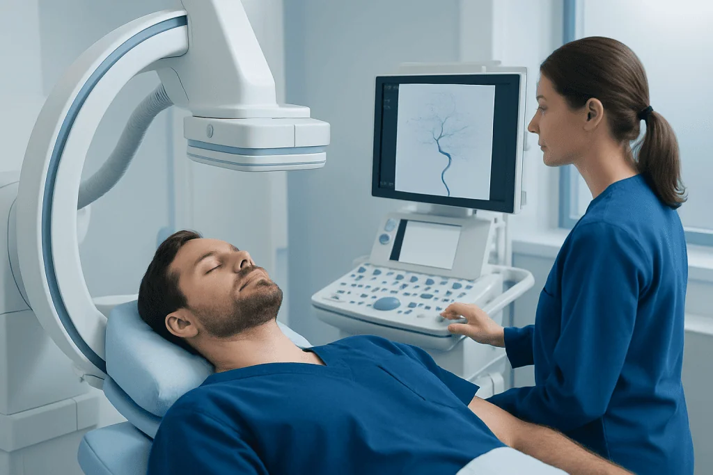
The brain depends on a constant blood supply. If one major artery must be closed during treatment, the most important question is whether other blood vessels can compensate well enough. Balloon test occlusion helps answer that question in real time by briefly simulating what would happen if the artery were permanently blocked.
Doctors may recommend this test before treating large or complex aneurysms, certain skull base tumors, traumatic vessel injuries, or carotid artery conditions that cannot be managed safely by preserving the vessel. It may also be considered before selected endovascular or surgical procedures when a vessel could need to be sacrificed to control bleeding or remove diseased tissue.
In some people, the circulation between the left and right sides of the brain and between front and back vessels is strong enough to provide backup flow. In others, those natural detours are limited. Balloon test occlusion helps identify which situation is present so that treatment planning is more individualized. For patients being evaluated for related conditions such as brain aneurysm or carotid artery disease, the test can be one part of a broader imaging and risk assessment process.
How the Procedure Is Performed

The test is usually done in an angiography suite. After the skin is cleaned and numbed, a catheter is inserted, often through an artery in the groin or wrist, and guided to the blood vessel being assessed. Contrast dye and X-ray imaging are used to confirm catheter position and study blood flow patterns. A small balloon at the catheter tip is then inflated to temporarily block the vessel.
While the balloon is inflated, the medical team closely monitors the patient for any neurological changes. The patient may be asked to speak, move the arms and legs, answer simple questions, or perform brief mental tasks. In some centers, additional monitoring may include blood pressure control, brain blood flow imaging, electroencephalography, or other supportive tests to improve assessment.
The balloon is typically kept inflated only for a limited period, often minutes rather than hours, though the exact approach depends on the vessel, the reason for testing, and institutional practice. Once the assessment is complete, the balloon is deflated and removed. The doctor then applies pressure or a closure device at the access site and observes the patient during recovery.
Balloon test occlusion is often performed alongside detailed cerebral angiography because the anatomy of the brain vessels needs to be clearly mapped. In selected cases, additional planning may involve endovascular aneurysm treatment or a surgical approach such as brain aneurysm surgery, depending on the final treatment decision.
What Doctors Look For During the Test
The main goal is to determine whether temporary vessel closure causes symptoms or signs of reduced blood flow to the brain. If a patient remains neurologically stable, that suggests the collateral circulation may be adequate. If weakness, speech difficulty, confusion, vision changes, or other neurological symptoms appear, the vessel may be too important to close permanently without further planning.
Doctors do not rely only on symptoms. They also consider imaging findings, the speed and direction of blood flow through other arteries, blood pressure response, and sometimes delayed venous filling patterns or perfusion studies. These details can reveal whether the brain is coping comfortably or only barely compensating.
In some cases, a patient may pass the basic test but still need additional evaluation if the planned procedure carries higher risk. This is because the brain’s tolerance can be affected by factors such as blood pressure changes, anesthesia, or the need for longer-term vessel closure. The final decision is therefore based on the full clinical picture rather than one single measurement.
- Favorable result: no neurological symptoms and reassuring flow patterns
- Borderline result: no obvious symptoms but less robust collateral flow on imaging
- Unfavorable result: neurological changes or clear evidence of inadequate blood supply
Who May Need It and Possible Risks
Not every patient having a brain vessel procedure needs balloon test occlusion. It is usually reserved for situations in which sacrificing an artery is a realistic possibility or when preserving the vessel may not be technically safe. The decision depends on the disease being treated, vessel anatomy, prior imaging results, and the experience of the multidisciplinary team.
Like other catheter-based vascular procedures, balloon test occlusion has potential risks. These can include bleeding or bruising at the access site, allergy or reaction to contrast material, temporary discomfort, blood vessel injury, blood clot formation, or stroke. Serious complications are uncommon but important, which is why the test is performed in specialized centers with continuous monitoring and strict safety protocols.
Doctors also consider whether a patient can cooperate with neurological testing during the procedure and whether kidney function, medications, or other medical conditions may affect planning. Before the test, patients are usually informed about the expected benefits, possible alternatives, and the reasons the team believes the information will help guide treatment safely.
Preparing for the Test and Recovery Afterward
Preparation instructions vary, but patients are commonly asked about allergies, blood-thinning medicines, kidney problems, previous reactions to contrast dye, and pregnancy status when relevant. Some may need blood tests or updated imaging before the procedure. Fasting for several hours may also be recommended, depending on sedation plans and local protocols.
During the procedure, sedation may be minimal or moderate because doctors often need the patient to respond to questions and commands. Afterward, the patient usually rests while the access site is monitored for bleeding. Nurses check blood pressure, pulse, and neurological status until it is safe to sit up, walk, and go home or move to a hospital room if further treatment is planned.
Recovery is often straightforward. Mild soreness or bruising at the catheter site can occur, and activity may be limited briefly. Patients are usually advised to drink fluids if appropriate, follow instructions about medicines, and seek medical help if they develop worsening headache, weakness, speech problems, chest pain, significant bleeding, or swelling at the puncture site.
What Happens After the Results
Once the test is complete, the team reviews the findings and explains what they mean for the next step. If the brain appears to tolerate temporary occlusion well, the planned procedure that involves permanent vessel closure may be considered safer. If tolerance is poor or uncertain, doctors may recommend preserving the artery, using a bypass strategy, choosing another endovascular technique, or reconsidering the timing of treatment.
The results are interpreted alongside MRI, CT, angiography, symptoms, and the specifics of the condition being treated. In many cases, treatment planning is discussed in a multidisciplinary meeting involving interventional neuroradiologists, neurosurgeons, neurologists, and anesthesiology teams. This collaborative approach helps balance treatment effectiveness with protection of brain function.
Patients often find it reassuring to know that balloon test occlusion is designed to reduce risk by gathering information before a permanent decision is made. Near the end of the care pathway, centers such as Acibadem International may support international patients through multidisciplinary evaluation and treatment in JCI-accredited hospitals when this type of advanced neurovascular assessment is needed.
Depending on the final plan, treatment may proceed with options such as carotid artery stenting or another open or endovascular procedure chosen for the patient’s anatomy and diagnosis. The care team will explain the expected benefits, possible risks, and follow-up schedule in clear terms before treatment moves forward.
Frequently asked questions
Is balloon test occlusion a surgery?
No. Balloon test occlusion is usually a minimally invasive catheter-based procedure rather than open surgery. It is performed through a blood vessel using imaging guidance to temporarily block blood flow and assess safety before another treatment.
Does balloon test occlusion hurt?
Most patients do not feel pain from the balloon itself. They may notice a brief sting from the numbing medicine and some pressure at the catheter insertion site, but the team works to keep the procedure as comfortable as possible.
How long does the test take?
The total appointment often lasts several hours because it includes preparation, the procedure, and recovery monitoring. The actual period of temporary vessel blockage is usually much shorter and is carefully limited by the medical team.
What does it mean if a patient 'passes' the test?
Passing the test generally means the patient had no concerning neurological symptoms and the blood flow patterns looked reassuring during temporary vessel closure. Even so, doctors still consider the full clinical picture before deciding on permanent artery closure.
Can someone fail the test and still be treated?
Yes. An unfavorable result does not mean treatment is impossible. It usually means the team may need a different strategy, such as preserving the artery, using another endovascular technique, or considering surgical reconstruction or bypass in selected cases.
Are there risks of stroke during balloon test occlusion?
There is a small risk, as with other procedures involving brain blood vessels. Because of that, the test is done in specialized settings with close neurological and imaging monitoring to reduce risk and respond quickly if a problem occurs.
This article is for general information only and is not a substitute for professional medical advice. Please consult a qualified doctor about your individual situation.
Explore treatments in Turkey — costs, top hospitals & a free quote
JCI-accredited · board-certified surgeons · reply within 24h
Add us as a Preferred Source to see more of our trusted health content across Google Search, AI Overviews and Discover.
Check your numbers in seconds
BMI, calories, due date, blood pressure and 30+ more clinical calculators — free, instant, doctor-reviewed ranges.

Neurosurgery
Surgical treatment of the brain, spine and nervous system, including minimally invasive and functional procedures.
70 specialists in this unitMore from the Health Library

Brain Aneurysm Symptoms: When Urgent Endovascular Treatment May Be Needed

Aneurysm Coiling: Who Is a Candidate and How Recovery Compares With Surgery

Stroke Thrombectomy Recovery: What to Expect After Clot Removal

Neurointerventional Radiology: What Happens During Diagnosis and Treatment Planning

Second Opinion for Brain Aneurysm: When Treatment Plans Need Review