Brain Aneurysm: How Neuroradiology Finds It and What Happens Next

Many brain aneurysms are found with imaging before they cause symptoms. CT, MRI, and cerebral angiography help doctors confirm the aneurysm’s size, shape, and location.
Key Takeaways
- Many brain aneurysms are found with imaging before they cause symptoms.
- CT, MRI, and cerebral angiography help doctors confirm the aneurysm’s size, shape, and location.
- Treatment depends on whether the aneurysm has ruptured, as well as its features and the person’s overall health.
- Options may include observation, blood pressure control, endovascular procedures, or neurosurgery.
- Sudden severe headache, new neurological symptoms, or loss of consciousness needs urgent medical evaluation.
A brain aneurysm is a weak, bulging area in a blood vessel in the brain. Neuroradiology plays a central role in finding aneurysms, assessing rupture risk, and helping doctors decide whether monitoring, endovascular treatment, or surgery is the safest next step.
Overview: What Is a Brain Aneurysm?
A brain aneurysm, also called a cerebral or intracranial aneurysm, is a weakened area in the wall of an artery in the brain that balloons outward. Some aneurysms remain small and stable for years, while others grow over time. The main concern is rupture, which can cause bleeding around the brain, known as a subarachnoid hemorrhage.
Not every brain aneurysm causes symptoms, and many are found incidentally during imaging done for headaches, dizziness, injury, or other unrelated reasons. Finding an aneurysm does not automatically mean emergency treatment is needed. The next steps depend on whether it has ruptured, how large it is, where it is located, and the person’s age, health, and risk factors.
Neuroradiology is especially important in this process. Specialists in this field use advanced imaging to detect aneurysms, describe their shape and blood flow, and help determine the most appropriate plan. In some cases, interventional neuroradiologists can also treat aneurysms from inside the blood vessels using minimally invasive techniques.
Symptoms and Warning Signs
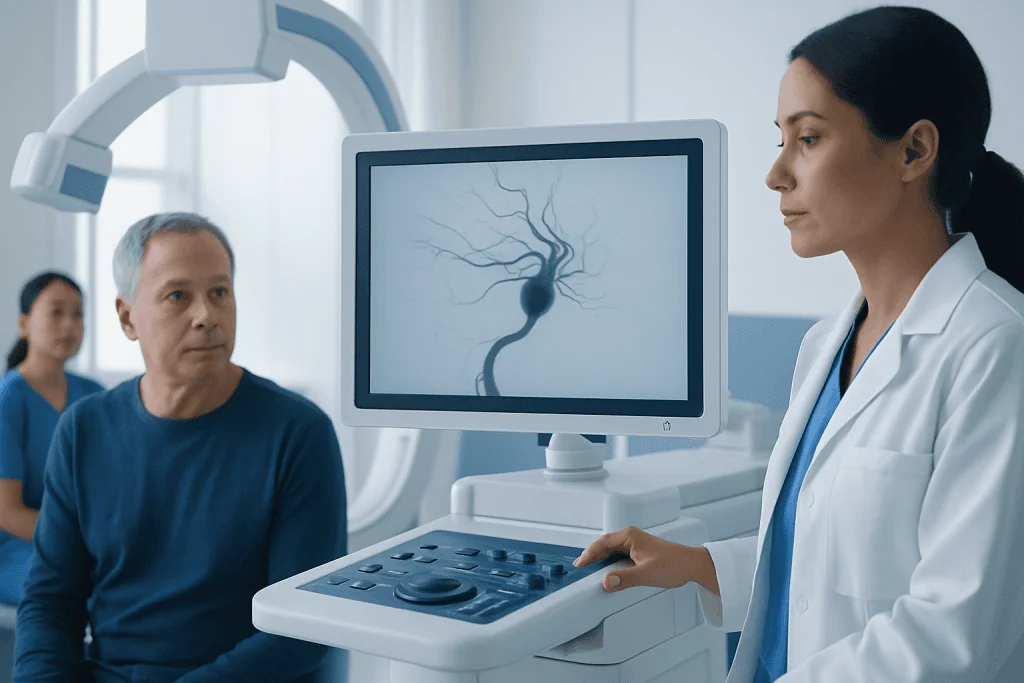
An unruptured brain aneurysm may cause no symptoms at all. When symptoms do occur, they are often related to the aneurysm pressing on nearby nerves or brain structures. Depending on its location, a person may notice headache, pain around or behind the eye, double vision, a drooping eyelid, or changes in vision. Less commonly, numbness, weakness, or balance problems may develop.
A ruptured aneurysm is a medical emergency. The classic symptom is a sudden, severe headache that is often described as the worst headache of a person’s life. This can happen along with nausea, vomiting, neck stiffness, sensitivity to light, confusion, seizure, fainting, or loss of consciousness.
Sometimes a small leak occurs before a major rupture. This may cause a sudden unusual headache or a severe new headache in the days before a larger bleed. Any abrupt, intense headache or new neurological symptom should be assessed urgently because rapid diagnosis can be lifesaving.
- Possible unruptured aneurysm symptoms: eye pain, vision changes, double vision, drooping eyelid, localized headache
- Possible rupture symptoms: sudden severe headache, vomiting, stiff neck, confusion, seizure, collapse
- Emergency help is important if symptoms start suddenly
Causes and Risk Factors

Brain aneurysms develop when part of an artery wall becomes weak. This weakness can be influenced by a combination of inherited factors, aging, blood flow stress, and medical conditions that affect the blood vessels. Most aneurysms form over time rather than being present from birth in a fully developed form.
Several factors are associated with a higher chance of developing or rupturing an aneurysm. These include high blood pressure, smoking, increasing age, and a family history of brain aneurysm or hemorrhagic stroke. Certain inherited disorders that affect connective tissue or blood vessel structure may also increase risk in some people.
Doctors also consider aneurysm-specific features when estimating risk. Larger aneurysms, irregularly shaped aneurysms, and aneurysms in certain parts of the brain circulation may be more concerning. Having had a previous aneurysm rupture, using stimulant drugs, or having poorly controlled vascular risk factors can further raise the level of concern.
How Neuroradiology Finds a Brain Aneurysm
Neuroradiology uses different imaging tools to look at the brain and its blood vessels. The choice of test depends on whether symptoms suggest an emergency, whether bleeding is suspected, and how much detail doctors need before making treatment decisions. In urgent settings, imaging is often performed quickly to look for bleeding and identify the source.
A non-contrast CT scan is commonly the first test when a ruptured aneurysm is suspected because it can rapidly show fresh bleeding around the brain. CT angiography adds contrast dye to outline the arteries and may reveal the aneurysm itself. MRI and MR angiography are also useful, especially for evaluating unruptured aneurysms or following them over time without radiation exposure in some cases.
The most detailed test is cerebral angiography, also called digital subtraction angiography. In this procedure, a thin catheter is guided through a blood vessel, usually from the wrist or groin, and contrast is injected while X-ray images are taken. This allows specialists to assess the aneurysm’s exact size, neck, shape, and relationship to nearby vessels. It is often used when treatment is being planned and may be part of interventional neuroradiology treatment for the aneurysm itself.
Imaging also helps distinguish a brain aneurysm from other conditions that can cause headache or neurological symptoms, such as vascular malformations or stroke. Careful interpretation by experienced neuroradiologists and neurologists is important because management may differ significantly from one condition to another.
What Happens After Diagnosis
After a brain aneurysm is found, the medical team first determines whether it has ruptured. A ruptured aneurysm requires urgent hospital care to stabilize the person, prevent rebleeding, manage blood pressure, and watch for complications such as vasospasm, hydrocephalus, or seizures. The aneurysm usually needs to be secured quickly with an endovascular procedure or surgery.
If the aneurysm is unruptured, the next step is a careful risk assessment. Doctors review its size, shape, and location, along with the patient’s symptoms, family history, blood pressure, smoking history, and overall health. This helps balance the natural risk of rupture against the risks and benefits of treatment.
In many cases, care involves a team approach that may include neuroradiology, neurology, neurosurgery, and critical care specialists. Some patients are advised to have periodic follow-up scans, while others are referred for more active treatment. The plan is individualized, and shared decision-making is an important part of choosing the next step.
Treatment Options
Treatment depends on whether the aneurysm has ruptured and how likely it is to rupture in the future. Small, low-risk unruptured aneurysms may be managed with observation, regular imaging, and control of risk factors such as high blood pressure and smoking. This approach aims to reduce strain on the blood vessel wall while monitoring for growth or shape changes.
When treatment is recommended, one common option is endovascular therapy. Through a catheter placed in a blood vessel, specialists can reach the aneurysm from inside the circulation and block it off from blood flow. Techniques may include coiling, stent-assisted coiling, or flow-diverting devices, depending on the aneurysm’s anatomy. These approaches are often part of aneurysm treatment and may offer a less invasive alternative to open surgery for selected patients.
Another option is microsurgical clipping, in which a neurosurgeon places a small clip across the neck of the aneurysm during an operation. Clipping may be preferred for some aneurysms based on their location, shape, or the patient’s age and medical needs. People with bleeding in the brain may also need treatment for related complications, and care can overlap with stroke treatment pathways when intensive neurological monitoring is required.
Doctors explain the benefits and limitations of each method, including the chance of complete aneurysm closure, need for follow-up imaging, and procedure-related risks. The goal is to choose the safest and most durable strategy for the individual patient rather than using the same approach for everyone.
Prevention, Self-care, and Recovery
It is not always possible to prevent a brain aneurysm, but managing vascular health can help reduce risk. Avoiding smoking is one of the most important steps. Good blood pressure control, regular medical follow-up, physical activity suited to the person’s health, and treatment of other cardiovascular risk factors may also help protect blood vessels.
For people living with a known unruptured aneurysm, self-care usually focuses on reducing strain on the circulatory system and following the imaging schedule recommended by the care team. Patients should ask their doctor about safe exercise, travel, work, and use of medications, especially blood thinners or drugs that affect blood pressure. It is also helpful to seek prompt advice if headaches change significantly or new neurological symptoms appear.
Recovery after aneurysm treatment varies. After endovascular therapy, many people recover relatively quickly, though they still need follow-up visits and imaging. Recovery after rupture can take much longer and may involve rehabilitation for strength, speech, memory, or daily functioning. If recovery needs are complex, doctors may coordinate with services related to neurosurgery and rehabilitation to support long-term function and quality of life.
When to See a Doctor
A person should seek medical advice if they have persistent unusual headaches, new visual symptoms, unexplained drooping of an eyelid, double vision, or a strong family history of brain aneurysm. These symptoms do not always mean an aneurysm is present, but they deserve evaluation, especially if they are new or worsening.
Emergency care is needed for sudden severe headache, sudden confusion, seizure, collapse, loss of consciousness, or any abrupt neurological deficit such as weakness, numbness, or trouble speaking. These symptoms may signal aneurysm rupture or another urgent neurological condition and should never be ignored.
Specialized centers can provide coordinated assessment with imaging, neurological examination, and treatment planning. Acibadem International’s multidisciplinary specialists and JCI-accredited hospitals diagnose and treat brain aneurysms for international patients, including advanced neuroradiology and endovascular care when appropriate.
Frequently asked questions
Can a brain aneurysm exist without symptoms?
Yes. Many brain aneurysms do not cause symptoms and are found incidentally during imaging for another reason. Symptoms are more likely if the aneurysm grows, presses on nearby structures, or ruptures.
What imaging test is best for a brain aneurysm?
There is no single best test for every situation. CT and CT angiography are often used in emergencies, while MRI or MR angiography may be used for further evaluation or follow-up. Cerebral angiography provides the most detailed view when treatment planning is needed.
Does every brain aneurysm need treatment?
No. Some small, unruptured aneurysms can be monitored safely with repeat imaging and control of risk factors. Treatment decisions are based on rupture status, size, location, shape, symptoms, and the person’s overall health.
Is a brain aneurysm the same as a stroke?
Not exactly. A ruptured brain aneurysm can cause bleeding in or around the brain, which is a type of hemorrhagic stroke. However, not all strokes are caused by aneurysms, and many strokes occur because of a blocked blood vessel instead.
What is the difference between coiling and clipping?
Coiling is an endovascular treatment performed from inside the blood vessels using a catheter, while clipping is an open surgical procedure done through the skull. Both aim to prevent blood from entering the aneurysm, but the best choice depends on the aneurysm’s anatomy and the patient’s condition.
Can a brain aneurysm come back after treatment?
Some treated aneurysms need long-term follow-up because blood flow can occasionally recur, especially after certain endovascular procedures. This is why doctors recommend repeat imaging at intervals based on the treatment type and aneurysm features.
References
- World Health Organization
- American Heart Association
- National Institute of Neurological Disorders and Stroke
- Radiological Society of North America
- Brain Aneurysm Foundation
This article is for general information only and is not a substitute for professional medical advice. Please consult a qualified doctor about your individual situation.
Neuroradiology in Turkey — costs, top hospitals & a free quote
JCI-accredited · board-certified surgeons · reply within 24h
Add us as a Preferred Source to see more of our trusted health content across Google Search, AI Overviews and Discover.
Check your numbers in seconds
BMI, calories, due date, blood pressure and 30+ more clinical calculators — free, instant, doctor-reviewed ranges.

More from the Health Library



Related Specialists

Dr. Veli Mehmet Kından
Emergency Service
Fzt. Ahsen Ercan
Physical Medicine & Rehabilitation
Dr. Murat Öksüz
Gastroenterology
Dr. İlke Karagöz
Aesthetic Plastic & Reconstructive Surgery