Breast MRI: What Is Normal, What Is Not, and When to Seek Care
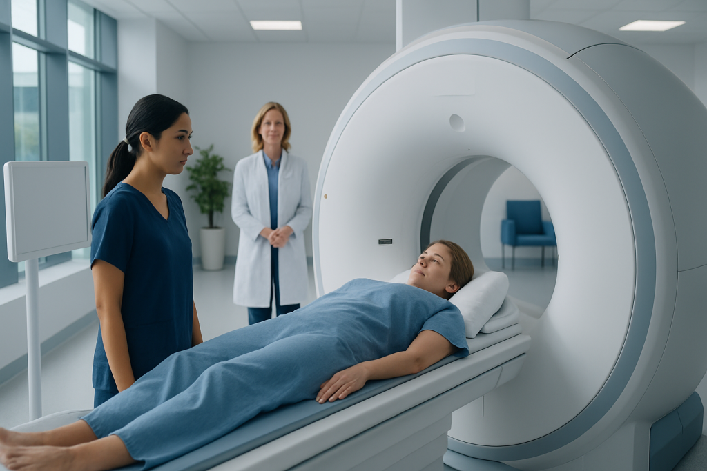
Breast MRI can detect subtle breast changes, but not every abnormal-looking area is dangerous. Normal breast MRI findings vary with age, hormones, breast density, and timing in the menstrual cycle.
Key Takeaways
- Breast MRI can detect subtle breast changes, but not every abnormal-looking area is dangerous.
- Normal breast MRI findings vary with age, hormones, breast density, and timing in the menstrual cycle.
- An abnormal breast MRI does not automatically mean cancer; additional imaging or biopsy may be needed.
- Breast MRI is often used for high-risk screening, evaluating unclear findings, and checking the extent of known disease.
- Medical care should be sought promptly for a new breast lump, nipple discharge, skin changes, or concerning MRI results.
A breast MRI is a highly sensitive imaging test that can show both normal breast tissue patterns and changes that may need closer evaluation. Many MRI findings are not cancer, but results should always be interpreted together with symptoms, breast exam findings, and other imaging such as mammography or ultrasound.
Overview: what a breast MRI can and cannot show
A breast MRI is a detailed imaging test that uses magnets, radio waves, and usually contrast dye to create pictures of the breast. In simple terms, a normal breast MRI may show background tissue enhancement, small benign cysts, and the natural differences between the two breasts. What is not normal depends on the pattern seen: some areas look suspicious enough to need more imaging or a biopsy, while many others turn out to be noncancerous.
Unlike mammography, which is especially good at showing calcifications, breast MRI is valued for its sensitivity. It can detect areas of increased blood flow and tissue activity that may not be visible on other tests. For that reason, doctors often use it to screen people at high risk, to investigate unclear findings, to assess breast implants, or to understand the extent of a known breast problem.
At the same time, MRI has limits. Because it is so sensitive, it can also find changes that look unusual but are not harmful. This is why breast MRI results are rarely interpreted alone. Radiologists compare the images with a person’s symptoms, physical examination, mammogram, ultrasound, and personal or family history before deciding whether a finding is reassuring, needs short-term follow-up, or requires tissue sampling.
What is considered normal on a breast MRI
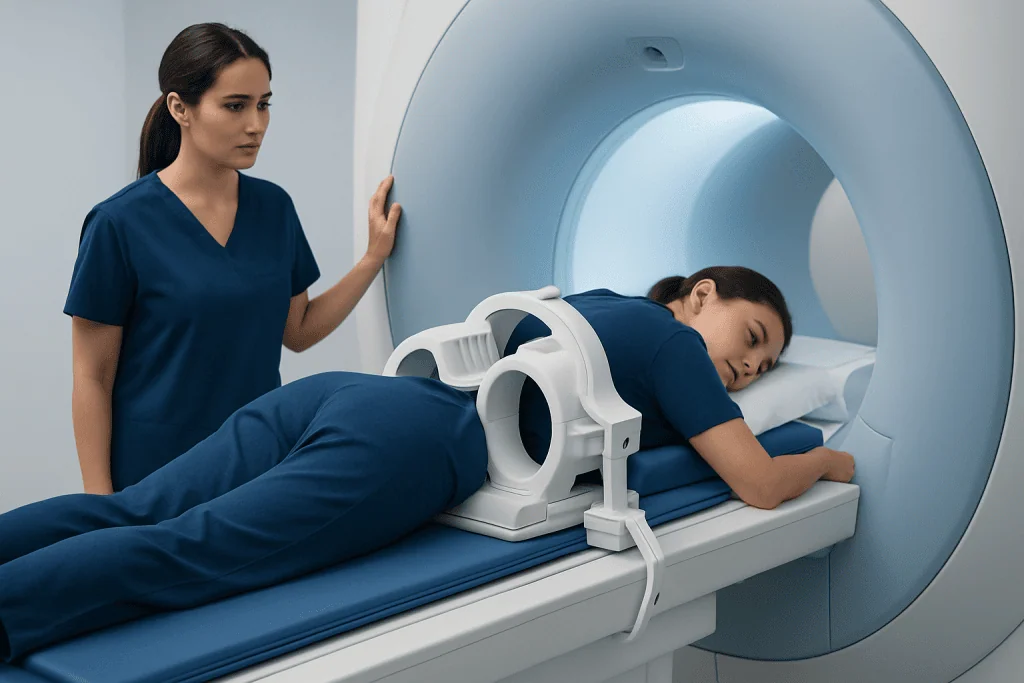
Normal on breast MRI does not always mean the breasts look completely uniform. Healthy breasts naturally contain a mix of fatty tissue, glandular tissue, ducts, blood vessels, and lymph nodes. These structures can appear differently from person to person. The breasts may also enhance differently after contrast because hormone levels affect breast tissue.
One common normal feature is background parenchymal enhancement, which describes how much normal fibroglandular tissue lights up after contrast. Mild or moderate enhancement can be a normal finding, especially in younger women or around certain times in the menstrual cycle. Small simple cysts, post-surgical scar tissue, and stable benign changes seen on earlier scans may also be considered normal or expected.
Radiologists also look for symmetry, shape, and the pattern of enhancement. Smooth, well-defined areas are more often benign than irregular or spiculated ones. MRI reports may describe findings using standard terminology, but the meaning often depends on context. A report that mentions benign features or recommends routine follow-up is generally reassuring, even if the wording sounds technical.
People with breast implants may have a breast MRI to assess implant integrity as well as breast tissue. In this setting, a normal result may show intact implants without leakage, along with no suspicious enhancement in the surrounding tissues.
What is not normal and why it may need follow-up

An MRI finding is considered not normal when it shows a pattern that cannot be confidently labeled benign. This might include a mass with irregular borders, non-mass enhancement in a suspicious distribution, rapid contrast uptake with certain washout patterns, enlarged abnormal lymph nodes, or signs of implant rupture. These findings do not confirm cancer, but they may justify more testing.
Abnormal does not always mean serious disease. Many noncancerous conditions can look concerning on MRI, including fibroadenomas, hormonal changes, inflammation, scar tissue, fat necrosis, and some benign proliferative breast changes. Infections, recent procedures, and even timing in the menstrual cycle can affect the appearance of the breasts.
Radiologists often classify MRI findings using the BI-RADS system. A BI-RADS 1 or 2 result is generally benign. BI-RADS 3 usually suggests that the finding is probably benign but should be checked again after a short interval. BI-RADS 4 or 5 means the finding is suspicious enough that biopsy is often recommended. If the result remains unclear, the next step may be targeted ultrasound, mammography, repeat MRI, or breast biopsy.
Some MRI-detected changes may relate to conditions such as breast cancer or fibrocystic breast changes, but imaging alone cannot always distinguish between them. That is why a stepwise evaluation is important.
When doctors recommend breast MRI
Breast MRI is not needed for everyone. It is usually recommended in specific situations where added detail can improve care. One common reason is screening in people at high risk for breast cancer, such as those with a strong family history, certain genetic mutations, or a history of chest radiation at a young age.
It may also be used when mammography or ultrasound does not fully explain a lump, nipple discharge, or another symptom. In people already diagnosed with breast cancer, MRI can help estimate the size of the tumor, look for additional areas in the same or opposite breast, and support treatment planning. In some cases, MRI is useful for evaluating implants or checking the response to treatment.
For the clearest images in menstruating women, the scan is often scheduled during a certain part of the cycle, commonly in the early follicular phase. This can reduce normal hormonal enhancement and make the images easier to interpret. A doctor will balance timing with the urgency of the clinical concern.
Although highly useful, breast MRI is not a replacement for routine mammography in most people. Instead, it is usually a complementary test, selected because it answers a specific clinical question.
How breast MRI is interpreted and what results may mean
A breast MRI report usually describes the amount of breast tissue, the background enhancement, and whether any masses or areas of non-mass enhancement are present. It may also mention lymph nodes, chest wall structures, and implants if relevant. Because the language is technical, a result can sound alarming even when the overall impression is reassuring.
If the MRI is normal or benign, the report may recommend routine screening based on age and risk. If the finding is probably benign, short-interval imaging follow-up may be advised rather than immediate biopsy. If the MRI is suspicious, doctors often correlate it with mammography and ultrasound to see whether the area can be identified using another method that may make biopsy easier.
When a suspicious MRI finding cannot be seen on other imaging, an MRI-guided biopsy may be needed. This helps determine whether the tissue is benign, high-risk, or cancerous. If cancer is diagnosed, the next steps may include consultations about breast cancer treatment and, when appropriate, breast surgery.
Patients often worry when the report mentions enhancement. However, enhancement simply means that tissue takes up contrast. What matters most is the pattern, location, shape, and clinical context, not the word itself.
Preparing for the scan and understanding possible limitations
Before a breast MRI, patients are usually asked about kidney health, allergies to contrast, pregnancy, implants, and any metal devices in the body. The scan is performed while lying face down with the breasts positioned in a dedicated coil. It is painless, although the machine can be noisy and some people find the enclosed space uncomfortable.
Contrast is often used because it improves the ability to see abnormal blood flow and tissue behavior. Most people tolerate contrast well, but the care team will review whether it is suitable in each case. It is important to tell the medical team about prior surgeries, biopsies, breastfeeding, or infections, since these can affect the images.
Breast MRI has some limitations. It can lead to false-positive results, meaning it finds something suspicious that is ultimately benign. It may also miss some abnormalities that are better detected by mammography, especially certain calcifications. In addition, normal hormone-related changes can make interpretation more difficult in some patients.
These limitations do not reduce the value of breast MRI; they simply explain why it is used thoughtfully and alongside other tests rather than as a stand-alone answer.
When to seek medical care
Medical care should be sought if a person notices a new breast lump, persistent breast swelling, skin dimpling, nipple inversion, bloody or spontaneous nipple discharge, redness that does not improve, or swollen lymph nodes under the arm. These symptoms do not always mean cancer, but they should be assessed promptly by a qualified clinician.
Follow-up is also important after an abnormal breast MRI result, even when there are no symptoms. Delaying the recommended ultrasound, mammogram, repeat MRI, or biopsy can postpone a clear diagnosis. If the report wording is confusing, it is reasonable to ask the doctor or radiologist to explain the BI-RADS category and the next step.
Urgent evaluation may be needed for signs of infection such as fever, warmth, increasing pain, and spreading redness, especially after a procedure or during breastfeeding. Immediate medical attention is also appropriate if there is a sudden change in an implant, such as a marked change in breast shape, new firmness, or significant swelling.
For people who need coordinated evaluation, Acibadem International’s multidisciplinary specialists and JCI-accredited hospitals diagnose and treat breast conditions for international patients, including advanced imaging and follow-up care when needed.
Frequently asked questions
Does an abnormal breast MRI mean cancer?
No. Breast MRI is very sensitive, so it can detect many changes that are ultimately benign, such as cysts, scar tissue, or hormone-related enhancement. An abnormal result means the finding needs interpretation in context and may require follow-up imaging or biopsy.
What does enhancement mean on a breast MRI?
Enhancement means that breast tissue takes up contrast dye and becomes more visible on the scan. Some enhancement is normal, especially in glandular tissue. Doctors focus on the pattern, shape, and distribution of enhancement to decide whether it looks benign or suspicious.
Is breast MRI better than mammography?
Breast MRI is not simply better; it serves a different purpose. It is more sensitive for some findings, especially in high-risk patients, but mammography remains important because it can detect changes like calcifications that MRI may not show as well. Many patients benefit from both tests used together.
When is the best time in the cycle to have a breast MRI?
For menstruating women, breast MRI is often scheduled during the early part of the menstrual cycle. This timing may reduce normal background enhancement and improve image interpretation. The exact timing should be guided by the doctor and the urgency of the reason for the scan.
Can a normal breast MRI rule out every breast problem?
No imaging test can rule out every possible breast condition with complete certainty. A normal breast MRI is reassuring, but symptoms such as a new lump, nipple discharge, or skin changes still deserve medical attention. Doctors always consider symptoms, examination, and other imaging together.
Why might a biopsy be needed after breast MRI?
A biopsy is recommended when the imaging pattern cannot be confidently labeled benign. The goal is to examine tissue directly and determine whether the change is harmless, high-risk, or cancerous. This helps guide the safest and most appropriate next steps.
References
- American Cancer Society
- Radiological Society of North America
- American College of Radiology
- National Cancer Institute
- World Health Organization
This article is for general information only and is not a substitute for professional medical advice. Please consult a qualified doctor about your individual situation.
Oncology care in Turkey — second opinion and treatment plan
JCI-accredited · board-certified surgeons · reply within 24h
Add us as a Preferred Source to see more of our trusted health content across Google Search, AI Overviews and Discover.