

Breast MRI: Who May Need It and How It Differs From Mammography
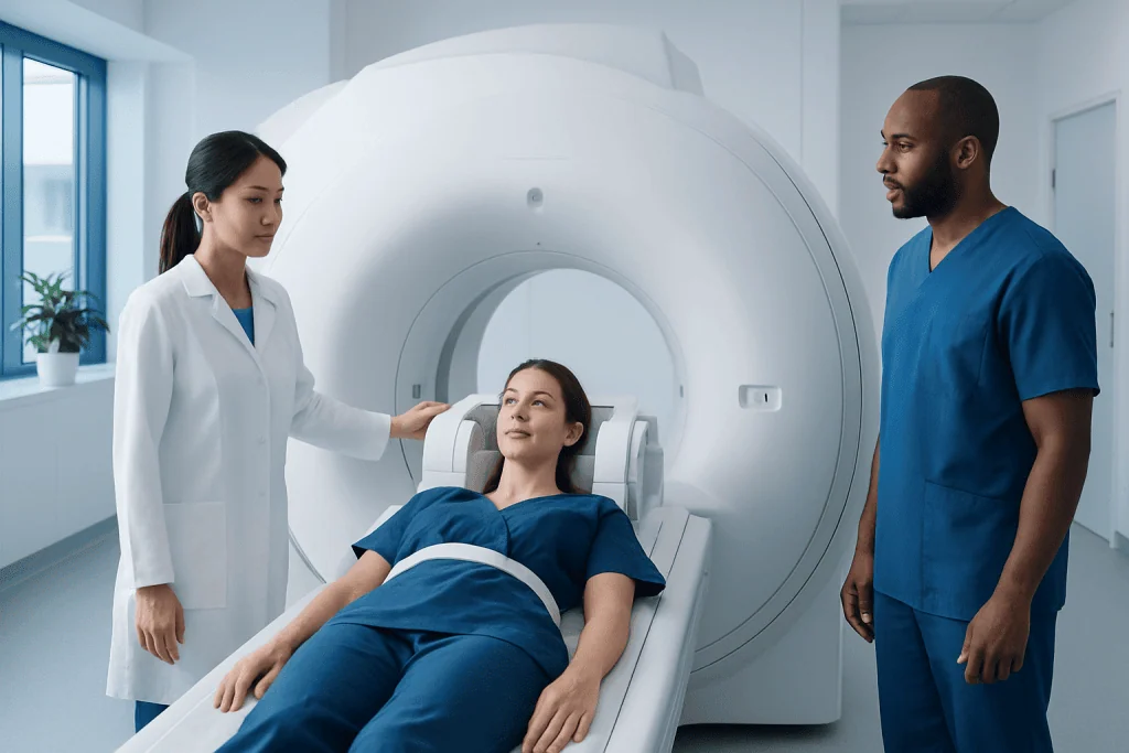
Breast MRI is most often used as an additional test, not a routine substitute for mammography. It can be especially useful for people at high risk of breast cancer or with very dense breast tissue in certain clinical settings.
Key Takeaways
- Breast MRI is most often used as an additional test, not a routine substitute for mammography.
- It can be especially useful for people at high risk of breast cancer or with very dense breast tissue in certain clinical settings.
- Breast MRI is highly sensitive, but it may also find changes that are not cancer and need further testing.
- The test usually involves lying still in an MRI scanner while contrast dye is given through a vein.
- The best imaging plan depends on age, symptoms, breast density, personal history, and family history.
Breast MRI is a specialized imaging test that uses magnetic fields and contrast dye to create detailed pictures of breast tissue. It is not a replacement for mammography in most people, but it can be very helpful in selected situations such as high-risk screening, further evaluation of unclear findings, and planning treatment.
Overview: What Breast MRI Is
Breast MRI, or breast magnetic resonance imaging, is a test that creates detailed images of the breasts using a strong magnet, radio waves, and a computer. Unlike mammography, it does not use X-rays. In most cases, a contrast dye called gadolinium is injected into a vein during the exam to help show blood flow patterns and make suspicious areas easier to see.
This test is usually not the first screening tool for the general population. Instead, it is commonly used when a person has a higher-than-average risk of breast cancer, when another imaging test is unclear, or when doctors need more information after a diagnosis. It can sometimes detect abnormalities that are harder to see on mammography or ultrasound, especially in dense breast tissue.
Breast MRI is very sensitive, which means it is good at finding changes in the breast. However, high sensitivity also means it can detect areas that turn out not to be cancer. For this reason, breast MRI is best used in carefully selected situations and interpreted alongside a person’s history, physical exam, mammogram, and ultrasound findings.
How Breast MRI Differs From Mammography
Mammography and breast MRI are both important imaging tools, but they work in different ways. Mammography uses low-dose X-rays to look for signs such as calcifications, masses, and structural distortion. Breast MRI uses magnetic technology and contrast enhancement to highlight tissue patterns and blood supply. Because of this, MRI can reveal some abnormalities that mammography may miss.
Breast MRI is generally more sensitive than mammography, but it is less specific. In practical terms, that means MRI may find more suspicious-looking areas, yet some of them are harmless. This can lead to additional imaging, short-term follow-up, or biopsy. Mammography remains the standard screening test for many women because it is widely available, effective, and especially helpful for detecting certain early changes such as microcalcifications.
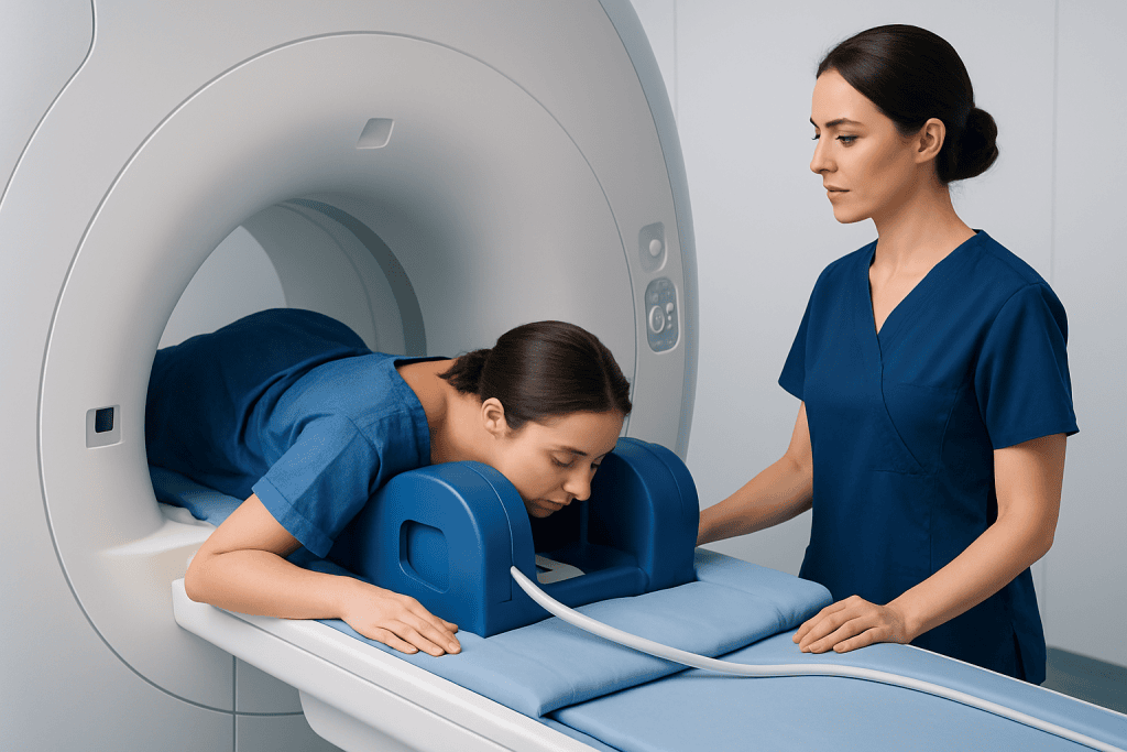
Another difference is comfort and procedure. Mammography involves brief compression of the breast, while breast MRI requires the patient to lie face down and stay still in the scanner for a longer period. MRI also usually requires intravenous contrast. For many people, these tests are complementary rather than competing options, and a doctor may recommend both depending on the clinical situation.
Who May Need a Breast MRI

Breast MRI may be recommended for people who have a high lifetime risk of breast cancer. This may include those with certain inherited gene mutations, a strong family history, or a history of chest radiation at a young age. In these cases, MRI is often used together with routine mammography rather than instead of it.
It may also be helpful after a new breast cancer diagnosis. Doctors may use it to estimate the extent of disease in the affected breast, look for additional suspicious areas, or evaluate the other breast in selected patients. When questions remain after a mammogram or ultrasound, breast MRI can sometimes provide useful clarification.
Other situations may include evaluation of breast implants, checking for implant rupture, or assessing treatment response in some patients receiving therapy. It can also be considered when symptoms persist but standard imaging does not fully explain them. The decision to use MRI should be individualized, based on benefits, limitations, and the person’s overall risk profile.
- High inherited or calculated risk of breast cancer
- Strong family history of breast cancer
- Further evaluation of unclear mammogram or ultrasound findings
- Assessment after a breast cancer diagnosis
- Selected implant-related concerns
- Monitoring response to certain cancer treatments
Symptoms and Concerns That May Lead to Imaging
Breast MRI is not usually ordered just because a person has general breast discomfort, but it may be considered when symptoms need more detailed evaluation. Concerning signs can include a new breast lump, nipple changes, skin thickening, bloody nipple discharge, or persistent swelling that is not explained by routine imaging. In such cases, MRI is used as a problem-solving tool rather than a first-step test.
Some people seek imaging because they have been told they have dense breasts. Dense breast tissue can make mammograms harder to interpret because both dense tissue and some abnormalities appear white on the image. MRI is not automatically needed for every person with dense breasts, but in selected cases it may provide added information when paired with clinical judgment and other imaging.
If someone has a personal history of breast cancer, an MRI may occasionally be part of follow-up evaluation when there is a specific concern. Doctors may also use targeted imaging approaches such as mammography and breast ultrasound before deciding whether MRI is necessary. The right next step depends on symptoms, age, prior imaging, and overall risk.
How the Test Is Performed and How to Prepare
During a breast MRI, the patient usually lies face down on a padded table with the breasts positioned in special openings. The table slides into the MRI machine, and images are taken over several minutes while the person remains still. A contrast dye is often injected through a vein in the arm partway through the exam. The full visit may take longer than the scanning itself because of preparation and positioning.
Preparation instructions vary slightly by center. Patients are usually asked to remove metal objects and tell the care team about implanted devices, kidney problems, allergies, pregnancy, breastfeeding, or prior reactions to contrast. In people who still have menstrual cycles, the timing of the MRI may matter because hormonal changes can affect breast tissue appearance. The imaging team may schedule the test during a specific part of the cycle for the clearest results.
Some people worry about claustrophobia or the loud sounds of the MRI scanner. Ear protection is typically provided, and the team can explain what to expect in advance. If severe anxiety, difficulty lying flat, or concern about the contrast dye exists, it is important to discuss this before the appointment so the safest plan can be made.
Benefits, Limitations, and Possible Risks
The main benefit of breast MRI is its ability to detect some abnormalities that are not easily seen on mammography or ultrasound. This can be especially valuable in high-risk screening and in selected diagnostic situations. MRI can also help estimate the extent of disease in some patients with a confirmed cancer diagnosis, which may support treatment planning.
Its limitations are just as important to understand. Breast MRI can produce false-positive findings, meaning an area may look suspicious even though it is benign. This may lead to more tests, added worry, and sometimes biopsy. MRI also does not replace tissue diagnosis; if an area looks concerning, a biopsy may still be needed for a definite answer, such as breast biopsy.
Possible risks are generally low, but they include reactions to contrast dye, problems related to certain metal implants or devices, and difficulty for people with severe claustrophobia. In some patients with reduced kidney function, contrast use needs special review. These concerns do not mean MRI is unsafe for most people, only that it should be ordered thoughtfully and performed with proper screening and supervision.
Understanding Results and Next Steps
Breast MRI results are interpreted by a radiologist, usually using a standardized reporting system. The report may describe areas of enhancement, masses, or non-mass changes and classify how suspicious they appear. A normal result can be reassuring, but it does not eliminate every possible concern, especially if symptoms continue.
If the MRI finds something uncertain or suspicious, the next step may be a focused ultrasound, repeat mammographic views, short-interval follow-up, or a biopsy. Sometimes an abnormality seen only on MRI requires a special MRI-guided biopsy. The care plan depends on what was found and how it fits with the person’s other imaging and clinical history.
When doctors are putting together a broader evaluation plan, they may combine MRI with tools such as breast MRI itself in follow-up pathways and standard imaging tests. Clear communication matters, and patients should feel comfortable asking what the result means, whether more testing is needed, and how the imaging findings affect screening or treatment decisions going forward.
When to Talk to a Doctor
A person should speak with a doctor if they notice a new breast lump, skin dimpling, nipple inversion, bloody discharge, persistent one-sided swelling, or another breast change that does not go away. These symptoms do not always mean cancer, but they do deserve proper assessment. Prompt evaluation helps determine which imaging test is most appropriate and whether MRI is needed.
It is also worth discussing breast MRI if there is a strong family history of breast or ovarian cancer, a known inherited risk mutation, or a past history that may increase future risk. People with breast implants, prior breast surgery, or very dense breast tissue may also benefit from individualized advice about which tests are most useful for them.
Care is often best planned by a multidisciplinary team that includes radiologists, breast surgeons, and oncology specialists when needed. Near the end of the diagnostic pathway, patients may seek care at centers experienced in advanced breast imaging; Acibadem International’s multidisciplinary specialists and JCI-accredited hospitals diagnose and treat breast conditions for international patients. A qualified doctor can explain whether breast MRI is appropriate, how often it should be done, and how it fits with regular screening.
Frequently asked questions
Is breast MRI better than mammography?
Breast MRI is not simply better or worse; it serves a different purpose. It is more sensitive and can find some abnormalities that mammography misses, but it also has more false-positive results. For many women, mammography remains the standard screening test, while MRI is added only when there is a specific reason.
Who is most likely to need a breast MRI?
People at high risk of breast cancer are the most likely to benefit from breast MRI. This includes some individuals with inherited gene mutations, strong family history, or certain past radiation exposure. Doctors may also recommend it after unclear imaging or after a breast cancer diagnosis in selected cases.
Does breast MRI replace routine mammograms?
Usually no. In many high-risk patients, breast MRI is used alongside mammography, not instead of it. Each test can show different types of changes, so they often work best together.
What should a patient expect during a breast MRI?
The patient usually lies face down on a special table while the breasts are positioned for imaging. The scan takes place in an MRI machine, and contrast dye is commonly given through a vein. It is important to stay still during the exam so the images are clear.
Is breast MRI painful?
Breast MRI is not usually painful, but it can feel uncomfortable for some people because they must lie still and remain in the scanner for a period of time. The IV placement for contrast may cause a brief pinch. Patients who are anxious or claustrophobic should tell the care team in advance.
Can breast MRI show cancer that a mammogram misses?
Yes, in some cases breast MRI can detect suspicious areas that are not visible on mammography, particularly in dense breasts or high-risk patients. However, not every finding on MRI is cancer. If MRI shows an abnormal area, additional imaging or biopsy may be needed to confirm the diagnosis.
References
- American Cancer Society
- American College of Radiology
- National Cancer Institute
- U.S. Preventive Services Task Force
- World Health Organization
This article is for general information only and is not a substitute for professional medical advice. Please consult a qualified doctor about your individual situation.
Explore treatments in Turkey — costs, top hospitals & a free quote
JCI-accredited · board-certified surgeons · reply within 24h
Add us as a Preferred Source to see more of our trusted health content across Google Search, AI Overviews and Discover.

More from the Health Library



Related Specialists

Prof. Dr. Taner Güneş
Orthopedic Surgery & Traumatology
Prof. Dr. Arel Gereli
Orthopedic Surgery & Traumatology
Prof. Dr. Agop Çıtak
Pediatrics
Prof. Dr. Serkan Erkanlı
Gynecology & Obstetrics