Broken Hand Xray: Preparation, Procedure and Results

Hand X-rays use a small amount of radiation to create images of the bones in the hand and wrist. Most people need no special preparation, and the imaging itself usually takes only a few minutes.
Key Takeaways
- Hand X-rays use a small amount of radiation to create images of the bones in the hand and wrist.
- Most people need no special preparation, and the imaging itself usually takes only a few minutes.
- X-ray results help determine whether a fracture can be treated with support and follow-up or needs realignment or surgery.
- A hand that is numb, pale, blue, cold, visibly deformed or affected by an open wound needs urgent medical assessment.
- Healing time varies by fracture type, alignment, treatment and overall health, but many hand fractures require several weeks of protection and follow-up.
A broken hand xray is a quick imaging test used to check whether a hand injury has caused a fracture, where the bone is broken and whether the pieces have moved out of position. It is usually performed soon after an injury and helps a clinician plan safe, appropriate treatment.
Broken Hand Xray: What It Shows
A broken hand xray is the main first imaging test used when a clinician suspects a fracture in one of the hand bones. It can show breaks in the metacarpals, which connect the wrist to the fingers, and the phalanges, which are the finger bones. The images help identify the fracture’s location, pattern, alignment and whether it reaches a joint.
Hand injuries can cause pain, swelling and bruising even when no bone is broken. An X-ray helps distinguish a fracture from a sprain, tendon injury, dislocation or soft-tissue bruise. It also provides a baseline image that can be compared with later X-rays while the injury heals.
Although people may search online for broken hand xray pictures, these images cannot reliably diagnose an individual injury. Fractures can be subtle, and image interpretation should be combined with an examination by a qualified clinician or radiologist.
When a Hand X-ray May Be Needed
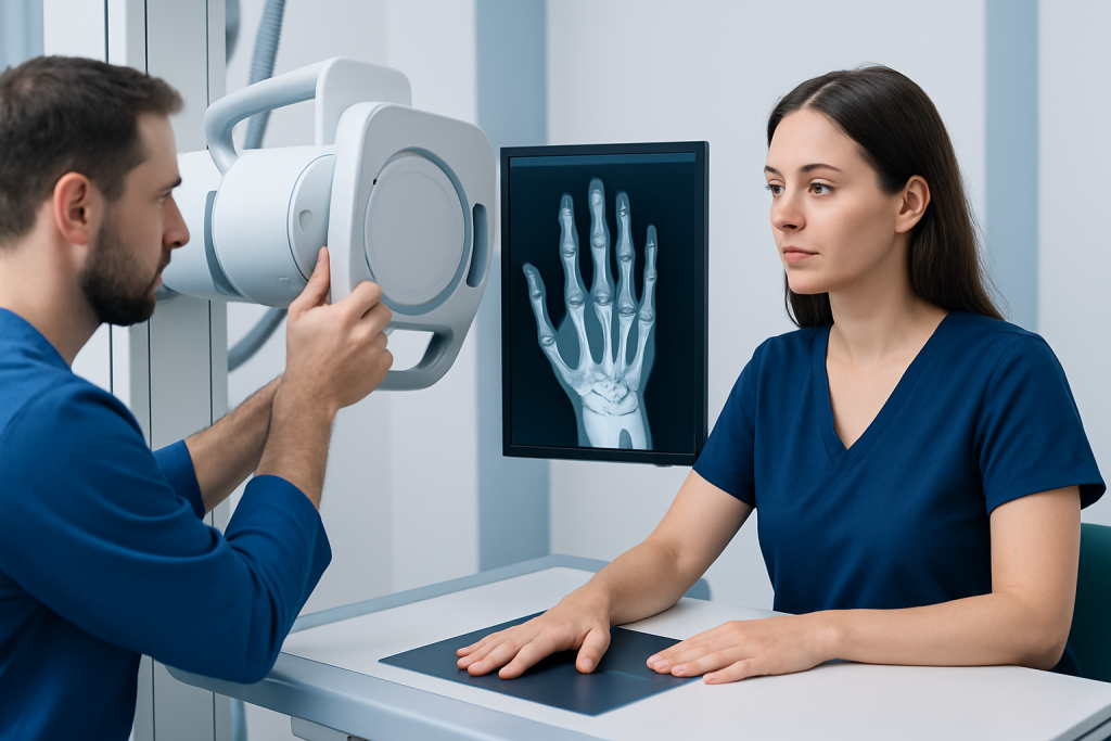
A clinician may recommend a hand X-ray after a fall, direct blow, crush injury, sports injury or twisting injury. It is particularly useful when pain is focused over a bone, there is swelling or bruising, movement is limited, or the hand looks misshapen. Pain when gripping, making a fist or moving a finger can also raise concern for a fracture.
Not every hand injury requires imaging immediately. However, it is important not to assume that an injured hand is only bruised, especially when symptoms persist or function is reduced. Delayed assessment can allow a displaced fracture to heal in a poor position, which may affect strength, movement or hand alignment.
Children, older adults and people with osteoporosis or other conditions that affect bone strength may have fractures after relatively minor trauma. The decision to image is based on the injury, symptoms, examination findings and a person’s medical history.
Preparation and Safety for a Broken Hand Xray

Most people do not need special preparation for a broken hand xray. They may be asked to remove rings, watches, bracelets or other jewellery from the affected hand and wrist because metal can obscure important details. It is helpful to tell the imaging team about any rings that cannot be removed due to swelling.
Hand X-rays use ionising radiation, but the dose for this type of examination is generally low. The radiographer positions the beam carefully and uses the lowest exposure needed to obtain clear diagnostic images. The benefits of identifying a fracture and planning treatment usually outweigh the small radiation risk.
People who are pregnant or think they may be pregnant should tell the radiographer before imaging. A hand X-ray is remote from the abdomen, but the team can consider the clinical need and use appropriate precautions. People should also mention severe pain, limited movement or a recent operation so positioning can be adapted safely.
How a Broken Hand Xray Is Performed
The test is performed by a radiographer in an X-ray department, urgent care centre or emergency setting. The person usually sits beside an X-ray table and places the hand on the detector. The radiographer will explain each position and support the hand as needed to reduce discomfort.
Several views are commonly taken, often from the front, side and an angled position. Different views are important because a fracture can be difficult to see from only one direction. Depending on the area of concern, images may include the fingers, hand, thumb or wrist.
The person needs to keep still for each image, usually for only a few seconds. The radiographer stands behind a protective screen while the image is taken, but can communicate throughout the procedure. No needles, contrast dye or sedation are usually required, and the scan itself is painless, although positioning an injured hand may be uncomfortable.
After the images are complete, the person can generally leave the imaging area immediately. If the injury is being assessed urgently, a clinician may review the images promptly; otherwise, a radiologist may issue a formal report for the referring doctor.
Understanding Results and the Broken Hand Protocol
A report may describe whether a fracture is present and identify the affected bone, such as a metacarpal or phalanx. It may also state whether the fracture is displaced, meaning the broken bone ends have shifted, or nondisplaced, meaning they remain well aligned. Other useful details include angulation, rotation, joint involvement and signs of a dislocation.
A broken hand protocol is not identical for every injury. It commonly includes pain assessment, examination of skin and circulation, checks of sensation and finger movement, imaging, protection with a splint or cast when appropriate, and follow-up with an orthopedic or hand specialist. Repeat X-rays may be arranged to make sure the bone remains aligned during healing.
Occasionally, an X-ray appears normal despite ongoing focal pain and a clinician still suspects a fracture. In this situation, the hand may be protected and reassessed, or further imaging such as CT or MRI may be considered. These tests can provide more detail for complex fractures or injuries not clearly visible on standard X-rays.
If treatment is needed beyond simple support, care may include hand surgery to restore alignment, stability and function in selected fractures. The right approach depends on the exact bone, fracture pattern, hand dominance, work or activity needs, and the condition of surrounding tendons, nerves and skin.
What Would a Doctor Do for a Broken Hand?
A doctor will first examine the hand for swelling, bruising, wounds, tenderness, deformity and changes in finger position. They will also assess circulation, skin colour and temperature, sensation and the ability to move the fingers. These checks are important because a fracture can sometimes occur alongside nerve, tendon or blood-vessel injury.
For a stable fracture in good alignment, treatment may involve a splint, cast or supportive brace to limit movement while the bone heals. The doctor may recommend elevation, cold packs wrapped in cloth and suitable pain relief, along with clear instructions about protecting the hand. Follow-up is important because swelling and bone alignment can change over time.
If the bone is out of position, the doctor may need to realign it, sometimes using local or regional anaesthesia. Some fractures need pins, plates, screws or other fixation to hold the bone in an appropriate position. Hand therapy may be recommended after the period of immobilisation to help restore movement, strength and everyday function.
How Long Does It Take for a Broken Hand to Heal?
Many uncomplicated hand fractures take around four to eight weeks for initial bone healing, but recovery is individual. The full return of comfort, strength, dexterity and confidence with activities may take longer, particularly after a displaced, joint-related or surgically treated fracture. Children may heal more quickly than adults, while smoking, diabetes, poor nutrition and some medicines can slow healing.
During recovery, clinicians may arrange repeat examinations and X-rays. These visits help confirm that the fracture is healing in a stable position and guide when it is safe to begin more movement. Starting activity too early can increase the chance of pain, loss of alignment or delayed healing, while keeping the hand still for too long may contribute to stiffness.
People should follow their individual instructions about work, driving, sport, lifting and hand exercises. A clinician or hand therapist can advise on a gradual return to normal activities. If pain, swelling, numbness or loss of movement worsens rather than improves, reassessment is appropriate.
When to Seek Medical Care
Prompt medical care is appropriate after a hand injury that causes severe pain, significant swelling, inability to use the hand, tenderness directly over a bone, finger deformity or persistent symptoms. A same-day assessment is especially sensible after a high-impact injury, crush injury or fall when a fracture is possible.
Emergency assessment is needed if the hand or fingers are pale, blue, cold, numb or increasingly weak; if there is uncontrolled bleeding; if bone is visible; or if there is a deep wound, severe deformity or rapidly worsening pain. These signs may indicate an injury requiring urgent treatment.
While awaiting assessment, the hand can be supported in a comfortable position, rings should be removed early if possible, and cold may be applied through a cloth for short periods. The hand should not be forced back into shape or tested repeatedly. Acibadem International’s multidisciplinary specialists and JCI-accredited hospitals can assess hand injuries and coordinate imaging, orthopedic care and rehabilitation for international patients.
Frequently asked questions
Can an X-ray show every broken bone in the hand?
Most hand fractures can be identified on standard X-rays, particularly when several views are taken. Very small, nondisplaced or early fractures may not be clear at first. If symptoms and examination findings still suggest a fracture, a clinician may recommend protection, repeat X-rays or another imaging test.
Does a broken hand X-ray hurt?
The X-ray itself does not hurt because it does not involve injections or contact with the inside of the body. However, placing an injured hand in the required positions may be uncomfortable. The radiographer can work carefully and adjust positioning as much as possible.
How long does a broken hand X-ray take?
The image-taking part usually takes only a few minutes. The full visit may take longer because of registration, positioning, image checks and waiting for clinical review. In urgent settings, timing also depends on how busy the department is and the seriousness of other cases.
Can a hand be broken if it can still move?
Yes. Some people can still move their fingers or hand with a fracture, especially if the break is stable or incomplete. Movement does not rule out a fracture, so persistent pain, swelling, bruising or tenderness after an injury should be assessed.
What happens if a broken hand is left untreated?
Some untreated fractures may heal out of alignment, leading to ongoing pain, stiffness, altered finger position, reduced grip strength or difficulty using the hand. Open fractures and injuries affecting circulation, nerves or joints need particularly urgent care. Early assessment gives clinicians the best opportunity to protect hand function.
Will a broken hand always need a cast?
No. Some stable fractures can be managed with a removable splint, brace or buddy taping, depending on the injured bone and fracture pattern. Other fractures require a cast, and displaced or unstable injuries may need realignment or surgery. The treatment plan should be based on clinical examination and imaging.
References
- American College of Radiology
- RadiologyInfo.org
- American Academy of Orthopaedic Surgeons
- National Institute for Health and Care Excellence
- NHS
This article is for general information only and is not a substitute for professional medical advice. Please consult a qualified doctor about your individual situation.
Joint and spine care in Turkey — expert assessment & treatment
JCI-accredited · board-certified surgeons · reply within 24h
Add us as a Preferred Source to see more of our trusted health content across Google Search, AI Overviews and Discover.