Can You Eat Before an Ultrasound: Preparation, Procedure and Results

Preparation instructions differ by ultrasound type, so the imaging center’s instructions should always take priority. Food and carbonated drinks can increase bowel gas and may reduce clarity during some abdominal ultrasounds.
Key Takeaways
- Preparation instructions differ by ultrasound type, so the imaging center’s instructions should always take priority.
- Food and carbonated drinks can increase bowel gas and may reduce clarity during some abdominal ultrasounds.
- Pelvic ultrasounds may require a full bladder because it can improve visualization of pelvic organs.
- Ultrasound uses sound waves rather than ionizing radiation and is generally painless and noninvasive.
- Regular prescribed medicines can often be taken with small sips of water, but patients should confirm this in advance.
Whether a person can eat before an ultrasound depends on the body area being examined. Many abdominal scans require fasting, while pelvic and early pregnancy ultrasounds commonly require drinking water and arriving with a comfortably full bladder.
Can You Eat Before an Ultrasound?
Can you eat before an ultrasound? It depends on the type of ultrasound being performed. For an ultrasound of the abdomen, gallbladder, liver, pancreas, or aorta, a person is often asked not to eat for several hours beforehand. For pelvic, bladder, and some pregnancy ultrasounds, eating is usually allowed, but drinking water and keeping a full bladder may be important.
The appointment letter or instructions from the radiology department should be followed exactly, because preparation can vary by the clinical question, the scanning method, and whether more than one area will be examined. If instructions are unclear, it is best to contact the imaging center before the appointment rather than guessing.
Ultrasound is a diagnostic imaging test that uses high-frequency sound waves to create real-time pictures of organs, blood vessels, tissues, and a developing pregnancy. It does not use X-rays or ionizing radiation and is commonly used to investigate symptoms, monitor known conditions, or guide further care.
Why Ultrasound Preparation Changes by Body Area
Sound waves travel differently through various tissues and fluids. Air or gas in the stomach and intestines can block or scatter ultrasound waves, making it more difficult to see deeper abdominal structures. Fasting may reduce stomach contents and bowel gas, and it also helps keep the gallbladder full enough to assess its size and contents.
In contrast, a full bladder can act as an acoustic window during some pelvic examinations. It can move bowel loops out of the pelvis and provide a clearer view of the uterus, ovaries, bladder, prostate, or early pregnancy. For a transvaginal ultrasound, patients are often asked to empty their bladder instead, so the test instructions are particularly important.
Ultrasound can be used in several specialties, including abdominal imaging, obstetrics, gynecology, urology, vascular medicine, and cardiology. It may help assess concerns such as gallstones, liver changes, urinary symptoms, pelvic pain, blood-flow concerns, or pregnancy-related questions. Results are interpreted alongside symptoms, medical history, physical examination findings, and sometimes blood or urine tests.
How It Works, Who May Need It, and What to Expect

An ultrasound examination is suitable for many people, including children, pregnant people, and adults with a range of medical conditions. A clinician may recommend it when symptoms need investigation, when an organ needs follow-up imaging, or when a procedure needs image guidance. In some situations, another imaging test may be more appropriate, depending on the body area and the information needed.
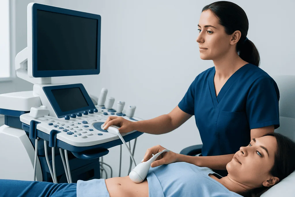
During the scan, the patient usually lies on an examination couch. A trained sonographer or doctor applies a water-based gel to the skin and moves a handheld device called a transducer across the relevant area. The transducer sends sound waves into the body and receives returning echoes, which are converted into images on a monitor.
The examination usually takes place in a quiet room and commonly lasts between 15 and 45 minutes, depending on the type of scan. The clinician may ask the patient to change position, hold their breath briefly, or report tenderness when gentle pressure is applied. The test is typically painless, although pressure over a sore area or a very full bladder may be uncomfortable.
When specialized assessment is needed, an ultrasound may be part of a broader diagnostic plan. For example, symptoms associated with the gallbladder may require evaluation for gallstones, while urinary symptoms may prompt assessment of the kidneys, bladder, or prostate.
What Happens If You Eat Food Before an Ultrasound?
If a person eats before an ultrasound that requires fasting, the scan may still be possible, but image quality can be reduced. Food can stimulate the gallbladder to contract, making it harder to assess. It may also increase digestive activity and gas, which can obscure organs such as the pancreas, gallbladder, liver, or abdominal blood vessels.
The imaging team may decide to continue with the scan, focus on areas that can still be evaluated, or reschedule it if the preparation makes the requested examination unreliable. This depends on the reason for the scan and the extent of the preparation issue. Patients should be honest with the team about what and when they ate or drank.
Fasting instructions often mean avoiding food for a set number of hours, but the exact period varies. Water may be permitted in small amounts, and medications may often be taken with a small sip of water. Diabetes medicines, insulin, blood-thinning medicines, and other essential treatments require individualized advice, so patients should ask their prescribing clinician or imaging department in advance.
For ultrasounds that do not require fasting, eating normally is usually acceptable. Pelvic, thyroid, breast, scrotal, musculoskeletal, and many soft-tissue ultrasounds generally do not require dietary restriction, unless the appointment includes an abdominal component.
How Many Ounces of Water to Drink Before an Ultrasound?
The appropriate amount of water depends on the scan and the patient’s age, health, and bladder capacity. For many transabdominal pelvic or bladder ultrasounds, adults may be asked to drink approximately 16 to 32 ounces (about 500 to 1,000 mL) of still water roughly one hour before the appointment and avoid urinating until the scan is complete.
These instructions are not universal. Some centers ask for a smaller or larger amount, and people with heart failure, kidney disease, fluid restrictions, urinary retention, or bladder problems may need modified guidance. Children are also given age-appropriate instructions rather than an adult volume.
Plain water is usually the best choice when a full bladder is required. Carbonated drinks can increase gas, while coffee and some teas may increase urination and make it harder to keep the bladder full. The goal is a comfortably full bladder, not severe pain or distress.
If the scan is a transvaginal pelvic ultrasound, the instructions may be the opposite: the patient may be asked to empty the bladder shortly before the examination. Calling ahead is worthwhile if the type of pelvic ultrasound is not clear.
What Happens If I Can't Hold My Pee Before an Ultrasound?
Patients should not force themselves to endure severe bladder pain. If a person cannot hold their urine, they should tell the receptionist or sonographer as soon as they arrive. Depending on the reason for the scan, the team may be able to perform it promptly, assess whether the bladder is sufficiently full, or advise drinking more water and waiting.
If the patient has already emptied their bladder completely, the ultrasound may need to be delayed until the bladder refills. This does not necessarily mean the appointment will be cancelled. The imaging team can explain the options based on the examination requested and the schedule that day.
People who have urinary urgency, incontinence, prostate enlargement, pregnancy-related bladder pressure, or known urinary conditions should mention this while booking. Modified preparation plans may be possible. For some pelvic concerns, a transvaginal approach may provide useful images without requiring a full bladder, although this is not appropriate for every patient or every clinical question.
There is no need to feel embarrassed about difficulty holding urine. It is a common practical issue, and accurate information helps the team obtain the best possible images while protecting the patient’s comfort.
What Not to Do the Night Before an Ultrasound?
The night before an ultrasound, patients should avoid changing or stopping prescribed medication unless a doctor specifically advises it. They should also avoid assuming that every ultrasound requires fasting or a full bladder. The most important step is reviewing the appointment instructions and confirming any uncertainty before the day of the scan.
For an abdominal ultrasound requiring fasting, it may be helpful to avoid a large late meal and foods or drinks that commonly cause bloating, such as carbonated beverages. Alcohol should generally be avoided before medical testing unless a clinician has said otherwise. Smoking and chewing gum may also increase swallowed air or digestive activity and may be discouraged by some imaging departments.
Patients should not drink excessive amounts of water in an attempt to prepare for a pelvic scan. Overfilling the bladder can cause significant discomfort and may not improve the images. People should also avoid wearing difficult-to-remove clothing; loose, comfortable clothing can make the examination easier.
Bringing the referral form, prior imaging reports if requested, a list of medicines, and relevant medical information can support an efficient appointment. The patient can normally return to usual food, drink, work, and daily activities immediately after a standard diagnostic ultrasound.
Results, Recovery, Benefits, Risks, and When to Seek Medical Care
There is usually no recovery period after an external ultrasound. The gel is wiped away, and patients can eat, drink, drive, and resume normal activities unless they have been given separate instructions. A radiologist or the clinician performing the study reviews the images, and results may be available the same day or sent to the referring doctor within several days.
The key benefits of ultrasound are that it is noninvasive, does not use ionizing radiation, and can show movement and blood flow in real time. Limitations include reduced image quality in the presence of bowel gas, difficulty viewing some deeper structures, and the possibility that additional tests are needed to confirm or clarify a finding. External ultrasound has no known significant risks when used appropriately; internal examinations may cause temporary mild discomfort.
When to seek medical care: A person should seek urgent medical assessment for severe or worsening abdominal or pelvic pain, fainting, heavy vaginal bleeding during pregnancy, chest pain, severe shortness of breath, fever with significant pain, yellowing of the skin or eyes, or inability to pass urine. These symptoms should not wait for a routine ultrasound appointment.
For planned diagnostic care, Acibadem International’s multidisciplinary specialists and JCI-accredited hospitals can evaluate symptoms and coordinate appropriate imaging for international patients. If ultrasound identifies a concern requiring further assessment, clinicians may discuss next steps such as general surgery evaluation or other specialty care based on the findings.
Frequently asked questions
Can you drink coffee before an ultrasound?
Coffee is often best avoided before an abdominal ultrasound, particularly when fasting instructions have been provided. It may stimulate digestive activity and, if it contains milk or sugar, it may break a fasting requirement. For a pelvic ultrasound, the imaging center may still prefer plain water rather than coffee because caffeine can increase urination.
Can I take my medication before an ultrasound?
Many medicines can be taken as usual with a small sip of water, but this depends on the examination and the medicine. People taking diabetes medicines, insulin, or medicines that must be taken with food should ask for individualized instructions before fasting. Prescribed medication should not be stopped without medical advice.
Do all abdominal ultrasounds require fasting?
No. Preparation depends on the organs being examined and the clinical reason for the scan. Gallbladder, liver, pancreas, and abdominal aorta examinations commonly require fasting, while other abdominal or kidney ultrasounds may have different instructions.
Can I brush my teeth before an ultrasound?
Brushing teeth is usually acceptable, but patients should avoid swallowing toothpaste or water if they have been instructed to fast. Some centers may advise against chewing gum or sucking sweets because these can stimulate digestive activity. The center’s specific instructions should be followed.
Can I eat after an ultrasound?
Yes, most people can eat and drink normally as soon as a standard diagnostic ultrasound is finished. There is typically no sedation or recovery restriction after an external ultrasound. Different instructions may apply if the ultrasound was part of another procedure.
Will an ultrasound show the cause of my pain?
Ultrasound can identify or help assess many possible causes of pain, including gallstones, ovarian cysts, kidney swelling, fluid collections, and some soft-tissue problems. However, not every condition is visible on ultrasound, and a normal result does not always rule out all causes. A clinician interprets the result together with symptoms and other tests.
References
- Radiological Society of North America
- American College of Radiology
- National Health Service
- U.S. Food and Drug Administration
- World Health Organization
This article is for general information only and is not a substitute for professional medical advice. Please consult a qualified doctor about your individual situation.
Ultrasound in Turkey — costs, top hospitals & a free quote
JCI-accredited · board-certified surgeons · reply within 24h
Add us as a Preferred Source to see more of our trusted health content across Google Search, AI Overviews and Discover.

Check-up & Preventive Medicine
Comprehensive health screenings designed for early detection and prevention.
2 specialists in this unit