Cardiac CT Before Bypass Surgery: Why It Is Done

Cardiac CT can provide a detailed map of the heart and blood vessels before coronary artery bypass surgery. It may help identify calcium, plaque, graft targets, aortic disease, and other anatomical details important for surgery.
Key Takeaways
- Cardiac CT can provide a detailed map of the heart and blood vessels before coronary artery bypass surgery.
- It may help identify calcium, plaque, graft targets, aortic disease, and other anatomical details important for surgery.
- Not every patient needs a cardiac CT; the decision depends on symptoms, prior test results, and surgical needs.
- The scan is usually quick and noninvasive, though contrast dye and radiation exposure are important considerations.
- Results from cardiac CT are reviewed together with other tests such as coronary angiography, echocardiography, and blood work.
Cardiac CT before bypass surgery is used to give the surgical team a detailed view of the heart, coronary arteries, and nearby structures before an operation. This imaging can improve planning, support safer decision-making, and help tailor treatment to the individual patient.
Overview: What is cardiac CT before bypass surgery?
Cardiac CT before bypass surgery refers to a computed tomography scan used during preoperative planning for coronary artery bypass grafting, often called CABG. The scan creates detailed cross-sectional images of the heart, coronary arteries, aorta, chest anatomy, and sometimes previously placed stents or bypass grafts. In selected patients, this information helps the care team understand the anatomy in a way that standard tests alone may not fully show.
Bypass surgery is performed to improve blood flow to the heart when coronary arteries are significantly narrowed or blocked. Before surgery, doctors need a clear picture of which vessels are affected, where suitable graft targets may be located, and whether there are additional concerns such as heavy calcification, an enlarged aorta, or chest wall anatomy that could influence the operation. Cardiac CT can add valuable details to this planning process.
It is important to know that cardiac CT does not replace a full clinical evaluation. Surgeons and cardiologists usually combine CT findings with a patient’s symptoms, physical examination, coronary angiography, echocardiography, laboratory tests, and overall health status. When used appropriately, cardiac CT is one of several tools that can support a well-prepared and individualized surgical plan.
Why doctors may order a cardiac CT before bypass surgery
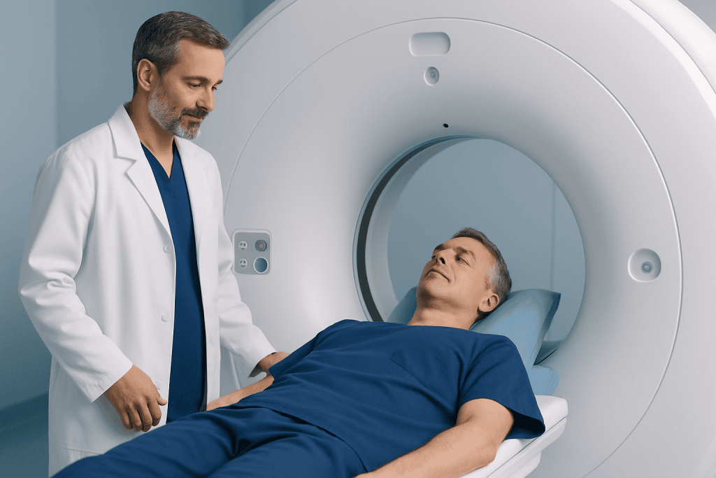
The main reason for a cardiac CT before bypass surgery is to help the team plan the safest and most effective approach. CT imaging may show the course and condition of the coronary arteries, the extent of calcification, and the exact relationship between the heart and surrounding structures. This can be especially useful when anatomy is complex or when earlier test results leave important questions unanswered.
Doctors may also use cardiac CT to evaluate the aorta, since severe calcification or plaque in the aortic wall can affect how the surgeon performs the procedure. In some patients, the scan can help identify whether there are technical challenges related to prior heart surgery, chest deformities, bypass grafts from earlier operations, or congenital variations in blood vessel anatomy. This information can reduce surprises during surgery and support better preparation.
Another common reason is to assess blood vessels that may be used in treatment planning. For example, the surgical team may want a clearer view of target vessels for grafting or an understanding of the surrounding anatomy before deciding between surgery, coronary angiography, medication, or another intervention. In this way, cardiac CT can contribute to a more complete picture of coronary artery disease and the best route forward.
What the scan can show

Cardiac CT can reveal several features that matter before CABG. These include the location and severity of coronary narrowing, calcium buildup in the coronary arteries, the size and shape of the heart, and the condition of the ascending aorta. It may also show whether there are anatomic variations that could affect access during surgery or change where grafts can be attached.
In patients who have had prior procedures, cardiac CT may help evaluate older grafts, surgical clips, scar tissue, or the relation of the heart to the breastbone. This can be important when repeat surgery is being considered. The scan may also help identify other chest findings, such as lung or mediastinal changes, that are relevant to anesthesia and operative planning.
Depending on the clinical question, doctors may request a coronary CT angiography study, which uses contrast dye to highlight blood flow inside the coronary vessels. In other cases, the focus may be on noncoronary details such as aortic calcification or preoperative chest anatomy. The exact type of CT depends on what the surgical team needs to know before coronary artery bypass grafting.
- Coronary artery narrowing or blockage
- Coronary and aortic calcification
- Heart size and surrounding chest anatomy
- Condition of previous grafts or prior surgical changes
- Potential technical issues that may affect the surgical approach
Who may benefit most from preoperative cardiac CT
Not every person scheduled for bypass surgery will need a cardiac CT. In many cases, standard coronary angiography and echocardiography already provide enough information for treatment planning. However, CT may be especially helpful when there are uncertainties about anatomy, a suspicion of significant aortic calcification, or a history of previous heart surgery.
Patients with complex coronary artery disease, unusual vessel anatomy, or concern for associated conditions may also benefit. For example, someone with known or suspected coronary artery disease and heavily calcified vessels may need more detailed imaging to guide the surgical approach. CT can also be useful in older adults or in patients whose surgeons want to avoid manipulating a heavily diseased aorta if possible.
People who have had prior chest surgery, radiation therapy, congenital heart differences, or unclear findings on other imaging may be candidates as well. The decision is always individualized. Doctors weigh the expected benefit of extra anatomical detail against factors such as kidney function, contrast allergy history, heart rhythm, and whether the results would actually change management.
How the test is performed and what to expect
A cardiac CT scan is usually a short outpatient imaging test. The patient lies on a table that moves through a CT scanner while multiple X-ray images are taken and reconstructed into detailed pictures. If a CT angiography study is planned, contrast dye is injected through a vein to make the coronary arteries and major blood vessels easier to see.
Before the scan, the team may review medications, kidney function, allergies, and heart rhythm. Some patients are asked to avoid caffeine for a period beforehand. In selected cases, medicine may be used to slow the heart rate so the images are clearer, but this depends on the type of scan and the person’s medical condition. The patient may also be asked to hold their breath briefly during image capture.
Most people tolerate the test well. The scan itself is painless, though the contrast dye may cause a warm sensation for a short time. Afterward, patients can usually return to normal activities unless their doctor gives different instructions. The images are then reviewed by radiologists and cardiologists, and the findings are discussed with the surgical team as part of the overall plan.
Benefits, risks, and limitations
The potential benefit of cardiac CT before bypass surgery is better preoperative knowledge. A more detailed understanding of coronary anatomy, calcification, and aortic disease may help surgeons choose the most suitable operative strategy. In some patients, it may also help refine the choice between surgery and other approaches such as angioplasty and stent treatment, depending on the broader clinical picture.
Like any imaging test, cardiac CT also has limitations. It uses ionizing radiation, although modern scanners and protocols aim to keep exposure as low as reasonably possible. When contrast dye is used, there is a small risk of allergic reaction or kidney-related problems, especially in people with reduced kidney function. Fast or irregular heart rhythms can sometimes reduce image quality.
Cardiac CT should also be understood as one part of a larger evaluation rather than a stand-alone answer. It may not fully replace invasive imaging in every case, and it cannot by itself determine whether bypass surgery is the right treatment. Doctors interpret the scan alongside symptoms, examination findings, the severity of other heart conditions, heart function, and the patient’s general health and goals.
How results affect surgical planning
The findings from a cardiac CT can directly influence how the operation is planned. Surgeons may use the images to decide where grafts are most likely to be effective, whether the aorta should be handled differently, or whether additional precautions are needed. In repeat operations, the scan may help show the safest way to reopen the chest and avoid injury to structures lying close to the breastbone.
CT findings can also support discussions within a multidisciplinary heart team. Cardiologists, cardiac surgeons, anesthesiologists, radiologists, and other specialists may review the images together to determine the best treatment plan. This team-based approach is especially valuable when disease is complex or when multiple treatment options are possible.
For international patients seeking advanced cardiac evaluation, Acibadem International’s multidisciplinary specialists and JCI-accredited hospitals diagnose and treat heart conditions using modern imaging and surgical planning methods. In many cases, the value of cardiac CT lies not only in the pictures themselves, but in how those pictures help the team prepare a safer, more personalized pathway to care.
Questions to ask the doctor and when to seek medical advice
Patients preparing for bypass surgery may feel more comfortable when they understand why a test is being ordered. It can be helpful to ask what the cardiac CT is expected to show, whether contrast dye will be used, and how the results may change the treatment plan. Patients may also ask about alternatives, radiation exposure, kidney safety, and whether any medicines should be stopped before the scan.
Anyone with a history of contrast allergy, asthma, kidney disease, diabetes, or an irregular heartbeat should tell the medical team before the test. These conditions do not always prevent a cardiac CT, but they may affect how the scan is prepared and performed. Clear communication helps the team reduce risk and plan appropriately.
Urgent medical advice is important if a person awaiting heart surgery develops worsening chest pain, fainting, severe shortness of breath, or other sudden concerning symptoms. These may need immediate assessment and should not wait for a scheduled scan. For routine questions about cardiac CT or bypass planning, the safest step is to speak with a qualified cardiologist or cardiac surgeon.
Frequently asked questions
Is cardiac CT always needed before bypass surgery?
No. Many patients can be planned for bypass surgery using coronary angiography, echocardiography, and other standard tests. Cardiac CT is usually reserved for situations where extra anatomical detail may improve planning or answer specific surgical questions.
What is the difference between cardiac CT and coronary angiography?
Cardiac CT is a noninvasive imaging test that uses X-rays to create detailed pictures of the heart and blood vessels. Coronary angiography is an invasive catheter-based test that directly visualizes the coronary arteries and is often central to diagnosing blockages. In some patients, both tests are used because they provide different but complementary information.
Does a cardiac CT scan hurt?
The scan itself is painless. If contrast dye is used, the person may feel a brief warm sensation or a metallic taste, but this usually passes quickly. The test is generally well tolerated.
Are there risks from the contrast dye?
Contrast dye is safe for most people, but it can occasionally cause an allergic reaction or affect kidney function, especially in those with existing kidney disease. Doctors reduce these risks by checking medical history, kidney function, and previous reactions before the scan.
Can cardiac CT replace bypass surgery or other treatment?
No. Cardiac CT is a diagnostic and planning tool, not a treatment. Its role is to help doctors better understand the anatomy and choose the most appropriate treatment, which may include medication, a catheter procedure, or surgery.
How should a patient prepare for a cardiac CT before surgery?
Preparation varies by the type of scan and the patient’s health. The care team may advise avoiding caffeine, reviewing medications, and checking kidney function before contrast is given. It is important to mention any allergies, diabetes, kidney problems, or abnormal heart rhythms in advance.
References
- American Heart Association
- American College of Cardiology
- Society of Cardiovascular Computed Tomography
- European Society of Cardiology
- National Heart, Lung, and Blood Institute
This article is for general information only and is not a substitute for professional medical advice. Please consult a qualified doctor about your individual situation.
Heart care in Turkey — expert evaluation and treatment
JCI-accredited · board-certified surgeons · reply within 24h
Add us as a Preferred Source to see more of our trusted health content across Google Search, AI Overviews and Discover.