Cardiac MRI: A Complete Medical Overview
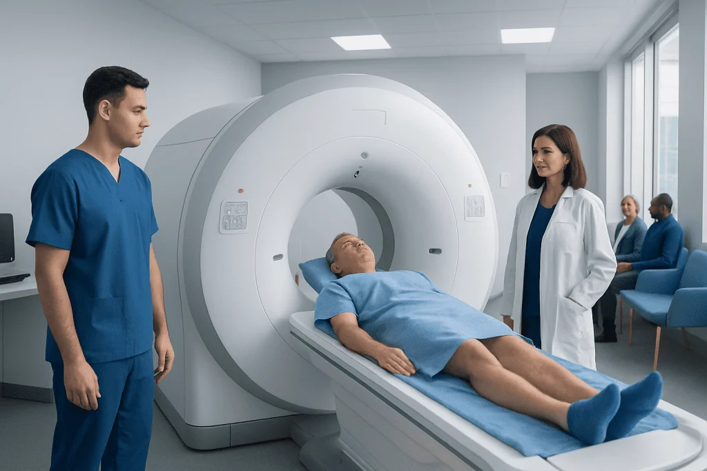
Cardiac MRI provides detailed images of the heart muscle, chambers, valves, and nearby blood vessels. It can help diagnose cardiomyopathy, myocarditis, congenital heart disease, scarring after heart injury, and other cardiac conditions.
Key Takeaways
- Cardiac MRI provides detailed images of the heart muscle, chambers, valves, and nearby blood vessels.
- It can help diagnose cardiomyopathy, myocarditis, congenital heart disease, scarring after heart injury, and other cardiac conditions.
- The test does not use radiation, but some people may need contrast dye to improve image quality.
- Preparation may include removing metal items, reviewing implants, and following instructions about food, drink, or medicines.
- Results are interpreted by specialists and are often used alongside ECG, echocardiography, CT, or blood tests.
Cardiac MRI is an advanced imaging test that creates detailed pictures of the heart using a strong magnet and radio waves. It helps doctors assess heart structure, pumping function, blood flow, inflammation, scarring, and some congenital or vascular conditions without using ionizing radiation.
Overview: what cardiac MRI is and why it is used
Cardiac MRI, also called a heart MRI, is a noninvasive imaging test that creates highly detailed pictures of the heart and surrounding blood vessels. It uses a strong magnetic field and radio waves rather than X-rays, so it does not expose the body to ionizing radiation. For many patients, it is one of the most informative ways to understand how the heart looks and how well it works.
Unlike some imaging tests that mainly show anatomy, cardiac MRI can evaluate both structure and function. It can assess the size of the heart chambers, the movement of the heart muscle, the pumping strength of the ventricles, blood flow patterns, and signs of inflammation or scar tissue. This makes it especially helpful when other tests leave unanswered questions.
Doctors may request cardiac MRI when they need more detail after an electrocardiogram, stress test, or echocardiogram. It is commonly used to investigate suspected cardiomyopathy, congenital heart abnormalities, heart muscle inflammation, prior heart damage, or masses around the heart. In some cases, it also helps guide planning for cardiology care or follow-up after treatment.
What cardiac MRI can show
One of the main strengths of cardiac MRI is its ability to characterize heart tissue, not just shape. It can show whether the heart muscle is thickened, weakened, stretched, inflamed, or scarred. This level of detail can help doctors distinguish between different causes of heart symptoms that may look similar on basic tests.
Cardiac MRI may be used to evaluate a wide range of concerns, including chest pain, shortness of breath, unexplained fainting, irregular heart rhythms, and reduced exercise tolerance. It can also help assess the impact of a previous heart attack, detect fluid or inflammation around the heart, and evaluate inherited or acquired diseases of the heart muscle.
It may provide information about:
- Heart chamber size and wall thickness
- Left and right ventricular pumping function
- Valve structure and blood flow across valves
- Scarring after reduced blood supply or heart injury
- Inflammation, such as in suspected myocarditis
- Congenital heart defects and major vessel anatomy
- Tumors, clots, or abnormalities involving the pericardium
For some patients, cardiac MRI complements tests such as advanced diagnostic evaluation or targeted treatment planning. It is often especially useful when a doctor wants a clearer explanation for symptoms but wishes to avoid unnecessary invasive procedures.
Who may need a cardiac MRI

A doctor may recommend cardiac MRI for many different reasons. Sometimes it is used to confirm or clarify a diagnosis that is already suspected. In other situations, it is ordered because symptoms or exam findings suggest a heart problem, but the cause is still uncertain.
Common reasons for referral include suspected heart muscle disease, possible inflammation after a viral illness, evaluation of congenital heart disease, assessment of heart function after a heart attack, or concern about structural abnormalities seen on echocardiography. It may also be used to monitor known conditions over time, especially when precise and repeatable measurements are important.
Patients with symptoms such as ongoing chest discomfort, unexplained breathlessness, palpitations, dizziness, or fainting may be considered for this test when initial evaluation points toward a cardiac cause. Cardiac MRI can also be useful in the assessment of arrhythmia when doctors need more information about underlying structural heart disease.
Not every person with heart symptoms needs an MRI. The choice depends on the clinical question, the patient’s medical history, and whether another test can answer the same question more simply. A cardiologist or radiologist helps determine when cardiac MRI is the most appropriate next step.
How to prepare and what happens during the scan
Preparation for cardiac MRI is usually straightforward, but exact instructions vary. Patients are commonly asked to remove jewelry, watches, hearing aids, and any other metal items before the scan. The imaging team will carefully review medical implants or devices, such as pacemakers, defibrillators, stents, clips, or implanted pumps, because some devices are MRI-compatible and some require special precautions.
Some cardiac MRI studies use a contrast agent, often gadolinium-based, to improve visibility of heart tissue and blood flow. If contrast is planned, the care team may ask about kidney disease, allergies, and prior reactions to contrast materials. In certain cases, patients may also be advised to avoid caffeine or heavy meals beforehand, especially if the study includes stress imaging.
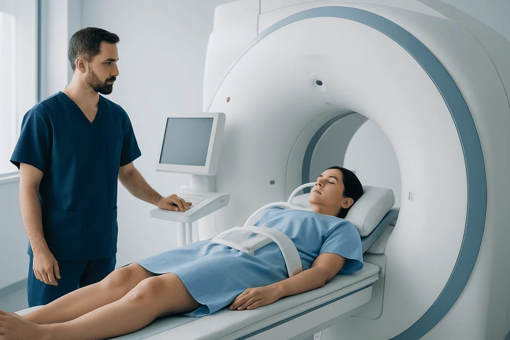
During the test, the patient lies on a table that moves into the MRI scanner. Electrodes may be placed on the chest to coordinate imaging with the heartbeat, and the patient may be asked to hold their breath briefly several times. The scanner makes loud tapping or knocking sounds, so ear protection is usually provided. The exam often lasts between 30 and 90 minutes, depending on the images needed.
Some people feel uneasy in enclosed spaces. If claustrophobia is a concern, it is helpful to mention it before the appointment. The team can often suggest strategies to make the test more comfortable. In specialized centers, including Acibadem International, multidisciplinary specialists in JCI-accredited hospitals assess international patients who may need heart imaging and related care such as cardiovascular surgery when appropriate.
Benefits, limitations, and possible risks
Cardiac MRI offers several important benefits. It provides excellent image quality, does not use radiation, and can assess both anatomy and tissue characteristics in a single examination. For many conditions, it helps reduce uncertainty by showing whether the heart muscle has edema, scar, fibrosis, or abnormal thickening.
It also has limitations. MRI is not always the best choice for people who cannot lie still, cannot follow breathing instructions, or have certain implants or medical devices. The test may be less suitable in emergencies when faster imaging is needed. In addition, access can be more limited than for ultrasound or CT in some settings.
The scan itself is generally safe, but there are a few considerations. The magnetic field means metal safety screening is essential. Contrast dye is usually well tolerated, though it may not be suitable for some people with severe kidney problems. A stress cardiac MRI, when used, may involve medicine that mimics exercise, and this requires additional monitoring.
Because the value of the test depends on the right indication and expert interpretation, results are best understood in the context of the patient’s symptoms, examination, and other investigations. Cardiac MRI often works as part of a broader diagnostic pathway rather than as a stand-alone answer.
Understanding the results and what comes next
After the scan, a radiologist or cardiologist with expertise in cardiac imaging reviews the images in detail. The report may describe chamber size, heart muscle thickness, ejection fraction, valve findings, blood flow patterns, and any signs of swelling, fibrosis, scar tissue, or congenital abnormalities. These findings help the referring doctor decide whether treatment, follow-up, or more testing is needed.
Normal results can be reassuring, especially when symptoms are mild or nonspecific. However, a normal cardiac MRI does not always rule out every cause of symptoms. For example, some rhythm problems, small vessel disease, or non-cardiac causes of chest discomfort may require different forms of evaluation.
If abnormalities are found, next steps depend on the diagnosis. A patient with signs of inflammation may need close monitoring and treatment of the underlying cause. Someone with scarring, reduced heart function, or a structural problem may be referred for medical therapy, procedures, or longer-term follow-up. In selected cases, the findings can support decisions about coronary angiography or other targeted investigations.
Patients should review the results with their doctor rather than trying to interpret the report on their own. The significance of a finding depends on the whole clinical picture, including age, symptoms, family history, physical examination, and previous tests.
When to seek medical care
Cardiac MRI is usually a planned outpatient test, not an emergency treatment. However, symptoms that suggest an urgent heart problem should never wait for a routine imaging appointment. Immediate medical attention is important if a person has severe chest pain, sudden shortness of breath, fainting, new weakness, or symptoms that rapidly worsen.
Medical review is also appropriate for ongoing but less urgent concerns such as repeated palpitations, reduced exercise tolerance, swelling in the legs, or unexplained fatigue. These symptoms do not always mean heart disease, but they deserve proper assessment, especially in people with known cardiac conditions or a strong family history of heart problems.
Patients should contact a qualified doctor promptly if they have been advised to have a cardiac MRI but are pregnant, have kidney disease, or have an implanted medical device and are unsure whether it is MRI-safe. Sharing this information in advance helps the imaging team plan the safest approach.
Anyone with persistent or worrying symptoms should seek individualized medical advice. Early evaluation often helps clarify the cause of symptoms and allows care to be tailored appropriately.
Frequently asked questions
What is the difference between cardiac MRI and echocardiography?
Echocardiography uses ultrasound and is often the first imaging test for heart symptoms because it is widely available and quick. Cardiac MRI usually provides more detailed information about heart tissue, scarring, inflammation, and complex structural abnormalities.
Does cardiac MRI use radiation?
No. Cardiac MRI uses a magnetic field and radio waves, so it does not expose the patient to ionizing radiation. This is one reason it is useful for some people who need detailed imaging or repeat follow-up studies.
How long does a cardiac MRI take?
The scan often takes about 30 to 90 minutes, depending on the type of study and whether contrast or stress imaging is included. Patients may need to hold their breath briefly several times to help produce clearer images.
Is cardiac MRI painful?
The scan itself is not painful. Some people find it uncomfortable to lie still or be inside the scanner, and those receiving contrast will usually have a small IV line placed in the arm.
Can someone with a pacemaker have a cardiac MRI?
Sometimes, yes, but it depends on the specific device and the center’s safety protocols. Many modern devices are MRI-conditional, which means scanning may be possible with careful planning and monitoring.
Why might contrast be used during cardiac MRI?
Contrast can help doctors see blood flow patterns and identify inflammation, scar tissue, or areas of damaged heart muscle more clearly. It is not needed for every patient, and the care team decides based on the clinical question and medical history.
References
- American Heart Association
- Radiological Society of North America
- National Heart, Lung, and Blood Institute
- Society for Cardiovascular Magnetic Resonance
- American College of Cardiology
This article is for general information only and is not a substitute for professional medical advice. Please consult a qualified doctor about your individual situation.
Heart care in Turkey — expert evaluation and treatment
JCI-accredited · board-certified surgeons · reply within 24h
Add us as a Preferred Source to see more of our trusted health content across Google Search, AI Overviews and Discover.

More from the Health Library

Perimenopause: An Evidence-Based Guide for Patients

Where Does Fertilization Occur: An Evidence-Based Guide for Patients

Scratch of Skin: An Evidence-Based Guide for Patients

COVID Quarantine Guidelines 2025: An Evidence-Based Guide for Patients

Amniotomy: An Evidence-Based Guide for Patients

Men’s Mental Health Month: What Patients Need to Know
Cardiology Specialists at Acibadem

Prof. Dr. Ahmet Tulga Ulus
Cardiovascular Surgery
Prof. Dr. Ahmet Ümit Güllü
Cardiovascular Surgery
Prof. Dr. Aleks Değirmencioğlu
Cardiology
Prof. Dr. Ali Aydınlar
Cardiology