Cardiac MRI vs Echocardiogram: Which Heart Imaging Test Answers What?
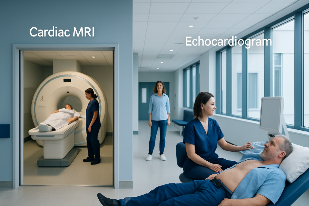
An echocardiogram uses ultrasound and is often the first-line heart imaging test. Cardiac MRI provides highly detailed images of heart structure, function, and tissue characteristics.
Key Takeaways
- An echocardiogram uses ultrasound and is often the first-line heart imaging test.
- Cardiac MRI provides highly detailed images of heart structure, function, and tissue characteristics.
- The best test depends on the clinical question, symptoms, and the information a doctor needs.
- These tests are often complementary rather than competing alternatives.
- A cardiologist may recommend one or both tests to clarify a diagnosis and guide treatment.
Cardiac MRI and echocardiography are both important heart imaging tests, but they are used for different purposes. An echocardiogram is often the first test because it is quick and widely available, while cardiac MRI can provide more detailed information about heart muscle, scarring, inflammation, and complex anatomy.
Overview: how these two heart imaging tests differ
When comparing cardiac MRI vs echocardiogram, the most helpful starting point is that both tests look at the heart, but they do so in different ways and answer different medical questions. An echocardiogram uses sound waves, or ultrasound, to create moving pictures of the heart. A cardiac MRI uses a strong magnetic field and radio waves to create very detailed images of the heart’s structure, movement, and tissue characteristics.
In everyday practice, an echocardiogram is often the first imaging test for many heart symptoms because it is fast, noninvasive, and can usually be done at the bedside or in an outpatient clinic. It helps doctors assess heart pumping function, heart valves, chamber size, and blood flow patterns. It is especially useful when a clinician needs an immediate overview of how the heart is working.
Cardiac MRI is typically used when more detail is needed. It can measure heart function very accurately and can also show information that ultrasound cannot always provide, such as inflammation, scarring, infiltration, or subtle damage to the heart muscle. For this reason, it is especially valuable in certain cardiomyopathies, myocarditis, congenital heart disease, and tissue-based evaluation after heart injury.
Rather than thinking of one test as “better” than the other, it is usually more accurate to think of them as complementary tools. The right choice depends on the person’s symptoms, medical history, physical findings, and the specific question the doctor is trying to answer.
What an echocardiogram shows best
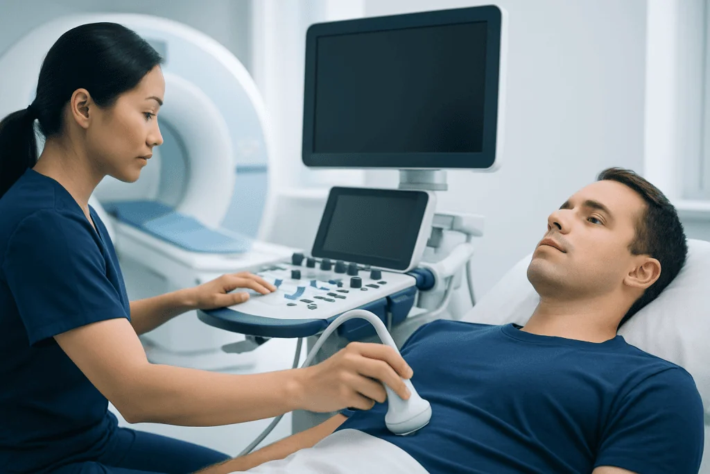
An echocardiogram is one of the most common heart tests because it gives real-time information about how the heart moves and pumps. It can show whether the left and right ventricles are squeezing well, whether the heart chambers are enlarged, and whether the valves open and close normally. It also helps estimate pressures inside the heart and lungs in many cases.
Doppler echocardiography adds information about the direction and speed of blood flow. This is particularly helpful for valve disease, such as narrowing or leakage, and for conditions involving abnormal blood flow between heart chambers. In people with shortness of breath, swelling, fatigue, murmurs, or suspected heart failure, echocardiography is often a key first step.
There are different types of echocardiograms. A transthoracic echo is the standard test performed with a probe placed on the chest. A transesophageal echo places a probe in the esophagus to provide closer views of some structures, especially valves, clots, and the back of the heart. Stress echocardiography combines ultrasound with exercise or medicine to see how the heart responds under strain.
An echocardiogram is often preferred when speed matters or when a doctor needs a practical, dynamic overview. It is widely used to evaluate suspected heart failure, valve disorders, fluid around the heart, and many common structural abnormalities. It can also help monitor known conditions over time without exposing the patient to radiation.
What cardiac MRI shows best

Cardiac MRI offers excellent image quality and can provide a very detailed assessment of the heart’s anatomy and function. It is considered one of the most accurate ways to measure ventricular size and pumping performance. It is particularly helpful when ultrasound images are limited by body shape, lung disease, chest wall anatomy, or other technical factors.
One of the biggest advantages of cardiac MRI is tissue characterization. This means it can help doctors distinguish healthy heart muscle from areas of inflammation, edema, fibrosis, scar, fat, or infiltration. In practical terms, this can help identify whether symptoms are related to myocarditis, prior heart attack, certain cardiomyopathies, or less common infiltrative conditions.
Cardiac MRI is also useful in more complex or less straightforward cases. It can clarify congenital heart anatomy, assess the aorta and nearby vessels, evaluate masses, and help determine the cause of reduced heart function when the diagnosis is uncertain. In some patients, contrast material may be used to provide additional detail about blood supply and tissue injury.
Because of these strengths, doctors may order cardiac MRI after an abnormal echocardiogram or when symptoms do not match the initial findings. It often plays an important role in evaluating cardiomyopathy and in detailed planning for ongoing heart care. In some cases, it may be part of a broader imaging pathway that also includes cardiac MRI as a dedicated advanced test.
When doctors choose one test over the other
The choice between these tests is guided by the clinical question. If the goal is to quickly assess heart function, look for valve disease, estimate pressures, or check for fluid around the heart, an echocardiogram is often the first and most practical option. It is portable, widely available, and can often be performed soon after symptoms begin.
If the goal is to understand the heart muscle in greater detail, define complex anatomy, or look for scar and inflammation, cardiac MRI may be more informative. It is especially useful when the initial ultrasound leaves unanswered questions or when the doctor suspects a condition that depends on tissue-level information.
Doctors also consider the patient’s circumstances. For example, some people cannot comfortably lie still in an MRI scanner, while others may have implanted devices that affect MRI eligibility, although many modern devices are MRI-conditional. Similarly, an echocardiogram may be less informative if image quality is poor, which can happen in some individuals.
In many situations, the two tests work together. An echocardiogram may identify a problem, and cardiac MRI may explain its cause more precisely. This stepwise approach helps avoid unnecessary testing while making sure important details are not missed.
How the tests are performed and what to expect
A standard transthoracic echocardiogram is simple and usually does not require special preparation. A technician places gel on the chest and moves a handheld transducer over the skin to create images. The test is painless for most people and generally takes less time than a cardiac MRI. Patients can usually return to normal activities right away.
Cardiac MRI usually takes longer and requires the patient to lie still inside the scanner. The team may ask the patient to hold their breath briefly at intervals while images are taken. Some examinations use intravenous contrast to improve visualization of blood flow and tissue features. The test is noninvasive, but the enclosed space and the need to remain still may be uncomfortable for some people.
Preparation for cardiac MRI may include screening for metal implants, kidney function review if contrast is planned, and discussion of claustrophobia or anxiety. For selected patients, additional imaging support may be coordinated through advanced diagnostic imaging services to tailor the exam to the clinical need.
Neither test uses ionizing radiation. That can be reassuring for patients who may need repeat heart imaging over time. The experience, timing, and level of detail differ, but both tests are established tools that help doctors make safer, more informed decisions.
Benefits, limitations, and safety considerations
Echocardiography has several important advantages: it is accessible, relatively quick, and excellent for evaluating motion and blood flow in real time. It is often the best first-line test for common cardiac symptoms and can be repeated easily. However, its image quality depends on the acoustic window, which means some patients may have limited views of important structures.
Cardiac MRI provides highly reproducible measurements and exceptional tissue detail. It is often the preferred test for characterizing heart muscle disease, identifying scar patterns, and evaluating complex structural conditions. Its limitations include longer scan time, higher cost in many settings, and less convenience compared with ultrasound. It is also not suitable for every patient.
Safety is an important consideration with both tests. Echocardiography is generally very safe, including for children and pregnant patients when clinically appropriate. Cardiac MRI is also considered safe for many patients, but screening is necessary to confirm that implanted devices, metal fragments, or other factors do not create risk. If contrast is used, doctors consider kidney health and any prior contrast reactions.
For some people, additional testing may still be needed. A patient with suspected blocked coronary arteries, for example, may need stress imaging, CT coronary imaging, or a procedure such as coronary angiography depending on the situation. Heart imaging is most useful when each test is matched carefully to the question at hand.
Common conditions these tests help evaluate
Both echocardiography and cardiac MRI are used in a wide range of heart conditions. These include shortness of breath, chest discomfort, palpitations, fainting, heart murmurs, suspected heart failure, congenital heart disease, and follow-up after heart attack or heart surgery. Imaging helps doctors understand whether symptoms are related to the heart and how severe the problem may be.
Echocardiography is especially valuable for assessing valve disease, chamber enlargement, weak heart pumping, and pressure changes that may suggest pulmonary hypertension or fluid overload. It is often the main test used to monitor valve conditions over time because it can be repeated regularly and compared with prior studies.
Cardiac MRI becomes particularly useful when the concern involves the heart muscle itself. It can help assess myocarditis, hypertrophic or dilated cardiomyopathy, right ventricular abnormalities, infiltrative disorders, and viability of the heart muscle after ischemic injury. It may also help evaluate unusual findings first seen on an echocardiogram.
In specialized centers, doctors may integrate echocardiography, MRI, blood tests, electrocardiography, and clinical examination into one coordinated plan. At the end of this process, the aim is not just to produce images, but to answer practical questions about diagnosis, prognosis, and treatment choices.
When to ask about advanced heart imaging
Patients may want to ask their doctor why a specific test is being recommended and what question it is meant to answer. That conversation can help set expectations and reduce uncertainty. In some cases, the doctor may explain that the echocardiogram is enough; in others, a cardiac MRI is the best next step to clarify the diagnosis.
It is sensible to seek medical advice for symptoms such as unexplained shortness of breath, chest discomfort, fainting, swelling of the legs, rapid or irregular heartbeat, or a newly detected heart murmur. Imaging may also be recommended when a person has a strong family history of heart muscle disease or when another test has shown an unexpected abnormality.
People who already know they have a heart condition can also ask whether imaging follow-up is needed and how often. The interval depends on the diagnosis, symptoms, and whether treatment decisions depend on changes in heart structure or function over time.
For international patients who need coordinated assessment, Acibadem International’s multidisciplinary specialists and JCI-accredited hospitals diagnose and treat heart conditions using modern imaging pathways, including echocardiography and advanced cardiac MRI when appropriate. A qualified cardiologist can explain which test, or combination of tests, is most useful in each individual situation.
Frequently asked questions
Is cardiac MRI better than an echocardiogram?
Neither test is universally better. An echocardiogram is often best for quick evaluation of heart function, valves, and blood flow, while cardiac MRI is better for detailed assessment of heart muscle and tissue changes. The best choice depends on the medical question being asked.
Why would a doctor order a cardiac MRI after an echocardiogram?
A doctor may order cardiac MRI when the echocardiogram shows an abnormality that needs further clarification or when ultrasound images are limited. MRI can provide more detail about scarring, inflammation, congenital anatomy, or the cause of reduced heart function.
Does either test use radiation?
No. Standard echocardiography and cardiac MRI do not use ionizing radiation. This makes them useful options for patients who may need repeat imaging over time.
Is a cardiac MRI painful or invasive?
Cardiac MRI is noninvasive and is not usually painful. The main challenges are lying still, hearing scanner noise, and being in an enclosed space for a period of time. Some patients also receive contrast through a vein for additional image detail.
Can an echocardiogram detect blocked arteries?
An echocardiogram does not directly show coronary artery blockages in most cases. However, it can show signs that suggest the heart muscle may not be getting enough blood, such as abnormal wall motion. Other tests are usually needed to evaluate the coronary arteries directly.
How long do these tests usually take?
A standard transthoracic echocardiogram is usually shorter and often completed within about 30 to 60 minutes, depending on the case. Cardiac MRI generally takes longer because it includes multiple image sequences and careful positioning. Exact timing varies by center and by the purpose of the exam.
References
- American Heart Association
- European Society of Cardiology
- Radiological Society of North America
- National Heart, Lung, and Blood Institute
- British Heart Foundation
This article is for general information only and is not a substitute for professional medical advice. Please consult a qualified doctor about your individual situation.
Heart care in Turkey — expert evaluation and treatment
JCI-accredited · board-certified surgeons · reply within 24h
Add us as a Preferred Source to see more of our trusted health content across Google Search, AI Overviews and Discover.