

Cataract Slit Lamp: An Evidence-Based Patient Guide
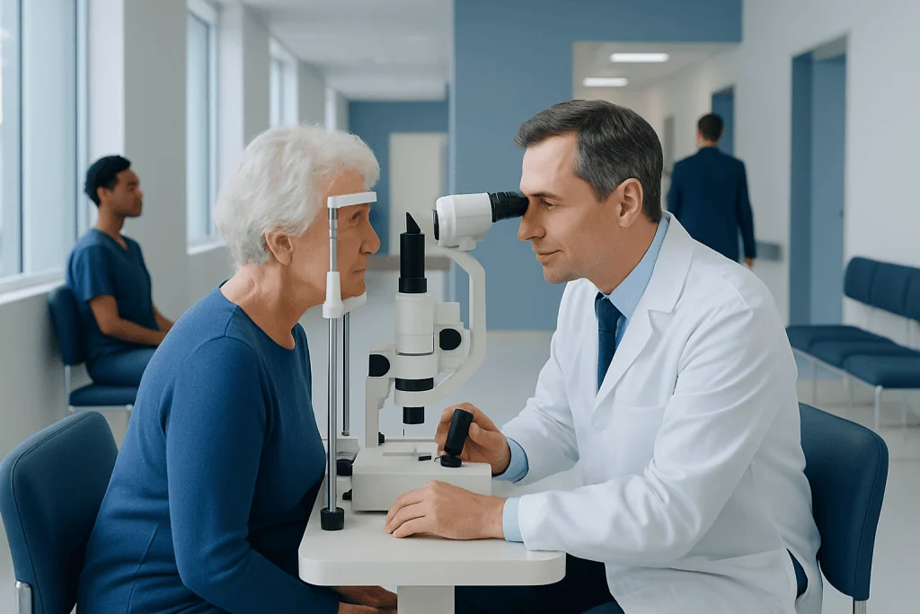
A slit lamp is a microscope combined with a narrow, adjustable beam of light used to examine the front and, with special lenses, parts of the back of the eye. Cataracts may appear as cloudy, yellow, brown, white, or spoke-like changes within the normally clear lens.
Key Takeaways
- A slit lamp is a microscope combined with a narrow, adjustable beam of light used to examine the front and, with special lenses, parts of the back of the eye.
- Cataracts may appear as cloudy, yellow, brown, white, or spoke-like changes within the normally clear lens.
- The examination is usually brief, noninvasive, and performed while the person is awake and seated.
- Pupil-dilating drops may be needed for a complete cataract assessment and can blur near vision temporarily.
- A slit lamp exam diagnoses and monitors cataracts, but cataract surgery is usually considered based on how vision affects daily life.
- Sudden vision loss, eye pain, marked redness, flashes, or a curtain-like shadow require prompt medical assessment.
A cataract slit lamp exam is a painless eye examination that lets an ophthalmologist view the eye’s lens in detail and identify clouding consistent with cataracts. It also helps assess other structures of the eye, determine whether cataracts may explain vision changes, and support treatment planning.
Overview: What Is a Cataract Slit Lamp Exam?
A cataract slit lamp exam is an eye examination in which an ophthalmologist or optometrist uses a slit lamp, a high-powered microscope with a focused beam of light, to inspect the eye. The narrow light beam creates an optical “slice” through transparent eye structures, allowing the clinician to assess the cornea, anterior chamber, iris, and natural lens in detail.
For cataracts, the examination helps show whether the natural lens has become cloudy and where the clouding is located. Cataracts can affect the center of the lens, the back surface, the outer edge, or more than one area. These patterns may affect vision differently and can help guide follow-up and treatment discussions.
A slit lamp examination does not remove or treat a cataract. It is a diagnostic assessment that is commonly combined with vision testing, eye pressure measurement, and often pupil dilation. The findings are considered alongside symptoms such as glare, reduced contrast, faded colors, blurred vision, or difficulty seeing at night.
How the Slit Lamp Works and What It Can Show
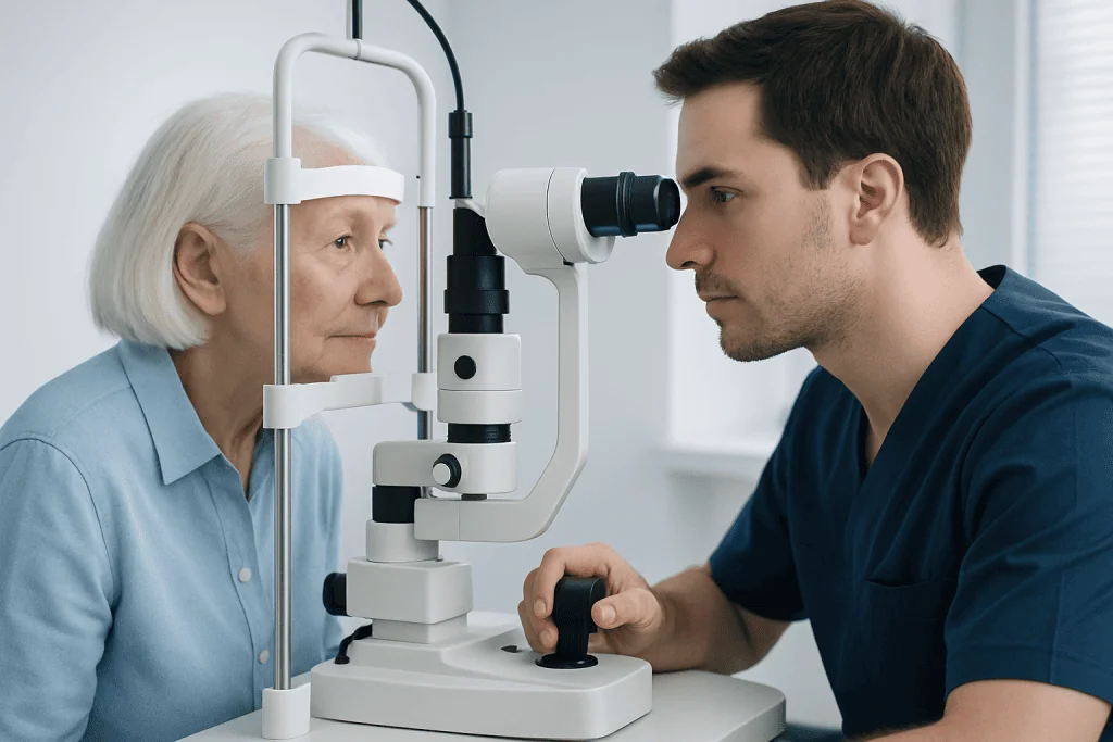
The slit lamp has two main components: a binocular microscope and a bright light source that can be narrowed, widened, angled, and adjusted in intensity. By changing the light beam and magnification, the clinician can view layers of the eye rather than seeing only the surface.
In a cataract slit lamp exam, the lens is assessed for opacity, color changes, and density. A normal lens is largely transparent, so light passes through it toward the retina. In a cataract slit lamp vs normal comparison, a cataractous lens has areas that scatter or block light, whereas a healthy lens looks clearer and allows a more unobstructed view.
A cataract slit lamp photo may show a dark background with pale or bright cloudy areas in the lens, or a spoke-like pattern extending from the edge. However, photographs do not replace an in-person evaluation. The clinical significance of a lens change depends on its location, severity, effect on vision, and the health of the rest of the eye.
The same examination can also identify signs of dry eye disease, corneal injury or infection, inflammation inside the eye, glaucoma-related changes, and problems involving the eyelids or conjunctiva. Additional lenses may be used to inspect the retina and optic nerve after the pupil is dilated.
What Eye Conditions Need a Slit Lamp?
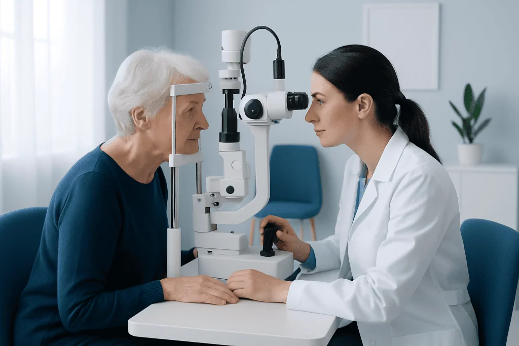
A slit lamp is used whenever a clinician needs a close examination of the eye’s front structures or a detailed view through the pupil. It is a routine part of many comprehensive eye examinations and is especially useful when someone has blurred vision, eye discomfort, redness, light sensitivity, discharge, floaters, flashes, or a change in vision.
Conditions that may require slit lamp assessment include cataracts, dry eye disease, conjunctivitis, corneal abrasions or ulcers, keratitis, uveitis, blepharitis, contact lens-related problems, and injuries from a foreign body or chemical exposure. It may also support assessment of glaucoma and retinal conditions when used with dilation and specialized lenses.
For people with diabetes, high blood pressure, autoimmune disease, prior eye surgery, or long-term steroid use, regular eye examinations may be particularly important. A slit lamp finding is only one part of diagnosis; clinicians may also use visual acuity testing, refraction, pressure testing, retinal imaging, optical coherence tomography, or visual field testing when appropriate.
How to Identify Cataracts on a Slit Lamp
During slit lamp examination, clinicians look for loss of clarity in the natural lens. Cataracts are described by their location and appearance. Nuclear cataracts develop in the central lens and may cause gradual yellowing or browning. Cortical cataracts often form wedge-shaped, spoke-like opacities around the lens edge. Posterior subcapsular cataracts develop near the back of the lens and may affect reading vision and glare disproportionately.
The clinician directs the light at different angles to determine whether an opacity is present, how dense it is, and whether it lies along the visual axis. An early cataract can be visible on examination without causing significant symptoms. Conversely, a relatively small opacity near the center of the lens may noticeably affect vision.
Symptoms and examination findings do not always progress at the same pace. The decision to observe or discuss treatment is therefore based on the person’s visual needs and daily functioning, not solely on the appearance of the lens. If cataracts interfere with activities such as driving, reading, work, or safe mobility, an ophthalmologist may discuss cataract surgery as a treatment option.
Other eye conditions can also cause blurred vision, including uncorrected refractive error, dry eye, retinal disease, glaucoma, and corneal disorders. A comprehensive assessment is important before attributing reduced vision entirely to cataracts.
What Should I Expect During a Slit Lamp Exam?
The person sits in front of the instrument and places their chin on a chin rest and forehead against a support. The clinician adjusts the equipment and asks the person to look in different directions while examining each eye. The room may be dimmed so the light beam is easier to use.
The bright light can feel intense for a few moments, but it does not normally hurt. The person should try to keep both eyes open and remain still, although blinking is usually permitted between views. The clinician may gently hold an eyelid open if needed, particularly when examining the lower lid, cornea, or tear film.
For a cataract assessment, dilating eye drops are often used to enlarge the pupil and improve the view of the lens and retina. These drops may sting briefly. Vision can become blurred and eyes may be more sensitive to light for several hours afterward, so it is sensible to bring sunglasses and arrange transportation if driving may not feel safe.
Some examinations involve yellow fluorescein dye to assess the ocular surface, anesthetic drops for a contact lens examination, or additional testing. These steps are selected according to the clinical question and are not required for every cataract slit lamp exam.
What Should I Avoid Before a Slit Lamp Exam?
Most people do not need special preparation before a slit lamp examination. They should continue prescribed medicines unless their own clinician has advised otherwise and should bring a current list of medicines, eye drops, allergies, and relevant medical conditions. It is also helpful to bring current glasses and contact lens information.
If possible, avoid wearing contact lenses immediately before an appointment for a red, painful, or irritated eye, unless the eye care professional has given different instructions. Contact lenses can affect the eye surface and may need to be removed for examination. A person should not use someone else’s eye drops or begin non-prescribed steroid drops before being assessed.
Heavy eye makeup, false eyelashes, or oils around the eyelids can sometimes make examination of the lid margins and tear film less straightforward, but they do not usually prevent an exam. The most important practical consideration is dilation: if drops are likely, people may wish to avoid driving themselves and should plan for temporary blurred vision and light sensitivity afterward.
Before the visit, it is useful to note when vision changes began, whether one or both eyes are affected, and whether glare, halos, pain, flashes, or floaters occur. This information helps the clinician interpret slit lamp findings in the right context.
Benefits, Limits, and Next Steps After Cataract Assessment
The main benefit of a slit lamp exam is detailed, real-time visualization of the eye. It can help detect cataracts early, document changes over time, distinguish lens clouding from other causes of visual symptoms, and identify conditions that may need treatment before cataract surgery is considered.
The examination itself is low risk. The main temporary effects are discomfort from bright light and, when dilation is used, short-lived blurred near vision and sensitivity to light. Rarely, dilating drops can trigger a sudden rise in eye pressure in people with anatomically narrow drainage angles; clinicians assess risk factors and advise patients about concerning symptoms.
If cataracts are mild and vision remains satisfactory, observation with regular eye care may be appropriate. Updated glasses, improved lighting, anti-glare strategies, and management of coexisting eye conditions can sometimes improve day-to-day comfort. Cataracts generally do not clear with drops, supplements, or exercises, but they may be monitored until treatment is needed.
When surgery is being considered, the ophthalmologist evaluates overall eye health, measurements for intraocular lens planning, and personal visual goals. At Acibadem International, multidisciplinary eye specialists in JCI-accredited hospitals assess cataracts and related eye conditions for international patients.
When to Seek Medical Care
Schedule an eye examination if vision becomes gradually blurred, glare worsens, colors appear less vivid, glasses no longer provide clear vision, or daily activities become more difficult. These changes may be caused by cataracts, but a full assessment is needed because other eye conditions can produce similar symptoms.
Seek urgent medical care for sudden or severe vision loss, a new curtain or shadow across vision, a sudden increase in floaters or flashes of light, severe eye pain, marked redness, nausea with eye pain, or vision changes after an eye injury. Cataracts usually develop gradually and are not typically associated with severe pain or abrupt vision loss.
People who have diabetes, glaucoma, previous eye surgery, a strong family history of eye disease, or long-term corticosteroid treatment should follow their clinician’s recommended eye examination schedule. Prompt evaluation is also important if an eye becomes painful or red after dilation or any other eye procedure.
Frequently asked questions
Is a cataract slit lamp exam painful?
No. The examination is usually painless and noninvasive, although the bright light may be briefly uncomfortable. If dilating drops are used, they may sting for a few seconds and can cause temporary blurred vision and light sensitivity.
Can a slit lamp detect an early cataract?
Yes. A slit lamp can identify subtle lens changes before they cause major visual symptoms. Whether treatment is needed depends on the effect on vision and everyday activities rather than the presence of an early cataract alone.
How long does a cataract slit lamp exam take?
The slit lamp portion often takes only a few minutes, but a complete appointment may take longer if vision testing, eye pressure checks, dilation, imaging, or further discussion is needed. Dilation can add waiting time while the pupil enlarges.
Do I need someone to drive me after a slit lamp exam?
Not always. If pupil-dilating drops are used, near vision can be blurred and light sensitivity can last for several hours, so driving may be uncomfortable or unsafe for some people. Arranging a ride is a practical precaution when dilation is expected.
Can a cataract slit lamp exam tell whether I need surgery?
It helps assess the cataract and rule out or identify other eye problems, but surgery decisions are individualized. An ophthalmologist considers visual symptoms, the impact on daily life, examination findings, and the overall health of the eye.
What does a normal lens look like on a slit lamp?
A normal lens appears largely clear, allowing the examiner’s light to pass through with minimal scattering. With cataracts, the lens has cloudy or discolored areas that scatter light and may reduce the clarity of the view through the eye.
References
- American Academy of Ophthalmology
- National Eye Institute
- Mayo Clinic
- National Health Service
- Merck Manual Consumer Version
This article is for general information only and is not a substitute for professional medical advice. Please consult a qualified doctor about your individual situation.
Explore treatments in Turkey — costs, top hospitals & a free quote
JCI-accredited · board-certified surgeons · reply within 24h
Add us as a Preferred Source to see more of our trusted health content across Google Search, AI Overviews and Discover.
Check your numbers in seconds
BMI, calories, due date, blood pressure and 30+ more clinical calculators — free, instant, doctor-reviewed ranges.

More from the Health Library



Related Specialists

Prof. Dr. Erkan Vardareli
Nuclear Medicine
Dr. Canan Savaş İyigün
Dermatology
Assoc. Prof. Dr. Mehmet Sağır
Aesthetic Plastic & Reconstructive Surgery
Dr. Seyfettin Aslan
Otorhinolaryngology