

Circle of Willis: A Complete Medical Overview
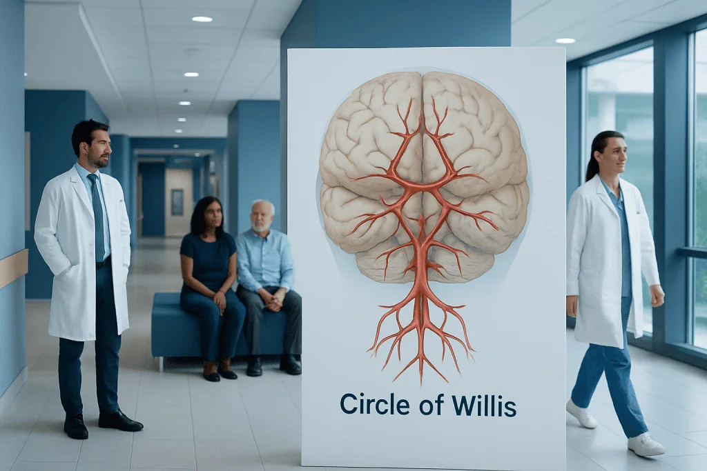
The circle of willis is an arterial connection at the base of the brain that can provide alternate routes for blood flow. Its exact shape varies from person to person, and these variations are common.
Key Takeaways
- The circle of willis is an arterial connection at the base of the brain that can provide alternate routes for blood flow.
- Its exact shape varies from person to person, and these variations are common.
- The structure itself usually causes no symptoms, but related problems such as aneurysm or stroke can be serious.
- Doctors evaluate the circle of willis with imaging tests such as CT angiography, MR angiography, or cerebral angiography.
- Treatment depends on the underlying problem and may include medicines, monitoring, endovascular procedures, or surgery.
The circle of willis is a ring-like network of arteries at the base of the brain that helps distribute blood between the front and back parts of the brain and between the right and left sides. It is medically important because natural differences in this structure can influence how the brain is protected during reduced blood flow and how conditions such as aneurysm or stroke are assessed.
Overview: what the circle of willis is and why it matters
The circle of willis is a ring of connected arteries located at the base of the brain. Its main role is to help balance blood supply between major arteries that feed the brain. In simple terms, it acts as a built-in connection system, so if blood flow is reduced in one area, another route may sometimes help deliver blood where it is needed.
This network is important in everyday brain function, but it is especially relevant in conditions that affect blood vessels, such as stroke, narrowing of arteries, or aneurysm formation. Many people learn about it only after a brain scan or after speaking with a neurologist, neurosurgeon, or vascular specialist.
One useful point for patients is that the circle of willis is not identical in every person. In fact, anatomical variation is very common. Some people have a complete and well-connected ring, while others have smaller segments, missing connections, or asymmetry. These differences do not automatically mean disease, but they can affect how doctors interpret risk and choose treatment.
How the circle of willis is built
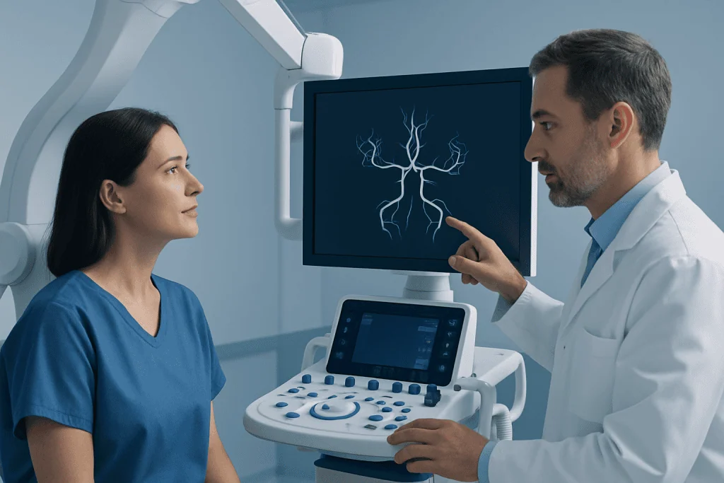
The circle of willis is formed by several arteries joining together near the underside of the brain. These include the internal carotid arteries, the anterior cerebral arteries, the anterior communicating artery, the posterior cerebral arteries, and the posterior communicating arteries. Together, they create a circular connection between the front and back circulation of the brain.
The front part of the brain is mainly supplied by the carotid system, while the back part is supplied by the vertebral and basilar arteries. The circle of willis links these two systems. It also connects the right and left sides, which is why it is often described as a collateral pathway, meaning a backup route for circulation.
Even though textbook diagrams show a neat, complete ring, real-life anatomy is often more complex. A vessel may be narrower than expected, one segment may be underdeveloped, or the connection may be absent. Radiologists and neurologists take these patterns into account because they can influence how blood moves during illness and how likely one area of the brain is to be protected if a blockage happens.
What the circle of willis does in the brain

The most important function of the circle of willis is to support stable brain perfusion, which means helping the brain receive a steady blood supply. The brain needs a constant delivery of oxygen and nutrients, and even brief interruptions can affect function. When one major artery becomes narrowed or blocked, this arterial ring may allow blood to reroute.
This protective effect is often called collateral circulation. It does not guarantee full protection, and its effectiveness depends on the size and openness of the connecting vessels. In some people, collateral flow through the circle of willis is strong, while in others it is limited because the natural connections are too small or incomplete.
Doctors consider this network when evaluating stroke risk, transient ischemic attacks, carotid artery disease, and some types of aneurysm. It is also relevant before certain vascular procedures because knowing the available backup circulation helps specialists plan treatment more safely and accurately.
Common variations and related medical conditions
Variation in the circle of willis is normal and common. A person may have a complete circle, a partially complete one, or one with smaller-than-usual arteries. Most of the time, these differences do not cause symptoms on their own and are found incidentally during brain imaging performed for another reason.
However, the circle of willis becomes clinically important when a blood vessel disorder is present. For example, if a major artery in the neck or brain narrows, an incomplete circle may offer less effective rerouting of blood. This can influence the impact of reduced circulation and may affect how symptoms appear.
Another key issue is aneurysm, a weak or bulging area in an artery wall. Aneurysms often occur near branching points around the circle of willis because these areas are exposed to ongoing blood-flow stress. An unruptured aneurysm may cause no symptoms, but if it ruptures, it can lead to bleeding around the brain, a medical emergency often discussed in relation to brain aneurysm.
In some cases, doctors also assess the circle of willis in people with dizziness, unusual headaches, or suspected vascular malformations, although these symptoms have many possible causes. The presence of a variation alone is not enough to explain symptoms without careful medical evaluation.
Symptoms: when the structure itself is silent but complications are not
The circle of willis itself usually does not produce symptoms. Most people do not feel any different because of their anatomy, even if their arterial pattern differs from the classic textbook version. Symptoms usually arise only when an associated condition affects blood flow or vessel integrity.
If reduced blood flow reaches the brain, symptoms may include sudden weakness, numbness, trouble speaking, facial drooping, confusion, vision changes, balance problems, or severe dizziness. These can be signs of a stroke or transient ischemic attack and require urgent medical attention. If a headache is sudden and severe, especially if described as the worst headache of a person’s life, bleeding from an aneurysm may be a concern.
Possible warning signs linked to vascular problems around this area can include:
- Sudden one-sided weakness or numbness
- Speech or understanding problems
- Loss of vision or double vision
- Severe, abrupt headache
- Fainting, seizure, or reduced alertness
- New difficulty walking or loss of coordination
Because these symptoms can overlap with several neurological emergencies, prompt assessment is essential. Early evaluation often includes brain imaging and vascular imaging to identify whether the cause involves blocked blood flow, bleeding, or another neurological condition.
How doctors diagnose problems involving the circle of willis
Diagnosis usually begins with a medical history and neurological examination. The doctor asks about the timing of symptoms, risk factors such as high blood pressure or smoking, and whether there is a family history of aneurysm or stroke. The physical exam may assess strength, speech, coordination, sensation, and eye movements.
Imaging is the main way to visualize the circle of willis. CT angiography and MR angiography are commonly used to show the arteries and identify narrowing, blockage, aneurysm, or anatomical variation. In selected cases, catheter-based cerebral angiography is recommended because it can provide very detailed images and may also be used as part of treatment planning.
Additional testing depends on the clinical situation. A standard CT or MRI of the brain may help detect stroke, bleeding, or tissue injury. Ultrasound of the carotid arteries, heart tests, and blood work may also be needed to look for the source of reduced brain blood flow.
Specialists may involve neurology, neurosurgery, interventional neuroradiology, and vascular teams. This multidisciplinary approach helps ensure that the anatomy of the circle of willis is interpreted in the context of the person’s actual symptoms, imaging findings, and overall vascular risk.
Treatment options and long-term management
Treatment is not aimed at the circle of willis itself unless there is a related vascular problem. If imaging shows a normal anatomical variation without disease, no treatment may be needed. In that situation, the finding mainly serves as useful background information for future care.
When the issue is reduced blood flow, treatment focuses on the cause. This may include medicines to lower stroke risk, careful management of blood pressure, cholesterol control, diabetes care, and lifestyle changes. In some patients, doctors may recommend procedures to improve blood flow or reduce the risk of future events. Depending on the diagnosis, care may include carotid endarterectomy or stroke treatment pathways.
For aneurysms located around the circle of willis, management depends on size, location, shape, symptoms, and rupture risk. Some aneurysms are monitored with periodic imaging, while others are treated with endovascular techniques or surgery. In selected patients, options may include brain aneurysm treatment or interventional neuroradiology procedures to secure the affected vessel.
Near the end of the care journey, patients may benefit from coordinated follow-up with specialists, especially if they have had a stroke, aneurysm, or vascular procedure. Acibadem International’s multidisciplinary specialists and JCI-accredited hospitals diagnose and treat these conditions for international patients, with care tailored to the individual diagnosis and imaging findings.
Protecting brain vessel health and when to seek medical care
It is not possible to change a person’s natural circle of willis anatomy, but general vascular health can often be improved. Steps that support brain blood vessels include not smoking, controlling high blood pressure, managing cholesterol and diabetes, staying physically active, eating a balanced diet, maintaining a healthy weight, and taking prescribed medicines as directed.
People with a known aneurysm, prior stroke, carotid disease, or strong family history of vascular disease should keep regular follow-up appointments. Imaging schedules vary depending on the condition. Patients should ask their doctor whether repeat scans are needed and what symptoms should prompt urgent reassessment.
Medical care should be sought urgently for sudden neurological symptoms such as weakness, numbness, speech difficulty, severe new headache, fainting, confusion, or vision loss. These symptoms should never be ignored, even if they improve quickly. Fast treatment can be important in limiting brain injury and improving recovery.
For non-urgent concerns, such as questions after an incidental imaging finding, a scheduled consultation with a neurologist, neurosurgeon, or vascular specialist can help clarify what the report means. In many cases, reassurance is appropriate once the anatomy is reviewed in context and no active disease is found.
Frequently asked questions
Is the circle of willis a disease?
No. The circle of willis is a normal part of brain anatomy. It becomes medically important because it can be involved in conditions such as stroke, artery narrowing, or aneurysm.
Can a person live normally with an incomplete circle of willis?
Yes, many people do and never know they have this variation. An incomplete or asymmetric circle of willis is common and often causes no symptoms by itself. Its significance mainly depends on whether another vascular problem is present.
Why is the circle of willis important in stroke?
It can sometimes provide alternate routes for blood flow if one artery becomes blocked or narrowed. This collateral circulation may help protect brain tissue, although the degree of protection varies from person to person based on the anatomy of the vessels.
How is the circle of willis seen on a scan?
Doctors usually assess it with CT angiography or MR angiography. In more complex cases, cerebral angiography may be used for a more detailed view, especially when planning treatment for aneurysm or other vascular disease.
Does a variation in the circle of willis need treatment?
Usually not, if it is only an anatomical variation and there is no associated disease. Treatment is considered only when there is an underlying problem such as reduced blood flow, aneurysm, or another vascular abnormality.
Can the circle of willis cause headaches?
The structure itself does not usually cause headaches. However, a vascular problem involving nearby arteries, especially an aneurysm or bleeding, can cause headache and needs medical evaluation.
References
- National Institute of Neurological Disorders and Stroke
- American Stroke Association
- National Health Service
- Radiological Society of North America
- World Federation of Neurosurgical Societies
This article is for general information only and is not a substitute for professional medical advice. Please consult a qualified doctor about your individual situation.
Explore treatments in Turkey — costs, top hospitals & a free quote
JCI-accredited · board-certified surgeons · reply within 24h
Add us as a Preferred Source to see more of our trusted health content across Google Search, AI Overviews and Discover.

More from the Health Library



Related Specialists

Prof. Dr. Mehmet Ziya Özüer
Otorhinolaryngology
Prof. Dr. Murat Yayla
Gynecology & Obstetrics
Dr. Ferhat Oto
Internal Medicine
Prof. Dr. Yüksel Ersoy
Physical Medicine & Rehabilitation