Corneal Topography Infusion: Procedure, Recovery and Results

Corneal topography is a quick, non-contact mapping test that measures the cornea's curvature and surface shape. It helps diagnose and monitor keratoconus, assess irregular astigmatism and plan selected refractive procedures.
Key Takeaways
- Corneal topography is a quick, non-contact mapping test that measures the cornea's curvature and surface shape.
- It helps diagnose and monitor keratoconus, assess irregular astigmatism and plan selected refractive procedures.
- Corneal cross-linking can help slow progressive keratoconus, while topography-guided ablation may improve surface regularity in carefully selected eyes.
- Recovery differs substantially between diagnostic topography, cross-linking and laser procedures.
- Blurred vision, increasing pain, marked redness or light sensitivity after an eye procedure should be assessed promptly by an eye specialist.
Corneal topography infusion is not a standard medical term; it is commonly used to describe corneal mapping that guides treatment for an irregular cornea, often alongside corneal cross-linking or topography-guided laser ablation. The right approach depends on the cause of corneal irregularity, the stability of vision, corneal thickness and overall eye health.
Overview: what does corneal topography infusion mean?
Corneal topography infusion is not a formal name for one specific eye procedure. In online searches, it may refer to using detailed corneal topography maps to guide treatment for an uneven or weakened cornea. These maps can support evaluation for keratoconus, irregular astigmatism, corneal scarring or changes following refractive surgery.
Corneal topography itself is a painless diagnostic examination, not an infusion and not surgery. A device projects illuminated rings or patterns onto the front surface of the eye and creates a color-coded map of corneal curvature. More advanced imaging, called corneal tomography, can also measure the front and back corneal surfaces and estimate corneal thickness.
When treatment is needed, the map may help an ophthalmologist decide whether monitoring, specialty contact lenses, corneal cross-linking, or selected laser treatment is appropriate. In some situations, clinicians may use the term cornea infusion informally when discussing a combined or customized approach, but patients should ask which exact test or treatment is being proposed.
How it works: corneal mapping and treatment planning
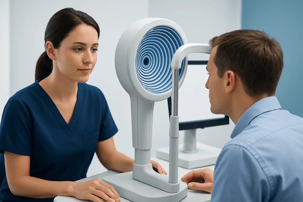
The cornea is the clear front window of the eye. Its smooth, rounded shape helps focus light on the retina. If the cornea becomes uneven, light may scatter or focus irregularly, causing blurred vision, glare, ghost images, halos or distortion. Topography identifies patterns that cannot always be seen during a routine eye examination.
For example, keratoconus causes progressive thinning and outward bulging of the cornea. Topography and tomography can reveal early asymmetry, steepening and changes over time. These measurements are also useful for assessing corneal topography after LASIK, particularly if vision has changed or a clinician needs to evaluate the cornea’s shape and stability.
In selected patients with a stable but irregular corneal surface, corneal topography-guided ablation may be considered. This laser approach is designed to smooth selected irregularities based on detailed measurements. If keratoconus is progressing, cross-linking may be recommended to strengthen corneal collagen and reduce the likelihood of further shape change. These treatments serve different purposes and are not interchangeable.
Who may benefit and what happens during the procedure?
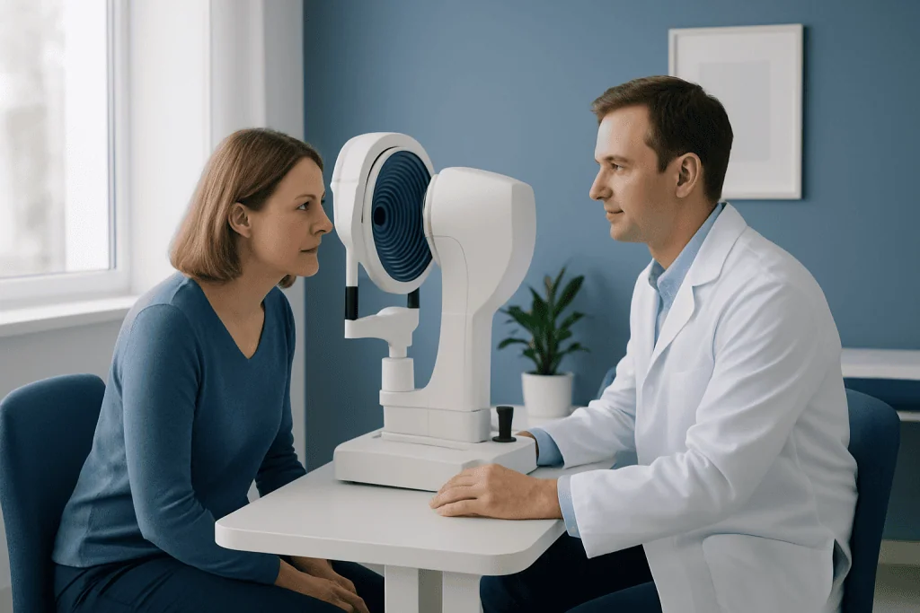
Topography is commonly performed for unexplained visual distortion, irregular astigmatism, suspected or known keratoconus, contact lens fitting and preoperative assessment before some refractive procedures. It may also be repeated over time to monitor whether the cornea is stable. The examination is generally suitable for most people who can briefly look at a fixation target.
People being considered for corneal cross-linking usually have evidence of progressive keratoconus or another corneal ectasia, adequate corneal thickness, and no active eye infection. Candidacy for topography-guided laser treatment is more selective. The ophthalmologist considers corneal stability, thickness, degree of irregularity, tear-film health, prescription, age and expectations for vision.
The topography test usually follows a simple sequence:
- Contact lenses may need to be stopped before testing, as advised, because they can temporarily alter corneal shape.
- The patient sits at an imaging device and looks at a central target.
- Several images are taken without touching the eye.
- An eye specialist reviews the maps alongside visual acuity, refraction, slit-lamp examination and, when indicated, tomography.
If cross-linking is planned, anesthetic eye drops are used, the corneal surface may be prepared, riboflavin drops are applied, and ultraviolet-A light is delivered under carefully controlled conditions. The exact technique varies. A protective contact lens is often placed after an epithelium-off procedure.
How long does corneal topography take?
Corneal topography usually takes only a few minutes per eye. The image capture itself is brief, although the overall appointment may take longer because the specialist may perform vision testing, corneal thickness measurements, pupil assessment or a dilated eye examination.
The test is non-contact in most modern systems and does not usually require anesthetic drops. It should not cause pain or require recovery time. People can generally return to normal activities immediately after the imaging test unless dilation or another examination performed during the same visit temporarily affects vision.
Corneal cross-linking takes considerably longer than mapping alone. Depending on the technique, preparation, riboflavin application and ultraviolet light exposure, patients should expect to spend several hours at the clinic. The care team can explain the planned technique and schedule before treatment.
Benefits, limitations and possible risks
The main benefit of corneal topography is precise information about corneal shape. It can help detect early disease, document changes, guide specialty lens fitting and support safer treatment planning. Repeated, comparable scans are especially valuable when a clinician is monitoring keratoconus progression.
Topography is highly useful, but it is not a diagnosis by itself. Dry eye, poor tear quality, blinking, eyelid position, recent contact lens wear and difficulty maintaining fixation can affect measurements. An ophthalmologist interprets the results together with symptoms, examination findings and other imaging rather than relying on one scan alone.
Topography has minimal physical risk because it is non-invasive. Risks relate instead to any treatment planned from the results. Cross-linking can involve temporary pain, light sensitivity, blurred vision, infection, haze or delayed epithelial healing. Laser ablation can involve dry eye, undercorrection, overcorrection, glare, haze or persistent visual irregularity. Careful selection and follow-up help reduce these risks but cannot eliminate them.
How do I know if my cornea is healing?
After an epithelium-off cross-linking procedure, the corneal surface typically heals over several days, although vision may remain blurry and fluctuate for weeks or longer. The strongest evidence of healing comes from follow-up examinations, where the ophthalmologist checks that the surface layer has closed, looks for signs of infection or inflammation, and assesses corneal clarity.
At home, gradual improvement in discomfort, tearing and light sensitivity can be reassuring. However, symptoms alone cannot confirm that the cornea is healing normally. Vision may temporarily worsen before it improves, and the final visual outcome after cross-linking may not be apparent for several months because the cornea needs time to stabilize.
Patients should use prescribed drops exactly as directed, avoid rubbing the eyes, attend all scheduled reviews and follow instructions about the protective contact lens. They should contact their eye team promptly if pain becomes worse rather than better, vision drops suddenly, discharge develops, or redness and light sensitivity increase.
Recovery timeline and how long until driving after corneal cross-linking surgery
Recovery after corneal topography requires no downtime. Recovery after corneal cross-linking is more variable. In the first few days after an epithelium-off procedure, discomfort, tearing, foreign-body sensation and light sensitivity are common. A bandage contact lens may remain in place until the surface has healed, as confirmed by the clinician.
Many people need several days away from work, school or close visual tasks, but the appropriate time depends on symptoms and the type of work. Vision often remains variable for weeks. Corneal remodeling and stabilization can continue for months, so follow-up topography is commonly used to assess the longer-term result.
How long until a person can drive after corneal cross-linking surgery depends on whether vision is clear enough to meet legal driving requirements and whether the individual feels comfortable driving safely. They should not drive themselves home on the day of treatment. Some may be able to resume driving after several days, while others need longer; the treating ophthalmologist should provide individualized clearance.
For patients needing assessment or treatment for a changing corneal shape, corneal cross-linking treatment may be discussed as part of a personalized ophthalmology plan.
Practical questions: accuracy, coverage and when to seek medical care
How accurate is corneal topography? Modern corneal topography is generally very accurate and reproducible when the eye surface is healthy and image quality is good. Accuracy improves when multiple high-quality scans are consistent. Contact lens wear, dry eye and unstable tears can distort results, so clinicians may repeat scans or recommend treating ocular surface dryness before making important decisions.
Corneal topography reimbursement varies by country, insurance plan and the clinical reason for testing. Coverage may differ when the test is used to evaluate a medical condition, such as suspected keratoconus, compared with elective refractive surgery planning. A corneal topography procedure code may also vary by healthcare system and billing rules. The clinic and insurer can clarify authorization and out-of-pocket responsibilities before care.
Medical review is important for new or worsening blurred vision, double or ghost vision, increasing glare, frequent prescription changes, or a family history of keratoconus. After cross-linking or any corneal procedure, urgent assessment is needed for severe or worsening pain, sudden loss of vision, increasing redness, discharge, marked swelling or intense light sensitivity.
Acibadem International’s multidisciplinary eye specialists and JCI-accredited hospitals diagnose and treat corneal conditions for international patients, with care plans based on detailed ophthalmic assessment and follow-up.
Frequently asked questions
Is corneal topography painful?
No. Corneal topography is usually a non-contact imaging test that does not touch the eye and should not cause pain. The patient simply looks at a target while the device captures images of the corneal surface.
Can corneal topography diagnose keratoconus?
Topography is an important test for detecting patterns that suggest keratoconus, such as localized corneal steepening and asymmetry. However, an ophthalmologist makes the diagnosis using the complete clinical picture, which may include tomography, corneal thickness measurements and repeat scans over time.
Can I wear contact lenses before corneal topography?
Contact lenses can temporarily change corneal shape and may affect the accuracy of measurements. The eye specialist will advise how long to stop wearing soft or rigid lenses before the appointment, as this varies by lens type and the purpose of testing.
Does corneal cross-linking improve eyesight?
The primary purpose of corneal cross-linking is to slow or stop progression of keratoconus or corneal ectasia. Some people notice visual improvement over time, while others mainly benefit from stabilization. Glasses or contact lenses may still be needed after treatment.
What is the difference between topography-guided ablation and cross-linking?
Topography-guided ablation uses a laser to reshape selected corneal irregularities in carefully chosen eyes. Cross-linking aims to strengthen corneal tissue and limit further weakening. In some cases they may be considered together or in sequence, but only after detailed assessment of corneal safety and stability.
When are repeat corneal topography scans needed?
Repeat scans may be used to monitor known or suspected keratoconus, evaluate changes after refractive surgery, assess the effect of contact lenses, or follow recovery after corneal treatment. The interval is individualized according to symptoms, findings and the risk of progression.
References
- American Academy of Ophthalmology
- National Eye Institute
- U.S. Food and Drug Administration
- Mayo Clinic
- International Society of Corneal, Contact Lens and Refractive Technologies
This article is for general information only and is not a substitute for professional medical advice. Please consult a qualified doctor about your individual situation.
Explore treatments in Turkey — costs, top hospitals & a free quote
JCI-accredited · board-certified surgeons · reply within 24h
Add us as a Preferred Source to see more of our trusted health content across Google Search, AI Overviews and Discover.

More from the Health Library

Related Specialists

Dyt. Zeynep Acar
Nutrition & Diet
Dr. Ali Koç
Orthopedic Surgery & Traumatology
Fzt. Busenur Sezer
Physical Medicine and Rehabilitation
Miray Karaoğlu
Physical Medicine & Rehabilitation