

Cryopexy vs Laser Retina: Differences Explained

Both cryopexy and laser retinopexy create a firm scar around a retinal tear to reduce the risk of retinal detachment. Cryopexy uses controlled freezing applied to the outside of the eye; laser retinopexy uses focused light energy inside the eye.
Key Takeaways
- Both cryopexy and laser retinopexy create a firm scar around a retinal tear to reduce the risk of retinal detachment.
- Cryopexy uses controlled freezing applied to the outside of the eye; laser retinopexy uses focused light energy inside the eye.
- The best option depends on the tear’s location, visibility, associated bleeding, and whether retinal detachment is already present.
- Most procedures are performed with local anaesthetic, and vision may remain blurred temporarily afterward.
- New flashes, a sudden increase in floaters, or a curtain-like shadow in vision need urgent eye assessment.
Cryopexy and laser retinopexy are treatments used to seal retinal tears and help prevent retinal detachment. Laser is often preferred when the retinal tear can be clearly reached from inside the eye, while cryopexy can be useful when laser treatment is difficult or not suitable.
Cryopexy vs Laser Retina: The Main Difference
In a cryopexy vs laser retina comparison, both procedures have the same broad goal: to create a scar that seals the retina around a tear, hole, or weak area. This scar helps anchor the retina to the tissue beneath it and can reduce the chance that fluid will pass through a tear and cause a retinal detachment.
Laser retinopexy, also called laser photocoagulation, delivers focused light through the pupil to place tiny burns around the retinal break. Retinal cryopexy uses a cooled probe placed on the outside surface of the eye to freeze the tissue overlying the break. The freezing response produces a similar protective scar as it heals.
Neither approach is automatically better for every person. An ophthalmologist, usually a retina specialist, selects the technique based on the retinal finding, the clarity of the eye’s internal structures, symptoms, and whether treatment is being performed alone or as part of retinal detachment repair.
How Cryopexy and Laser Retinopexy Work
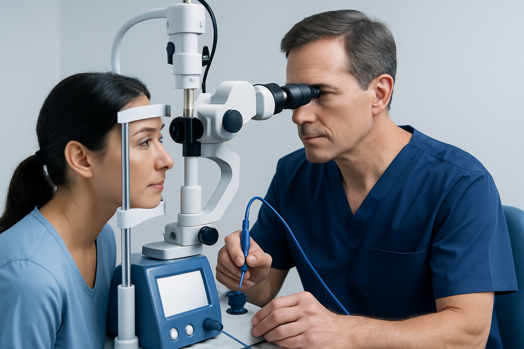
Laser retinopexy works by directing laser energy through a dilated pupil and a special contact lens or viewing system. The laser creates a controlled series of small spots around the retinal tear. Over the following days to weeks, healing creates a chorioretinal adhesion, meaning the retina becomes more firmly attached to the layer beneath it.
Cryopexy is sometimes described as retinal cryotherapy. In the comparison of cryopexy vs cryotherapy, these terms are commonly used to describe the same freezing-based technique when it is applied to the retina. A cryoprobe is positioned externally on the white part of the eye, known as the sclera, over the area of the retinal break. Cooling reaches the retina through the eye wall and creates a localized freeze-thaw injury that later scars.
In practical terms, cryopexy vs photocoagulation means freezing versus laser energy. Both can be effective, but cryopexy may cause more short-term inflammation than laser. Laser may be less suitable if the view of the retina is blocked by bleeding, cataract, corneal opacity, or certain peripheral tear locations.
Who May Be a Candidate for Each Procedure

Laser retinopexy is often considered for a symptomatic retinal tear, particularly when the specialist can see the tear clearly and apply laser spots around it. It may also be used for selected retinal holes or areas of lattice degeneration when there are features suggesting a higher risk of detachment. Not every retinal hole or area of lattice degeneration needs preventive treatment.
Cryopexy may be chosen when the tear is very far in the retinal periphery, when the view through the eye is limited, or when it is being used during surgery for retinal detachment. It can also be useful when the eye’s anatomy makes laser placement difficult. The decision is individualized after a dilated retinal examination and, when needed, imaging such as ocular ultrasound.
Cryopexy vs retinopexy can be confusing terminology because retinopexy means creating an attachment between the retina and underlying tissue. Both laser and cryopexy are forms of retinopexy. If a detachment is present, treatment may also involve procedures such as a gas-bubble repair, scleral buckle, or vitrectomy rather than a stand-alone office treatment.
What Happens During the Procedure
Before either treatment, drops are usually used to dilate the pupil. Numbing eye drops, an injection of local anaesthetic around the eye, or both may be used depending on the procedure, the patient’s comfort, and the treatment setting. The retina specialist examines the retina carefully to identify the precise margins of the tear.
For laser retinopexy, the patient sits at a laser machine or lies back while the specialist views the retina through a lens. Laser spots are applied around the break in a pattern designed to surround it. The actual laser treatment may take minutes, although preparation and examination make the visit longer.
For cryopexy, the specialist places the cryoprobe on the external eye surface and applies brief freezing applications at planned points. The eye may feel pressure or pulling. Cryopexy can be performed in an office setting or in an operating room, particularly if it is combined with retinal detachment surgery.
If a gas bubble is used to treat a retinal detachment, the procedure may be called pneumatic retinopexy. This is different from cryopexy or laser alone, although one of these sealing treatments is often performed alongside it.
Are You Awake During Pneumatic Retinopexy?
Most people are awake during pneumatic retinopexy. The eye is numbed with drops and sometimes a local anaesthetic injection, so the person should not feel sharp pain, although pressure, brightness, and some discomfort can occur.
During pneumatic retinopexy, a retina specialist injects a small gas bubble into the eye and uses laser or cryopexy to seal the retinal break. The bubble helps press the retina back into position while the treatment scar develops. Specific head positioning is usually essential after the procedure, and flying or travelling to high altitude is unsafe while a gas bubble remains in the eye.
Some people may require sedation or a different surgical approach depending on their medical needs, anxiety level, retinal anatomy, and the complexity of the detachment. The treating team explains the anaesthetic plan before treatment.
Recovery Timeline, Benefits and Possible Risks
After laser retinopexy, vision can be blurred and light sensitivity is common for several hours. Mild aching or a headache may occur. Many people return to usual light activities within a day or two, but the retina takes time to form a secure scar, commonly around one to two weeks. The specialist may recommend limiting strenuous activity for a short period.
How long does it take to recover from retinal cryopexy? Surface discomfort, redness, swelling, blurred vision, and light sensitivity can last several days and sometimes longer than after laser treatment. The treated area generally needs one to two weeks to develop a strong scar, while complete symptom improvement may take several weeks depending on the amount of freezing, inflammation, and any accompanying retinal detachment repair.
The main benefit of both procedures is reducing the risk of a retinal tear progressing to detachment. However, neither treatment removes the need for follow-up. New retinal tears can occur elsewhere, and a treated tear may occasionally need additional laser or cryopexy.
Possible risks include temporary inflammation, eye pressure changes, bleeding, new floaters, incomplete sealing of the tear, or retinal detachment despite treatment. Cryopexy can produce more inflammation and may increase the risk of scarring in some situations. Serious complications are uncommon but should be discussed with the retina specialist.
Is Laser Retinopexy Considered Surgery?
Laser retinopexy is generally considered a retinal procedure rather than open surgery. It does not usually require an incision, stitches, or general anaesthesia, and it is often performed as an outpatient treatment in a clinic or laser suite.
Even though it is less invasive than operations such as vitrectomy or scleral buckling, it is still a medical treatment for a potentially sight-threatening retinal problem. It requires specialist assessment, accurate placement of laser burns, and follow-up to confirm that the tear is sealed.
When a retinal tear has already led to a significant detachment, laser alone may not be enough. In these circumstances, the specialist may recommend surgical retinal repair based on the extent and location of the detachment.
Is Laser Retinopexy Painful?
Laser retinopexy is usually tolerable with numbing drops, but it can be uncomfortable. People may notice bright flashes of light, brief stinging, pressure from the contact lens, or an ache around the eye. Discomfort varies according to the location of the tear and the amount of laser required.
The specialist can pause treatment if needed and may use additional local anaesthetic for comfort. Pain that becomes severe after treatment, especially if accompanied by worsening vision, increasing redness, nausea, or headache, should be reported promptly.
In the cryo vs laser retina decision, comfort is only one consideration. Cryopexy may involve more pressure and post-procedure soreness, while laser may be more comfortable for suitable tears. The retinal specialist prioritizes the approach most likely to adequately surround and seal the break.
When to Seek Medical Care
Urgent assessment by an ophthalmologist or emergency eye service is important for new flashes of light, a sudden shower of floaters, a dark curtain or shadow, reduced side vision, or a sudden decrease in vision. These symptoms can occur with a posterior vitreous detachment, retinal tear, or retinal detachment and cannot be reliably distinguished without a dilated eye examination.
After cryopexy, laser retinopexy, or pneumatic retinopexy, patients should follow the scheduled review plan even if they feel well. They should contact their eye team sooner if vision worsens, pain increases substantially, there is marked redness or discharge, or they develop new symptoms in either eye.
Acibadem International’s multidisciplinary specialists and JCI-accredited hospitals diagnose and treat retinal conditions for international patients. A retina specialist can explain whether observation, laser retinopexy, cryopexy, or surgical repair is the most appropriate next step.
Frequently asked questions
Which is better, cryopexy or laser for a retinal tear?
Neither treatment is best in every case. Laser is often used when the retinal tear is clearly visible and accessible through the pupil, while cryopexy may be useful for very peripheral tears or when the view inside the eye is limited. A retina specialist chooses the method based on the individual retinal finding.
Does cryopexy permanently fix a retinal tear?
Cryopexy creates a lasting scar around the treated retinal tear, helping secure that particular area. However, it does not prevent a new tear from developing elsewhere in the retina. Follow-up examinations remain important, especially if new flashes or floaters appear.
How soon can vision improve after laser retinopexy?
Blurred vision from dilating drops, the examination lens, and treatment effects often improves within hours to a few days. If symptoms were caused by a retinal tear or vitreous changes, floaters may persist longer. Recovery can take more time if there was retinal detachment or another eye condition.
Can a retinal tear become detached after laser treatment?
Yes, retinal detachment can still occur despite treatment, although laser retinopexy is intended to lower this risk. A treated tear may need further laser, and new tears can develop in other areas. Worsening flashes, floaters, shadows, or vision loss should be assessed urgently.
Can someone drive after cryopexy or laser retinopexy?
Driving immediately afterward is usually not advised because the pupil remains dilated and vision may be blurred or sensitive to light. It is sensible to arrange transport home. The eye specialist can advise when driving is appropriate based on vision and the treatment performed.
Is cryopexy used only for retinal detachment?
No. Cryopexy can be used to treat selected retinal tears or holes before a detachment develops, as well as during repair of an established retinal detachment. It may also be used when laser treatment cannot be safely or effectively applied.
References
- American Academy of Ophthalmology
- National Eye Institute
- Royal College of Ophthalmologists
- Mayo Clinic
This article is for general information only and is not a substitute for professional medical advice. Please consult a qualified doctor about your individual situation.
CRYO in Turkey — costs, top hospitals & a free quote
JCI-accredited · board-certified surgeons · reply within 24h
Add us as a Preferred Source to see more of our trusted health content across Google Search, AI Overviews and Discover.

More from the Health Library



Related Specialists

Dt. Emre Orhon Kuybulu
Oral & Dental Health
Prof. Dr. Nesliar Eser Kutsal
Gastroenterology
Dr. Harun Ayangil
Pediatric Surgery
Dr. İpek Betül Özçivit Erkan
Gynecology & Obstetrics