

Diagnostic Mammogram: Who Needs It and What Results Mean

A diagnostic mammogram is different from a screening mammogram because it focuses on a specific breast concern. It may be recommended for a lump, breast pain, nipple discharge, skin changes, or an abnormal screening result.
Key Takeaways
- A diagnostic mammogram is different from a screening mammogram because it focuses on a specific breast concern.
- It may be recommended for a lump, breast pain, nipple discharge, skin changes, or an abnormal screening result.
- Results often show benign changes, but some findings need additional imaging or biopsy to confirm the cause.
- Common result terms include calcifications, asymmetry, mass, and distortion.
- A radiologist usually assigns a BI-RADS category to guide follow-up recommendations.
- Prompt follow-up helps clarify findings and supports early diagnosis when needed.
A diagnostic mammogram is a more detailed breast X-ray used when there is a breast symptom or a finding on a screening mammogram that needs a closer look. It helps radiologists study a specific area and decide whether more imaging, follow-up, or a biopsy may be needed.
Overview
A diagnostic mammogram is a specialized type of mammogram used to examine a specific area of the breast in greater detail. Unlike a screening mammogram, which is done routinely in people without symptoms, a diagnostic mammogram is ordered when there is a concern that needs closer evaluation. This concern may come from a new breast symptom or from a finding seen on a screening mammogram.
During a diagnostic mammogram, the technologist takes additional images from different angles and may use magnified or spot-compression views. These extra images help the radiologist look more closely at an area that is unclear or appears different from normal breast tissue. In many cases, the radiologist may review the images during the visit and request more views right away if needed.
A diagnostic mammogram does not by itself diagnose cancer. Instead, it helps clarify whether a finding is likely benign, needs short-term follow-up, or should be investigated further with tests such as ultrasound, MRI, or a biopsy. Most abnormal mammogram findings are not cancer, but careful evaluation is important so the right next step can be chosen.
Who May Need a Diagnostic Mammogram

A doctor may recommend a diagnostic mammogram for people who have a new breast symptom. Common reasons include a lump, focal breast pain, nipple discharge, skin thickening, nipple inversion, or a visible change in breast shape. It may also be used to evaluate a breast change found during a physical exam by a healthcare professional.
Another common reason is an abnormal screening mammogram. Screening images sometimes show an area that is not clearly benign, such as asymmetry, calcifications, or a possible mass. In this situation, a diagnostic mammogram is used to take a closer look and determine whether the finding is harmless overlap of normal tissue or something that needs more evaluation.
Diagnostic mammography may also be used for follow-up of a probably benign finding, to monitor changes after breast treatment, or to assess certain breast implants and postoperative changes. For some patients, ultrasound is performed on the same day because mammography and ultrasound provide complementary information, especially when evaluating lumps or dense breast tissue.
What Happens During the Test
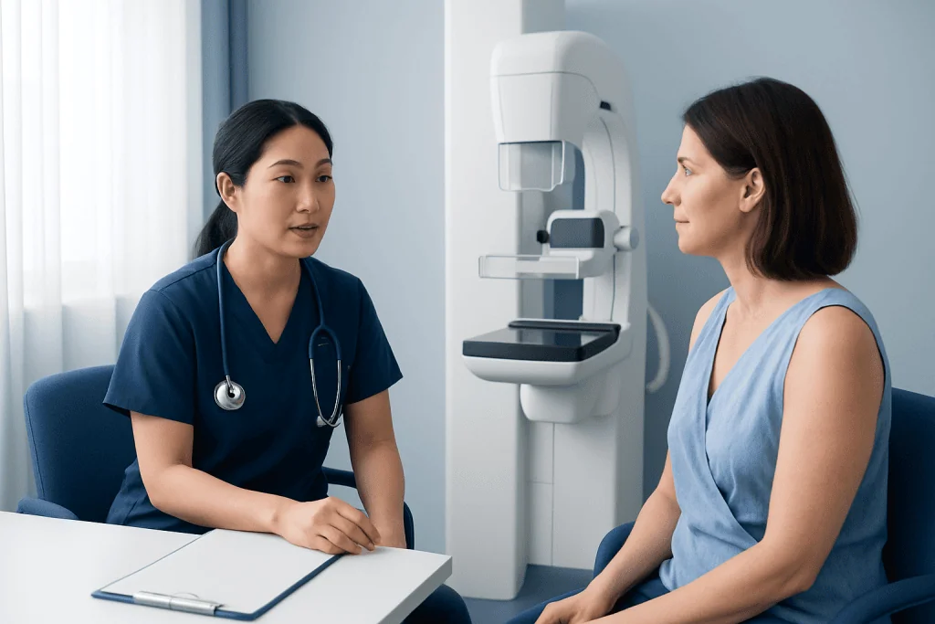
The test is performed using the same basic mammography equipment used for screening, but the appointment usually takes longer because more images may be needed. The breast is positioned on the machine and gently compressed for a few seconds while each image is taken. Compression can feel uncomfortable, but it is brief and helps produce clearer images with less motion.
The technologist may take views of one breast or both breasts depending on the reason for the exam. Special views, such as spot compression or magnification, may be used to examine calcifications, asymmetry, or a suspected mass. In some centers, digital breast tomosynthesis, also called 3D mammography, may be included to improve detail in certain cases.
Many patients are able to return to normal activities immediately after the test. If an ultrasound is also needed, it may happen during the same visit. A diagnostic mammogram uses a low dose of radiation, and for most people the benefits of accurate evaluation outweigh the small risk from radiation exposure.
To prepare, it is often helpful to avoid applying deodorant, powders, or creams on the chest and underarm area on the day of the exam because these can sometimes affect the images. Bringing prior mammograms, if done at another facility, can also be very helpful because comparison with earlier images often makes interpretation more accurate.
What the Results May Mean
After reviewing the images, the radiologist looks for patterns that suggest normal tissue, benign breast changes, or findings that require more attention. Common terms in reports include mass, asymmetry, calcifications, and architectural distortion. These words describe how the breast tissue looks on imaging, but they do not automatically mean cancer.
Calcifications are tiny calcium deposits within the breast. Many calcifications are benign and related to aging, prior inflammation, or harmless breast changes. Some patterns of calcifications can be more suspicious and may lead to short-term follow-up or biopsy. A mass refers to a space-occupying area, which may be a cyst, fibroadenoma, or another type of lesion. Asymmetry means one area looks denser than expected, and additional views often show that it is simply overlapping tissue.
Radiologists usually classify the result using the BI-RADS system. A BI-RADS 1 result means the exam is negative, and BI-RADS 2 indicates a benign finding. BI-RADS 3 means probably benign and often leads to short-term follow-up imaging, while BI-RADS 4 or 5 means the finding is suspicious enough that a biopsy is usually recommended. BI-RADS 0 means more imaging is needed before a final assessment can be made.
If a biopsy is advised, this does not mean that cancer is certain. It means imaging alone cannot fully explain the finding. Depending on the situation, the next step may include a breast biopsy or further MRI imaging to provide more information and help guide care.
Common Causes of Abnormal Findings
There are many noncancerous reasons why a diagnostic mammogram may be abnormal. Benign breast cysts, fibroadenomas, dense breast tissue, postsurgical scar tissue, and fat necrosis can all create findings that need closer evaluation. Hormonal changes may also affect breast tissue and alter the way it appears on imaging.
Calcifications are especially common and are often harmless. In some cases, their shape or distribution may suggest the need for closer review. Architectural distortion, which means the normal breast tissue pattern looks pulled or disrupted, may be caused by prior surgery, benign radial scars, or less commonly malignancy.
When a patient has a palpable lump, ultrasound is often added because it can show whether the area is fluid-filled or solid. If the imaging suggests a suspicious breast lesion, the care team may discuss the possibility of breast cancer while emphasizing that only tissue sampling can confirm the diagnosis. This stepwise approach helps avoid unnecessary procedures while making sure important findings are not missed.
Next Steps After a Diagnostic Mammogram
The next step depends on what the images show. If the result is normal or clearly benign, the patient may simply return to routine screening at the interval recommended by the doctor. If the finding is probably benign, follow-up imaging in a few months may be advised to confirm that the area remains stable over time.
When more evaluation is needed, the doctor may recommend breast ultrasound, breast MRI, or a biopsy. Ultrasound is especially useful for characterizing lumps and for guiding needle procedures. In some cases, suspicious calcifications or subtle distortions are best sampled with image-guided biopsy after mammographic localization.
Waiting for results can feel stressful, but clear communication with the care team is important. Patients should ask what BI-RADS category was assigned, whether any further test is needed, and when follow-up should occur. Understanding the plan can make the process more manageable and help ensure that recommended care is not delayed.
For people receiving care internationally, coordinated imaging and follow-up can be helpful. Near the end of the care pathway, patients may choose centers such as Acibadem International, where multidisciplinary specialists in JCI-accredited hospitals evaluate breast imaging findings and arrange further testing when appropriate.
Prevention, Breast Awareness, and Self-care
A diagnostic mammogram is not a preventive test on its own, but it plays an important role in evaluating changes early. Following age- and risk-based screening advice from a doctor remains one of the best ways to support breast health. People at higher risk because of family history, genetic factors, or prior chest radiation may need a more personalized screening plan.
Breast awareness is also valuable. This means becoming familiar with how the breasts normally look and feel and reporting any new change promptly. Changes worth mentioning include a new lump, persistent focal pain, nipple discharge, skin dimpling, swelling, redness, or a change in the nipple.
General self-care supports overall health as well. Maintaining a healthy lifestyle, limiting alcohol, staying physically active, and attending regular checkups may all contribute to better long-term breast health. If previous imaging showed dense breasts or benign changes such as breast cysts, patients can ask their doctor how this affects future screening and follow-up recommendations.
When to See a Doctor
A person should contact a doctor if there is a new breast lump, bloody or spontaneous nipple discharge, skin thickening, breast swelling, dimpling, or a persistent change in breast shape. Ongoing focal pain in one area of the breast, especially if it does not improve, also deserves medical attention. Many symptoms turn out to have benign causes, but it is safest to have them assessed.
It is also important to follow up promptly after an abnormal screening mammogram. Delaying a diagnostic mammogram can prolong uncertainty and may postpone treatment if treatment is needed. If a report is hard to understand, patients should ask for the findings and next steps to be explained in plain language.
Urgent evaluation may be needed if breast changes are accompanied by fever, increasing redness, or significant swelling, as these can suggest infection or inflammation. A qualified healthcare professional can decide whether diagnostic mammography, ultrasound, or another test is the most appropriate starting point.
Frequently asked questions
What is the difference between a screening mammogram and a diagnostic mammogram?
A screening mammogram is a routine test for people who do not have breast symptoms. A diagnostic mammogram is done when there is a symptom or when a screening mammogram shows an area that needs a closer look. It usually includes extra images focused on a specific part of the breast.
Does a diagnostic mammogram mean cancer is likely?
No. A diagnostic mammogram means more detailed evaluation is needed, not that cancer is already diagnosed. Many findings turn out to be benign, such as cysts, overlapping tissue, or harmless calcifications.
How long does a diagnostic mammogram take?
It often takes longer than a screening mammogram because additional views may be needed. Many appointments last around 30 to 60 minutes, especially if the radiologist reviews images during the visit or if an ultrasound is added.
Is a diagnostic mammogram painful?
Some people feel pressure or brief discomfort because the breast must be compressed to get clear images. The sensation usually lasts only a few seconds for each image. If the breasts are tender, scheduling the exam for a less sensitive time in the menstrual cycle may help.
What does BI-RADS mean on a mammogram report?
BI-RADS is a standardized system radiologists use to describe mammogram findings and recommend next steps. Lower categories usually mean normal or benign findings, while higher categories suggest that closer follow-up or biopsy may be needed. It helps doctors and patients understand the level of concern and the plan.
Can a diagnostic mammogram be normal even if a lump is felt?
Yes, sometimes a lump is not clearly seen on mammography, especially in dense breasts. That is why ultrasound is often used as well when there is a palpable lump. If the physical finding remains concerning, the doctor may recommend further evaluation even if mammogram images are not clearly abnormal.
References
- American College of Radiology
- Radiological Society of North America
- National Cancer Institute
- U.S. Centers for Disease Control and Prevention
- World Health Organization
This article is for general information only and is not a substitute for professional medical advice. Please consult a qualified doctor about your individual situation.
Oncology care in Turkey — second opinion and treatment plan
JCI-accredited · board-certified surgeons · reply within 24h
Add us as a Preferred Source to see more of our trusted health content across Google Search, AI Overviews and Discover.

More from the Health Library



Related Specialists

Prof. Dr. Ercan Karaarslan
Radiology
Prof. Dr. Bora Peynircioğlu
Interventional Radiology
Prof. Dr. Koray Güven
Interventional Radiology
Assoc. Prof. Dr. Deniz Can Aliş
Radiology