Does PET Scan Have Radiation: How It Works, Results and What to Expect
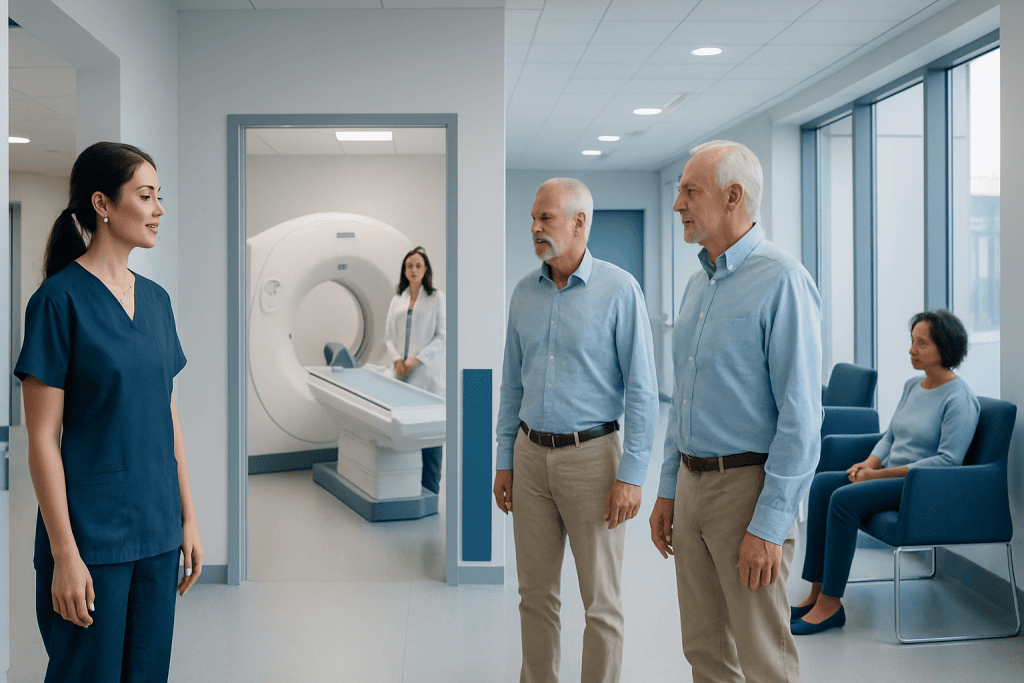
PET scans use a short-lived radioactive tracer, often combined with CT or MRI imaging. Most of the tracer leaves the body through urine within hours, although small amounts may remain briefly.
Key Takeaways
- PET scans use a short-lived radioactive tracer, often combined with CT or MRI imaging.
- Most of the tracer leaves the body through urine within hours, although small amounts may remain briefly.
- The main limitations are radiation exposure, possible false-positive or false-negative findings, cost and availability.
- Pregnancy, breastfeeding, diabetes and kidney problems may require special planning before a PET scan.
- Following preparation and aftercare instructions helps improve image quality and reduce unnecessary exposure.
Does PET scan have radiation? Yes. A PET scan uses a small amount of a radioactive tracer to show how tissues and organs are functioning, with exposure that is generally low and carefully considered against the value of the information obtained. PET imaging can help clinicians diagnose, stage and monitor several conditions, particularly cancer, heart disease and some neurological disorders.
Does PET Scan Have Radiation? An Overview
Yes, a PET scan has radiation. Positron emission tomography (PET) uses a small quantity of radioactive material, called a radiotracer, to create images of metabolic activity inside the body. The tracer gives off signals that are detected by the scanner, allowing clinicians to see how actively cells and tissues are using energy.
The radiation dose is usually limited and is used only when the expected clinical benefit outweighs the possible risk. Many PET scans are performed as PET/CT scans, meaning that the examination includes radiation from both the radiotracer and the CT portion. PET/MRI uses the PET tracer but replaces CT with MRI, which does not use ionizing radiation.
PET imaging is especially useful when structural imaging alone does not fully explain what is happening in the body. For example, it may help evaluate suspected or known cancer, assess response to treatment, investigate certain heart conditions, or support assessment of selected neurological conditions.
How a PET Scan Works and Why It Is Used
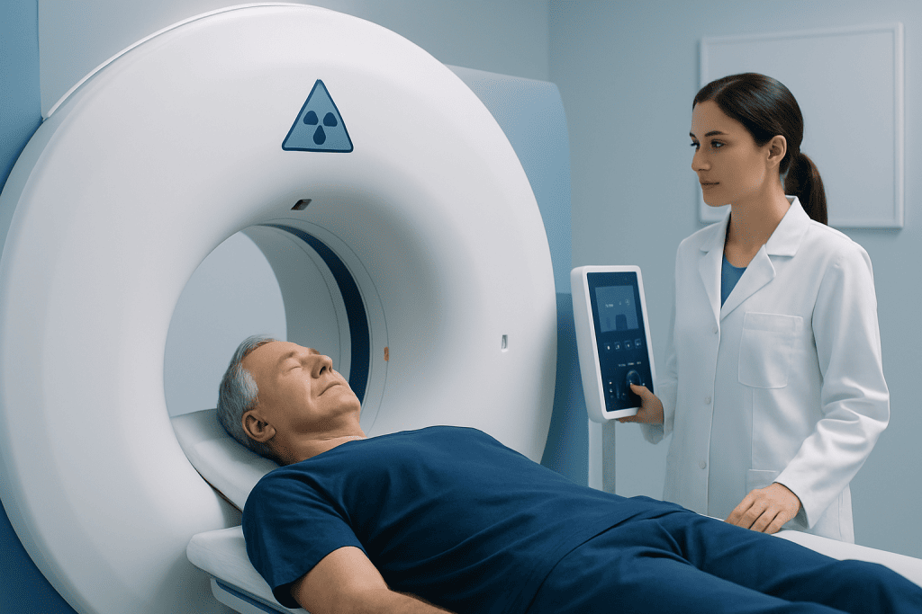
Before the scan, a healthcare professional injects a radiotracer into a vein, most commonly a glucose-like substance called fluorodeoxyglucose (FDG). Cells that use more glucose may absorb more tracer. This can include many cancer cells, but it can also include healthy tissues and areas affected by inflammation or infection.
After an uptake period, the patient lies still on a padded table that moves through the scanner. A PET camera detects signals from the tracer and a computer creates detailed images. When PET is combined with CT or MRI, the images provide both functional information and anatomical detail.
Clinicians may use PET to help locate active disease, determine the extent of a condition, guide biopsy planning, assess whether treatment is working, or look for recurrence. In oncology, PET may be part of the assessment of conditions such as lung cancer, lymphoma, colorectal cancer and other malignancies when clinically appropriate.
Who May Need a PET Scan and How to Prepare

A doctor may recommend a PET scan when it is likely to answer a specific clinical question that cannot be answered adequately by examination, laboratory tests, CT, MRI or other imaging. It is not routinely needed for every symptom or every cancer diagnosis. The decision is individualized according to the person’s medical history, symptoms and previous test results.
Patients should tell the imaging team about pregnancy, a possible pregnancy, breastfeeding, diabetes, kidney disease, allergies, recent illness, surgery, infection and all medicines or supplements. Pregnancy requires particular consideration because ionizing radiation can affect a developing fetus. Breastfeeding advice depends on the tracer used, so the nuclear medicine team should provide specific guidance.
Preparation varies by examination. For many FDG-PET scans, patients are asked to avoid strenuous exercise beforehand and to fast for several hours, while drinking plain water may be encouraged. Blood glucose is checked in many patients because high glucose can affect tracer distribution and image interpretation. The imaging center will provide personalized instructions.
- Wear comfortable clothing and remove metal items if requested.
- Bring prior imaging reports or scans when available.
- Ask in advance about diabetes medication, insulin timing and dietary instructions.
- Inform the team if lying flat or remaining still may be difficult.
What Happens During and After the Procedure
The appointment commonly begins with a review of medical information and a small injection of tracer. The injection may feel similar to a routine blood test. There is then a quiet waiting period, often about 30 to 60 minutes, while the tracer circulates and is taken up by tissues. Resting during this time is important because muscle activity can alter the images.
The scanning portion usually takes about 20 to 45 minutes, depending on the body area being examined and whether CT or MRI is included. The scan itself is painless, although remaining still can be uncomfortable for some people. The team can communicate with the patient throughout the examination.
Most people return to normal activities soon after the scan. Drinking extra fluids, unless a clinician has advised fluid restriction, can help the body clear the tracer through urine. Patients should follow the imaging team’s instructions about toilet hygiene and close contact with infants, young children or pregnant people during the first several hours after the scan.
Images are interpreted by a radiologist or nuclear medicine physician and sent to the referring clinician. Results should be discussed in context, because PET findings are not interpreted in isolation. Further imaging, blood tests or a biopsy may sometimes be needed to confirm the cause of an abnormal area.
What Is the Downside of a PET Scan?
The principal downside is exposure to ionizing radiation from the radiotracer and, when used, the CT component. The amount varies according to the tracer, the scan protocol and the patient’s size. Clinicians aim to use the lowest radiation dose needed to obtain useful images, and they recommend PET only when it can meaningfully influence care.
PET scans can also produce findings that are not definitive. Inflammation, infection, recent surgery, healing tissue and some normal organs may show increased tracer uptake and resemble cancer. Conversely, some small tumors or slower-growing cancers may not be clearly visible. This is why PET results need expert interpretation alongside clinical information and other tests.
Practical drawbacks may include fasting, a lengthy appointment, the need to remain still and temporary anxiety while awaiting results. Reactions to the radiotracer are uncommon. If contrast material is used for the CT portion, there may be additional considerations, including allergy history and kidney function.
How Long Do I Stay Radioactive After a PET Scan?
After a typical PET scan, the tracer loses radioactivity quickly because it has a short physical half-life and because the body removes it in urine. For the commonly used FDG tracer, much of the radioactivity decreases over the first several hours, and most people do not need special isolation.
Imaging centers may recommend simple temporary precautions, especially around pregnant people and very young children. These may include limiting prolonged close contact for several hours, washing hands carefully after using the toilet, flushing twice when instructed, and drinking fluids if medically appropriate. The exact advice should come from the nuclear medicine team because different tracers have different properties.
Patients should not assume that every PET scan has identical aftercare requirements. Anyone who is breastfeeding, provides close personal care to an infant, or is scheduled for another medical procedure should ask the care team for tracer-specific guidance before leaving the imaging center.
What Are the Odds of Getting Cancer From a PET Scan?
It is not possible to calculate an exact personal chance of cancer from one PET scan. Radiation exposure is understood to carry a small theoretical long-term risk, but the risk from a medically necessary single examination is considered low. It must be weighed against the potential benefit of finding, characterizing or monitoring a serious condition accurately.
Risk is influenced by the type of PET examination, whether CT is included, the total number of radiation-based tests over time, age and individual circumstances. Children and younger adults are generally more sensitive to radiation, so imaging teams take particular care to justify scans and tailor protocols when possible.
Patients can support informed decision-making by asking why the scan is needed, whether another test could provide the same information, and whether prior imaging can be used to avoid duplicate examinations. A clinician should not delay an important PET scan solely because of concern about the small radiation-related risk when the scan could guide appropriate treatment.
What Is the Biggest Disadvantage in Using a PET Scan?
The biggest disadvantage is that PET is highly sensitive but not always specific. It can identify areas of increased metabolic activity, but it cannot always determine why that activity is present. Cancer, infection, inflammation and tissue healing can sometimes look similar on PET images, leading to uncertainty or additional testing.
Another important limitation is resolution. Very small abnormalities may be difficult to detect, and some conditions do not take up the tracer strongly. PET is therefore often combined with CT or MRI and interpreted with pathology results, laboratory findings and the patient’s symptoms rather than used as a stand-alone diagnostic answer.
Despite these limitations, PET can provide valuable information that changes clinical decisions. In patients undergoing cancer evaluation, it may help specialists select the most appropriate next step, including cancer treatment planning or further diagnostic procedures. Acibadem International’s multidisciplinary specialists and JCI-accredited hospitals diagnose and treat complex conditions for international patients.
When to Seek Medical Care
A PET scan itself does not usually cause delayed symptoms. However, patients should contact the imaging center or seek medical advice promptly if they develop signs of a possible allergic reaction after contrast or an injection, such as widespread hives, swelling of the face or throat, wheezing, severe dizziness or trouble breathing. Emergency care is appropriate for severe breathing difficulty or swelling.
Patients should also contact their referring clinician if they are pregnant or learn they may have been pregnant at the time of the examination, if they did not receive clear breastfeeding instructions, or if diabetes preparation instructions were difficult to follow. These situations can be addressed calmly and individually by the medical team.
For questions about results, the best next step is a follow-up discussion with the clinician who ordered the scan. They can explain whether any finding requires observation, additional imaging, laboratory tests, biopsy or referral to a specialist.
Frequently asked questions
Does a PET scan have more radiation than a CT scan?
A PET/CT scan includes radiation from both the PET radiotracer and the CT scan, so its total exposure is generally higher than that of a CT scan alone. The amount varies by protocol and body area. The care team recommends the examination when the expected diagnostic value is greater than the potential radiation risk.
Is a PET scan painful?
A PET scan is usually not painful. There is a brief needle stick for the tracer injection, and some people find it uncomfortable to lie still during the scan. The scanner does not typically touch or compress the body.
Can I be around my family after a PET scan?
Most people can go home and be around others after a PET scan. The imaging team may recommend reducing prolonged close contact with infants, young children and pregnant people for several hours. The advice depends on the tracer used, so patients should follow their center's instructions.
Why do I need to fast before a PET scan?
Many PET scans use a glucose-like tracer, and fasting helps improve the distinction between normal and abnormal tissue uptake. Food intake, exercise and high blood sugar can affect image quality. The exact fasting instructions differ by scan type and individual health needs.
Can PET scans show inflammation as well as cancer?
Yes. Areas of infection, inflammation, injury or healing can absorb PET tracer and appear active on the scan. This is one reason an abnormal PET finding does not automatically mean cancer. Doctors assess the images alongside symptoms, medical history and other tests.
How soon will PET scan results be available?
The images are reviewed by a trained radiologist or nuclear medicine physician, and a report is sent to the clinician who ordered the scan. Timing varies between centers and depends on the complexity of the study. The referring clinician can explain the results and their relevance to the individual's care.
References
- International Atomic Energy Agency
- Radiological Society of North America
- Society of Nuclear Medicine and Molecular Imaging
- National Cancer Institute
- U.S. Food and Drug Administration
This article is for general information only and is not a substitute for professional medical advice. Please consult a qualified doctor about your individual situation.
Heart care in Turkey — expert evaluation and treatment
JCI-accredited · board-certified surgeons · reply within 24h
Add us as a Preferred Source to see more of our trusted health content across Google Search, AI Overviews and Discover.