Doppler Ultrasound: What Patients Need to Know

Doppler ultrasound measures blood flow and can help detect blockages, narrowing, or blood clots. The test is painless, does not use radiation, and is often done in an outpatient setting.
Key Takeaways
- Doppler ultrasound measures blood flow and can help detect blockages, narrowing, or blood clots.
- The test is painless, does not use radiation, and is often done in an outpatient setting.
- Different types of Doppler ultrasound are used for arteries, veins, pregnancy, and heart-related assessments.
- Preparation depends on the body area being examined, but many Doppler scans need little or no special preparation.
- Results are interpreted together with symptoms, physical examination, and other tests when needed.
Doppler ultrasound is a safe, noninvasive imaging test that shows how blood moves through blood vessels and the heart. It helps doctors investigate symptoms such as leg swelling, pain, dizziness, or suspected circulation problems without using radiation.
Overview: what a Doppler ultrasound shows
Doppler ultrasound is an imaging test that uses sound waves to evaluate blood flow in the body. Unlike a standard ultrasound, which mainly shows structures such as organs or blood vessels, Doppler ultrasound adds information about movement. In practical terms, it helps a doctor see whether blood is flowing normally, too slowly, too quickly, or in the wrong direction.
This test is commonly used to assess circulation in the legs, arms, neck, abdomen, and sometimes the heart. It can help investigate concerns such as swelling, pain, numbness, suspected blood clots, narrowed arteries, or reduced blood supply to tissues. In pregnancy, Doppler techniques may also be used selectively to evaluate blood flow in the placenta or umbilical cord when medically indicated.
For many patients, one of the most reassuring aspects of doppler ultrasound is that it is noninvasive and does not use ionizing radiation. A handheld device called a transducer is placed on the skin with gel, and the reflected sound waves are converted into images and flow information. The test is widely used because it is generally quick, accessible, and informative.
How Doppler ultrasound works and the different types
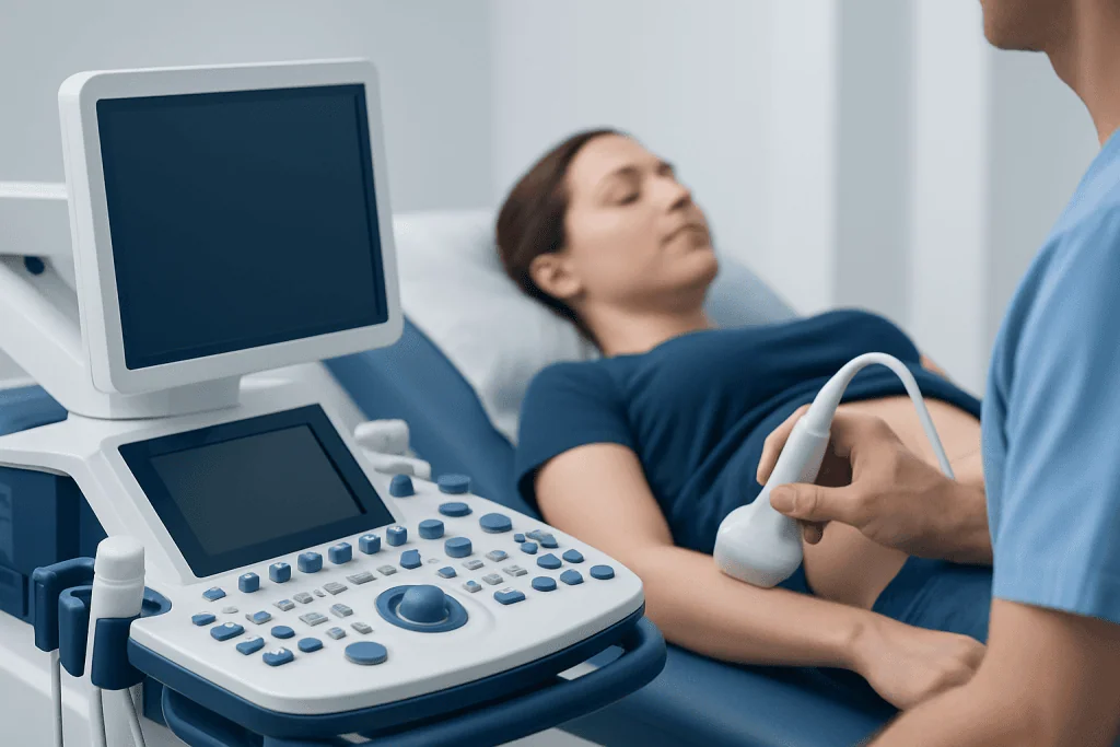
Doppler ultrasound is based on the Doppler effect, which refers to how sound waves change when they bounce off moving blood cells. The ultrasound machine analyzes these changes and displays them as visual signals, waveforms, or color patterns that help the clinician judge the direction and speed of blood flow.
There are several types of Doppler ultrasound, and the choice depends on the clinical question. Color Doppler adds color overlays to show the direction and pattern of flow. Spectral Doppler creates a waveform that can help estimate flow speed. Power Doppler is more sensitive to slower blood flow and can be helpful in some settings. Duplex ultrasound combines standard imaging with Doppler analysis, while triplex ultrasound includes color flow as well.
Because it can examine both structure and circulation, this test is often part of a broader vascular assessment. For example, doctors may use it when looking for problems related to peripheral artery disease or when checking for reduced flow after symptoms suggest a blockage. It may also support evaluation before or after procedures involving blood vessels.
Why doctors order a Doppler scan

A Doppler scan is ordered when a doctor needs more information about circulation. Symptoms that may lead to this test include leg pain when walking, one-sided swelling, skin color changes, cold feet or hands, dizziness, bruit sounds heard with a stethoscope, or wounds that are slow to heal. It may also be used when a doctor suspects a blood clot, narrowed artery, varicose vein problem, or impaired blood flow to an organ.
Common clinical uses include looking for deep vein thrombosis in the legs, checking carotid arteries in the neck, assessing circulation in the limbs, and evaluating blood flow in abdominal vessels. In heart care, specialized Doppler ultrasound is often part of an echocardiogram to examine blood flow through heart valves and chambers. If symptoms relate to the heart, a physician may recommend echocardiography as the most appropriate Doppler-based test.
Doppler ultrasound can also help guide treatment decisions and monitor known vascular conditions over time. It may be used before surgery, after vascular procedures, or during follow-up for people who have stents, bypass grafts, or chronic circulation disorders. In some cases, the findings suggest the need for additional imaging or procedures such as angiography for a more detailed view of blood vessels.
What to expect before, during, and after the test
Preparation depends on the area being examined. Many Doppler ultrasound tests require little preparation beyond wearing comfortable clothing. For abdominal vessel studies, a patient may be asked to avoid eating for several hours to reduce gas in the intestines, which can affect image quality. Patients should follow the specific instructions given by their imaging center or doctor.
During the test, the patient usually lies on an examination table while a sonographer applies gel to the skin. The transducer is moved over the area being studied, and mild pressure may be used to improve contact. The machine may produce whooshing or pulsing sounds, which reflect blood flow and are a normal part of the examination. The test is generally painless, although pressing on a tender area can be briefly uncomfortable.
Most Doppler ultrasound scans take about 30 to 60 minutes, though some are shorter or longer depending on the body region and complexity. After the test, patients can usually return to normal activities right away. A radiologist, vascular specialist, or other trained physician interprets the findings and sends a report to the referring doctor, who explains what the results mean in the context of symptoms and medical history.
What the results can help diagnose
Doppler ultrasound does not by itself provide a complete diagnosis in every case, but it is very useful for identifying patterns that suggest a vascular problem. It can show reduced blood flow through a narrowed artery, absent flow in a blocked vessel, reversed flow through an incompetent vein, or turbulent flow caused by vessel narrowing. These findings help doctors decide whether the circulation issue is mild, significant, or urgent.
Conditions commonly investigated with Doppler ultrasound include deep vein thrombosis, chronic venous insufficiency, carotid artery disease, peripheral artery disease, aneurysms in selected settings, and blood flow problems affecting transplanted organs or abdominal vessels. In some patients, Doppler techniques also help evaluate concerns related to varicose veins or suspected complications from long-standing vein disease.
It is important to remember that a normal result can also be helpful. A normal Doppler study may rule out certain circulation problems and direct the doctor to look for other causes of symptoms, such as muscle strain, nerve compression, arthritis, or fluid retention from a nonvascular cause. When the picture remains unclear, the doctor may combine Doppler findings with blood tests, physical examination, and other imaging studies.
Benefits, limitations, and safety
The main benefits of doppler ultrasound are that it is noninvasive, does not use radiation, and can provide real-time information about blood flow. It is often available quickly and can be repeated safely when follow-up is needed. For many patients, it is an excellent first-line test because it can answer important questions without requiring contrast dye, needles, or hospital admission.
Like any medical test, it has limitations. Image quality can be affected by body habitus, swelling, dressings, bowel gas, or the depth and location of a blood vessel. Some vessels may be difficult to visualize, and very small or complex abnormalities may require another imaging method. In addition, the quality of the study can depend on the equipment used and the experience of the sonographer and interpreting clinician.
Overall, Doppler ultrasound is considered very safe when performed appropriately. There is no routine recovery time, and complications are rare because the test is external and noninvasive. If results suggest a more serious circulation problem, the next step may involve consultation with specialists in vascular medicine, cardiology, or radiology, and in selected cases further evaluation such as cardiology care or another vascular procedure.
When to seek medical care
Patients should seek medical attention promptly if they develop symptoms that may suggest a circulation problem. These include sudden swelling in one leg, new calf pain, chest pain, shortness of breath, sudden weakness, trouble speaking, severe dizziness, or a limb that becomes pale, cold, or painful. These symptoms do not always mean a serious vascular condition, but they should be assessed without delay.
For nonurgent concerns, it is reasonable to arrange a routine medical review for recurring leg cramps when walking, gradually increasing swelling, visible vein changes, cold extremities, or slow-healing wounds. A doctor can decide whether Doppler ultrasound is the right next step or whether another evaluation would be more useful.
Near the end of the care pathway, patients may benefit from a coordinated review if an abnormal Doppler result needs specialist input. Acibadem International’s multidisciplinary specialists and JCI-accredited hospitals diagnose and treat circulation-related conditions for international patients, including assessment pathways that may involve vascular imaging and follow-up care when appropriate.
Frequently asked questions
Is Doppler ultrasound the same as a regular ultrasound?
Not exactly. A regular ultrasound mainly shows body structures, while Doppler ultrasound adds information about how blood is moving through vessels or the heart. Many exams combine both methods in one study.
Does a Doppler ultrasound hurt?
Doppler ultrasound is usually painless. The transducer may press on the skin, which can feel slightly uncomfortable if the area is already tender, but the test does not involve surgery or radiation.
How long does a Doppler ultrasound take?
Most Doppler ultrasound exams take about 30 to 60 minutes. The exact time depends on the body area being studied and whether the doctor needs a more detailed vascular assessment.
Can Doppler ultrasound detect blood clots?
Yes, it is commonly used to help detect blood clots, especially in the leg veins. It can also show whether blood flow is reduced or blocked, although results are always interpreted together with symptoms and examination findings.
Do patients need to prepare for a Doppler scan?
Many Doppler scans need little or no preparation. Some abdominal studies may require fasting for several hours, so patients should follow the instructions provided by their doctor or imaging center.
Is Doppler ultrasound safe in pregnancy?
When used appropriately by trained professionals, ultrasound techniques are generally considered safe in pregnancy. Doppler may be used when there is a medical reason to assess blood flow, such as concerns about placental or fetal circulation.
This article is for general information only and is not a substitute for professional medical advice. Please consult a qualified doctor about your individual situation.
Ultrasound in Turkey — costs, top hospitals & a free quote
JCI-accredited · board-certified surgeons · reply within 24h
Add us as a Preferred Source to see more of our trusted health content across Google Search, AI Overviews and Discover.

Check-up & Preventive Medicine
Comprehensive health screenings designed for early detection and prevention.
1 specialists in this unitMore from the Health Library

Study Music Music for Studying: What Patients Need to Know

Hammer Curls: An Evidence-Based Guide for Patients

Pennis Swelling — Explained by Medical Evidence, Not Myths

Ebglyss — Explained by Medical Evidence, Not Myths

Tranexamic Acid: A Complete Medical Overview