Ectopia Lentis: A Complete Medical Overview
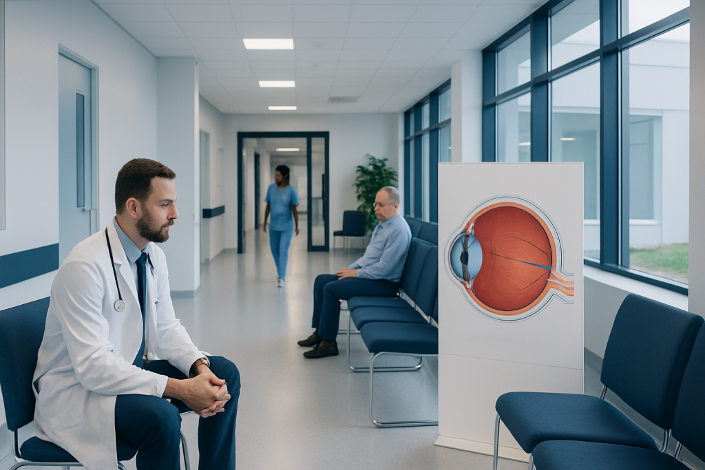
Ectopia lentis means the eye’s lens is partially or completely displaced. Symptoms often include blurred vision, double vision in one eye, glare, and frequent changes in glasses prescription.
Key Takeaways
- Ectopia lentis means the eye’s lens is partially or completely displaced.
- Symptoms often include blurred vision, double vision in one eye, glare, and frequent changes in glasses prescription.
- It may be inherited or linked to conditions such as Marfan syndrome, homocystinuria, trauma, or other eye diseases.
- Diagnosis relies on a detailed eye examination, often after the pupil is dilated.
- Treatment depends on severity and may include glasses, contact lenses, monitoring, or surgery.
- Because ectopia lentis can be associated with whole-body conditions, some patients need evaluation beyond the eyes.
Ectopia lentis is a condition in which the eye’s natural lens is out of its normal position. It can cause blurred vision, glare, and focusing problems, and it may occur on its own or as part of a broader connective tissue or metabolic disorder.
Overview of ectopia lentis
Ectopia lentis is a medical term for displacement of the eye’s natural lens. The lens normally sits behind the iris and helps focus light onto the retina. When it shifts out of place, vision can become blurred or distorted because light is no longer being focused properly.
The condition may be mild, with only a small shift in lens position, or more severe, with marked lens instability or complete dislocation. Some people are born with a tendency toward lens displacement, while others develop it after eye injury, inflammation, or certain eye diseases. In some cases, ectopia lentis is an important clue to an underlying inherited condition that also affects the heart, skeleton, or metabolism.
A helpful way to think about ectopia lentis is that it is both an eye problem and, at times, a diagnostic sign. For that reason, care often involves not only an ophthalmologist but sometimes pediatricians, genetic specialists, cardiologists, or metabolic disease experts, depending on the person’s age and overall health.
How the lens becomes displaced
The lens is held in place by tiny fibers called zonules, also known as suspensory ligaments. These delicate fibers attach the lens to surrounding structures inside the eye. If the zonules are weak, stretched, broken, or abnormally formed, the lens can move away from its central position.
This movement may be partial, called lens subluxation, or complete, called lens dislocation. In partial displacement, the lens still remains partly supported but tilts or shifts enough to affect vision. In complete dislocation, the lens may move farther within the eye, creating a more urgent situation because it can raise eye pressure or damage other structures.
The direction of lens movement can also offer clues about the cause. In inherited connective tissue disorders, the pattern of displacement may be typical, while traumatic ectopia lentis may be more irregular. Although patients do not need to recognize these details themselves, specialists use them as part of the diagnostic picture.
Symptoms and possible complications
Symptoms vary depending on how far the lens has moved and whether one or both eyes are affected. Some people notice gradual blurred vision, while others suddenly become aware of visual distortion after an injury. Children may not describe symptoms clearly, so a squint, poor school vision screening, or unusual head posture may be the first sign.
Common symptoms can include:
- Blurred or fluctuating vision
- Glare or light sensitivity
- Double vision in one eye
- Difficulty focusing at different distances
- Frequent changes in glasses prescription
- A visible edge of the lens in some cases
Ectopia lentis can sometimes lead to complications if not recognized and monitored. These may include refractive errors, amblyopia (lazy eye) in children, cataract changes, or increased pressure inside the eye. If the lens blocks fluid drainage, it may contribute to glaucoma, which requires prompt evaluation to protect vision. In severe cases, the displaced lens may irritate internal eye tissues or shift into a position that causes pain and sudden vision loss.
Causes and risk factors
Ectopia lentis has several possible causes. In many patients, especially children and young adults, it is related to inherited disorders that affect connective tissue or metabolism. Marfan syndrome is a well-known cause, but other conditions may also be involved, including homocystinuria, Weill-Marchesani syndrome, sulfite oxidase deficiency, and some rarer genetic disorders.
Not every case is inherited. Eye trauma is a common acquired cause and can damage the zonules directly. Other acquired causes include previous eye surgery, chronic inflammation inside the eye, high myopia, advanced cataract changes, or disorders that weaken the lens support over time. In older adults, lens instability may occasionally appear as part of age-related changes, though true ectopia lentis still requires specialist assessment.
Risk factors and associated situations include:
- Family history of lens displacement or connective tissue disease
- Known genetic syndromes
- History of blunt or penetrating eye trauma
- Previous intraocular surgery
- Long-standing eye inflammation
- Marked nearsightedness or other structural eye abnormalities
Because lens displacement may be linked to conditions beyond the eye, a doctor may ask about body build, joint flexibility, developmental history, blood clotting history, and cardiovascular symptoms. This wider assessment helps identify people who may benefit from tests or referrals outside ophthalmology.
How ectopia lentis is diagnosed
Diagnosis begins with a comprehensive eye examination. An ophthalmologist will ask about vision changes, age at symptom onset, previous injuries, family history, and any known medical conditions. The examination usually includes visual acuity testing, refraction, slit-lamp examination, and measurement of eye pressure.
The most important part of the assessment is often the dilated eye exam. After the pupil is widened with drops, the doctor can better see the lens position, signs of zonular weakness, and related changes in the retina or optic nerve. Imaging or additional tests may be used when the view is limited or when other eye problems are suspected. In some settings, detailed eye examination and ocular imaging help document lens position and plan follow-up.
If an inherited syndrome is suspected, the workup may extend beyond the eye. Depending on the clinical picture, doctors may recommend cardiac imaging, laboratory studies, metabolic testing, or genetic consultation. This is especially important in children and young adults, because early identification of an associated systemic condition can improve long-term care.
Treatment options and long-term care
Treatment depends on the degree of lens displacement, the quality of vision, the age of the patient, and whether complications are present. Mild cases may be managed conservatively with glasses or contact lenses if acceptable vision can be achieved. Regular monitoring is important, since the lens position may change over time and children are at risk of developing amblyopia if clear vision is not maintained.
When vision cannot be corrected well, when the lens becomes increasingly unstable, or when complications such as raised eye pressure occur, surgery may be recommended. Surgical management is individualized and may involve removing the displaced lens and, in selected cases, implanting an intraocular lens using a technique suited to the eye’s anatomy. For patients with associated cataract changes, specialists may discuss cataract surgery as part of the treatment plan when appropriate.
Patients with coexisting retinal or pressure-related concerns may need additional care. For example, if lens displacement contributes to retinal traction or there are other structural problems, a retinal specialist may be involved. Depending on the findings, this may overlap with services used in retinal treatment pathways, although management remains tailored to the underlying cause.
Long-term follow-up matters even after treatment. Vision correction may need adjustment, children may need monitoring for visual development, and adults may require ongoing checks for glaucoma, retinal disease, or recurrent instability. In complex or inherited cases, coordinated multidisciplinary care provides the safest overall approach.
Prevention, self-care, and living with the condition
There is no guaranteed way to prevent inherited ectopia lentis, but early detection can reduce the risk of avoidable vision problems. Families with a known genetic condition should keep routine eye appointments and ask whether genetic counseling or screening of relatives is appropriate. Children who seem to struggle with vision, reading, or eye alignment benefit from timely eye assessment.
For acquired cases, prevention focuses mainly on eye safety and management of underlying disease. Protective eyewear during sports, high-risk work, or activities involving impact can help lower the risk of trauma-related lens displacement. People with inflammatory eye disease or high myopia should attend recommended follow-up visits so structural changes can be recognized early.
Daily self-care does not replace treatment, but it can support comfort and function. Good lighting, updated prescriptions, and following contact lens instructions carefully may help some people see more comfortably. Those who have sudden pain, marked blur, or a rapid change in vision should not wait for a routine visit, because these symptoms can signal a complication.
When to seek medical care
Medical evaluation is important whenever a person develops unexplained blurred vision, monocular double vision, or frequent prescription changes, especially if there is a family history of connective tissue disease or previous eye trauma. A child who fails a vision screening, squints often, or tilts the head to see more clearly should also be assessed by an eye specialist.
Urgent care is needed if symptoms begin suddenly or are accompanied by eye pain, redness, halos around lights, nausea, a shadow in the vision, or a rapid drop in sight. These features may suggest rising eye pressure, retinal complications, or complete lens dislocation. Prompt examination helps reduce the risk of permanent visual damage.
In people with suspected inherited ectopia lentis, follow-up may involve more than eye care alone. If the clinical picture raises concern for a broader syndrome, doctors may recommend heart, metabolic, or genetic evaluation. Near the end of the care pathway, some patients may choose centers such as Acibadem International, where multidisciplinary specialists in JCI-accredited hospitals evaluate and treat complex eye and systemic conditions for international patients, including related eye pressure disorders when present.
Frequently asked questions
Is ectopia lentis the same as a cataract?
No. Ectopia lentis means the natural lens has moved out of its normal position, while a cataract means the lens has become cloudy. A person can have one condition without the other, although they may sometimes occur together.
Can ectopia lentis affect both eyes?
Yes, it can affect one or both eyes. Bilateral involvement is more common in inherited forms, while a traumatic case may involve only the injured eye. The degree of displacement can also differ between eyes.
Does ectopia lentis always need surgery?
No. Mild cases may be managed with glasses, contact lenses, and regular monitoring if vision is acceptable and the lens is stable. Surgery is considered when vision remains poor, the lens becomes increasingly unstable, or complications develop.
Is ectopia lentis hereditary?
It can be. Some cases are linked to inherited disorders such as Marfan syndrome or homocystinuria, while others happen after trauma or eye disease. A doctor may recommend further evaluation if the history suggests a genetic cause.
What tests are done for ectopia lentis?
The main test is a detailed eye examination, usually including a dilated exam so the lens position can be seen clearly. Doctors may also measure eye pressure, assess refraction, and order other studies if they suspect related eye or systemic conditions.
Can children with ectopia lentis develop normal vision?
Many children do well when the condition is recognized early and managed carefully. The key is to provide clear visual correction, monitor for amblyopia, and treat complications promptly. Regular pediatric ophthalmology follow-up is especially important during visual development.
This article is for general information only and is not a substitute for professional medical advice. Please consult a qualified doctor about your individual situation.
Explore treatments in Turkey — costs, top hospitals & a free quote
JCI-accredited · board-certified surgeons · reply within 24h
Add us as a Preferred Source to see more of our trusted health content across Google Search, AI Overviews and Discover.

More from the Health Library

Qulipta Uses for Preventing Migraine in Adults

Broccoli Sprouts: A Complete Medical Overview

Testosterone for Women: A Complete Medical Overview

CHF: An Evidence-Based Guide for Patients

Mushroom — Explained by Medical Evidence, Not Myths