Eye Nevi Removal: Procedure, Recovery and Results
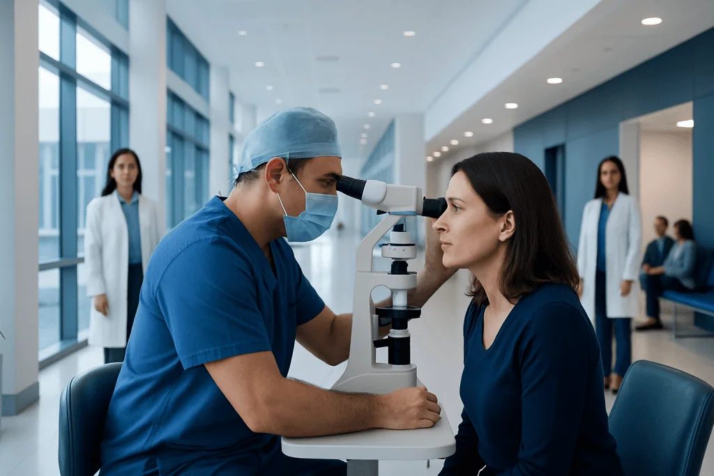
Most eye nevi are benign and can be safely observed with regular eye examinations and photographs. A changing, elevated, symptomatic or suspicious lesion may require specialist assessment, biopsy or treatment.
Key Takeaways
- Most eye nevi are benign and can be safely observed with regular eye examinations and photographs.
- A changing, elevated, symptomatic or suspicious lesion may require specialist assessment, biopsy or treatment.
- The eye nevus removal procedure varies greatly by location; surface lesions are treated differently from lesions inside the eye.
- Recovery is often days to weeks for superficial procedures, while treatment involving the inside of the eye may need longer follow-up.
- Removing a nevus does not always mean removing the eye; eye removal surgery is reserved for uncommon, serious situations such as certain eye cancers.
Eye nevi removal is not needed for most eye moles, which are usually benign and monitored over time. When removal or biopsy is advised, an ophthalmologist chooses the safest approach based on whether the nevus is on the eyelid, conjunctiva, iris or deeper inside the eye.
Overview: What Is Eye Nevi Removal?
Eye nevi removal refers to treatment for a nevus, commonly called an eye mole, that affects the eyelid, the clear membrane over the white of the eye (conjunctiva), the iris, or less commonly the choroid at the back of the eye. Most nevi are harmless pigment-cell growths and do not need to be removed. Instead, an eye specialist may document their appearance with photographs and monitor them at scheduled visits.
Removal, biopsy or other treatment may be considered when a lesion changes in size, colour, shape or thickness, causes discomfort, affects vision, or has features that need assessment for melanoma or another condition. The goal is not simply cosmetic: it is to establish an accurate diagnosis, protect eye function and manage symptoms when present.
Importantly, eye nevi removal is different from removal of the eyeball. In the vast majority of cases, treatment is directed at the lesion while preserving the eye. Removal of the eye is an uncommon procedure considered only in specific serious circumstances, such as advanced eye cancer that cannot be controlled with eye-preserving treatment.
Where an Eye Nevus Is Located Matters
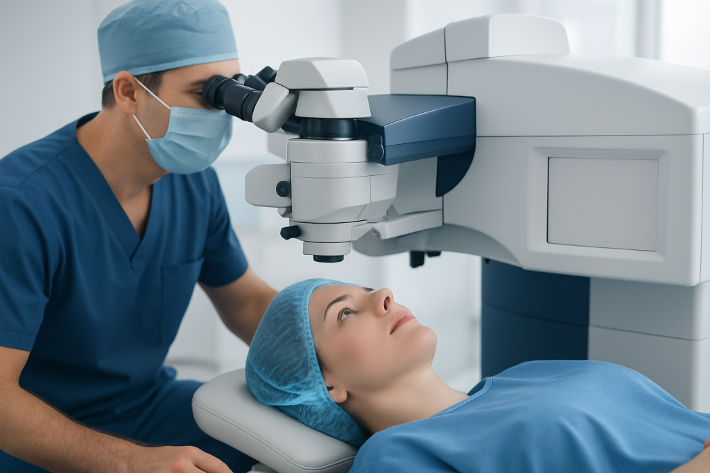
The treatment plan depends first on location. A nevus on the eyelid behaves more like a skin mole and may be assessed by an ophthalmologist, oculoplastic surgeon or dermatologist. A conjunctival nevus is visible on the surface of the eye and can often be examined closely using a slit lamp. It may appear as a brown, tan or lightly pigmented spot, sometimes with small cyst-like spaces.
An iris nevus is a pigmented spot in the coloured part of the eye. A choroidal nevus lies beneath the retina and cannot be seen without a dilated eye examination and specialised imaging. Because each location has different possible diagnoses and risks, self-diagnosis from a mirror or photograph is not reliable.
Specialist imaging may include slit-lamp photography, optical coherence tomography, ultrasound, fundus photography and, when appropriate, fluorescein imaging. These records allow clinicians to identify genuine change over time rather than normal differences in lighting or image quality.
Who May Be a Candidate for Removal or Biopsy?
An ophthalmologist may discuss an eye nevus removal procedure when there is uncertainty about the diagnosis, documented growth, a new change in pigment or contour, persistent irritation, recurrent bleeding, interference with eyelid movement, or concern that the lesion could be malignant. Some people also request treatment for a visible lesion after it has been assessed as benign, although cosmetic removal is considered only when it can be done safely.
Not every changing spot is dangerous, and not every nevus can or should be removed. For example, a stable choroidal nevus is usually monitored rather than excised because surgery inside the eye can carry significant risks. A specialist will weigh the likely benefit of intervention against the possibility of affecting vision, eye pressure, tear production, eyelid position or appearance.
Before treatment, the clinician reviews medical history, medicines that affect bleeding, allergies, previous eye surgery and vision needs. People should tell the team if they are pregnant, use anticoagulant or antiplatelet medication, have dry eye disease, glaucoma, diabetes or a history of skin or eye cancer. Medication should never be stopped without advice from the prescribing clinician.
How the Eye Nevus Removal Procedure Works
For a small eyelid or conjunctival nevus, the procedure is commonly performed as an outpatient treatment using local anaesthetic, sometimes with light sedation. The surgeon identifies the lesion, removes it carefully with a small margin when clinically appropriate, and may send the tissue to a pathology laboratory. Depending on the size and site, the area may heal naturally, be closed with fine sutures, or be covered with a tissue graft.
For a conjunctival lesion, protecting the cornea and maintaining a smooth ocular surface are important parts of the operation. The procedure may involve removing the pigmented tissue and reconstructing the surface if needed. A pathology report can help distinguish a benign nevus from other pigmented conditions and guide follow-up.
Deeper lesions, including iris or choroidal lesions, require a different pathway. A suspicious choroidal lesion is generally evaluated by an ocular oncology specialist. If treatment is needed, options may include focused radiation, laser-based treatment in selected cases, surgery or other cancer-directed care depending on the diagnosis. This is not the same as simple surface nevus removal.
Patients considering surgical treatment can discuss eye surgery options with an ophthalmology team. The most appropriate technique is based on the lesion’s location, size, features on imaging and the person’s visual health.
Eye Nevus Removal Recovery and Expected Results
Eye nevus removal recovery varies with the procedure. After a minor eyelid or conjunctival excision, many people experience redness, mild swelling, tearing, foreign-body sensation or blurred vision for several days. Surface redness can remain noticeable for a few weeks while the tissue heals, even when comfort and vision improve sooner.
The eye nevus removal recovery time may be longer if sutures, a graft or extensive surface reconstruction is needed. The care team may prescribe lubricating drops, antibiotic drops or ointment, and anti-inflammatory treatment. Follow the individual instructions about bathing, makeup, contact lenses, driving, exercise and returning to work; these recommendations depend on the operation and visual symptoms.
For treatment involving the iris, retina or choroid, recovery and monitoring can take weeks to months. Visual recovery may be gradual and depends on the lesion’s position and the treatment used. Follow-up appointments are essential to check healing, vision, eye pressure and any signs of recurrence or new pigment changes.
Most patients undergoing removal of a benign superficial nevus can expect the lesion to be removed or reduced with preservation of the eye. However, no procedure can guarantee a particular cosmetic result, and pigmentation can occasionally recur or remain in surrounding tissue. The pathology result and follow-up examination provide the clearest information about long-term outlook.
Benefits, Risks and Safe Aftercare
Potential benefits of treatment include obtaining a definitive tissue diagnosis, relieving irritation, addressing a lesion that interferes with eyelid function or vision, and improving the appearance of an appropriate benign lesion. Monitoring rather than surgery also has benefits when a nevus appears low risk, because it avoids procedure-related effects while still allowing changes to be detected early.
Possible risks depend on the site and extent of treatment. They can include pain, redness, dry eye symptoms, infection, bleeding, scarring, delayed healing, visible blood vessels, changes in eyelid position, recurrence of pigment and temporary or lasting visual changes. Surgery inside the eye has additional risks that the ophthalmologist will discuss in detail before consent.
After a surface procedure, patients should use prescribed medicines as directed, avoid rubbing the eye, wash hands before applying drops, and attend all follow-up visits. They should not use leftover drops or non-prescribed eye products. Contact the surgical team promptly if pain becomes severe, vision declines suddenly, redness rapidly worsens, discharge develops, or there is a new curtain, flash or shower of floaters in vision.
- Use protective eyewear if advised after the procedure.
- Wear sunglasses for comfort in bright light during early healing.
- Do not resume contact lens use until the clinician confirms that the surface has healed.
- Keep scheduled monitoring visits even after a benign pathology result.
When to Seek Medical Care
A person should arrange an eye examination if a known eye mole appears to grow, darken, become raised, develop new blood vessels, bleed, or cause persistent irritation. Medical review is also appropriate for a new pigmented spot on the eye, especially if it is changing or associated with blurred vision.
Urgent assessment is needed for sudden loss of vision, severe eye pain, marked light sensitivity, a new field-of-vision shadow, flashes of light, or a sudden increase in floaters. These symptoms do not necessarily mean a nevus is the cause, but they require timely evaluation because they can be linked to other eye conditions.
Questions about a suspicious or changing lesion may involve general ophthalmology, oculoplastic surgery, retinal care and ocular oncology. Acibadem International’s multidisciplinary specialists and JCI-accredited hospitals diagnose and treat eye conditions for international patients, with referral to the appropriate eye subspecialist when needed.
Frequently asked questions
Do all eye nevi need to be removed?
No. Most eye nevi are benign and remain stable, so observation with regular examinations and photographs is often the preferred approach. Removal or biopsy is usually considered only when there are concerning changes, symptoms, diagnostic uncertainty or a safe, appropriate cosmetic reason.
How long is eye nevus removal recovery?
Recovery after removal of a small eyelid or conjunctival nevus often involves a few days of discomfort and several weeks of visible redness or surface healing. Recovery can be longer when a graft, sutures or deeper eye treatment is required. The treating ophthalmologist can give the most accurate timeline for the individual procedure.
Is eye nevus removal painful?
The procedure is commonly performed with anaesthetic so pain during treatment is minimised. Mild soreness, scratchiness, tearing or light sensitivity can occur afterward and is usually managed with prescribed treatment and protective measures. Severe or worsening pain should be reported promptly.
Will removing an eye nevus affect vision?
Many superficial lesions can be treated without lasting effects on vision, but temporary blur may occur during healing. The risk is higher when a lesion is close to the cornea, eyelid margin, iris, retina or other structures important for vision. A specialist explains the expected visual risks before treatment.
Is eye nevus removal the same as eye removal surgery?
No. Eye nevus removal usually means removing or treating a localised pigmented lesion while keeping the eye intact. Eye removal surgery recovery time applies to enucleation or related procedures, which are rare and used for selected severe eye diseases, including certain advanced cancers.
Can an eye nevus come back after removal?
Pigment may occasionally recur or remain in nearby tissue, particularly if complete removal would risk harming the eye surface or vision. Regular follow-up helps identify recurrence and confirms that healing is progressing as expected. Pathology findings also help determine the appropriate monitoring plan.
References
- American Academy of Ophthalmology
- National Eye Institute
- American Cancer Society
- Royal College of Ophthalmologists
This article is for general information only and is not a substitute for professional medical advice. Please consult a qualified doctor about your individual situation.
Oncology care in Turkey — second opinion and treatment plan
JCI-accredited · board-certified surgeons · reply within 24h
Add us as a Preferred Source to see more of our trusted health content across Google Search, AI Overviews and Discover.

More from the Health Library


Related Specialists

Prof. Dr. Ilgaz Doğusoy
Thoracic Surgery
Dilem Kadıoğlu
Physical Medicine & Rehabilitation
Prof. Dr. Aziz Sümer
General Surgery
Prof. Dr. Ahmet Tayyar
Gynecology & Obstetrics