Harmonious Hemianopia: What Patients Need to Know

Harmonious hemianopia means the same half of the visual field is reduced or absent in both eyes. It is commonly linked to conditions affecting the brain, especially stroke, trauma, tumors, or inflammation.
Key Takeaways
- Harmonious hemianopia means the same half of the visual field is reduced or absent in both eyes.
- It is commonly linked to conditions affecting the brain, especially stroke, trauma, tumors, or inflammation.
- Diagnosis usually combines visual field testing, a neurologic and eye examination, and brain imaging such as MRI or CT.
- Treatment focuses on the underlying cause and may also include visual rehabilitation and practical adaptation strategies.
- Sudden visual field loss is a medical emergency and should be assessed promptly.
Harmonious hemianopia is a type of visual field loss in which the same side of vision is missing in both eyes, most often because of damage in the brain's visual pathways rather than a problem inside the eyes themselves. Patients often notice bumping into objects on one side, difficulty reading, or trouble navigating, and assessment usually involves eye testing, brain-focused examination, and imaging to find the cause.
Overview: what harmonious hemianopia means
Harmonious hemianopia is a pattern of vision loss in which the same side of the visual field is affected in both eyes. In everyday terms, a person may lose awareness of what is on the left side or the right side of their world, even though the eyes themselves may still be able to see normally in the remaining field. This usually happens because the visual pathways in the brain have been affected.
The word “harmonious” is often used to describe a highly similar or matching visual field defect between the two eyes. In many clinical settings, it overlaps with the term homonymous hemianopia. The important practical point for patients is that the missing vision follows a brain-based pattern, not a simple problem with one eye.
This condition can affect safety, reading, driving, work, and confidence with daily movement. Some people notice the change immediately, especially if it appears suddenly. Others only realize something is wrong when they begin bumping into doorframes, missing words at the start or end of lines, or feeling disoriented in crowded places.
How it can affect daily life
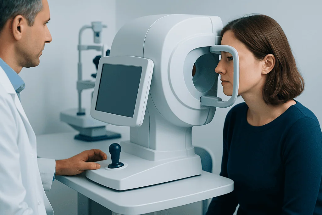
Harmonious hemianopia does not usually cause complete blindness, but it can make normal tasks more challenging because one side of the visual environment is not fully perceived. A person may leave food on one side of the plate, overlook faces approaching from one direction, or have trouble crossing streets safely. Reading can become slower because the eyes need to make more deliberate scanning movements.
Symptoms vary depending on which side is affected and how extensive the loss is. Some people have a complete half-field loss, while others have only part of that side missing. The brain may gradually adapt to some extent, but adaptation is often incomplete without targeted strategies or rehabilitation.
Because the eyes themselves may seem normal, the condition can be confusing at first. Family members may notice changes before the patient does, such as turning the head repeatedly to one side or appearing inattentive to people or objects on the affected side.
- Bumping into furniture or walls on one side
- Missing words or parts of sentences while reading
- Difficulty driving or noticing traffic from one direction
- Problems finding objects in busy visual environments
- Feeling off balance or less confident when walking outdoors
Symptoms and related signs
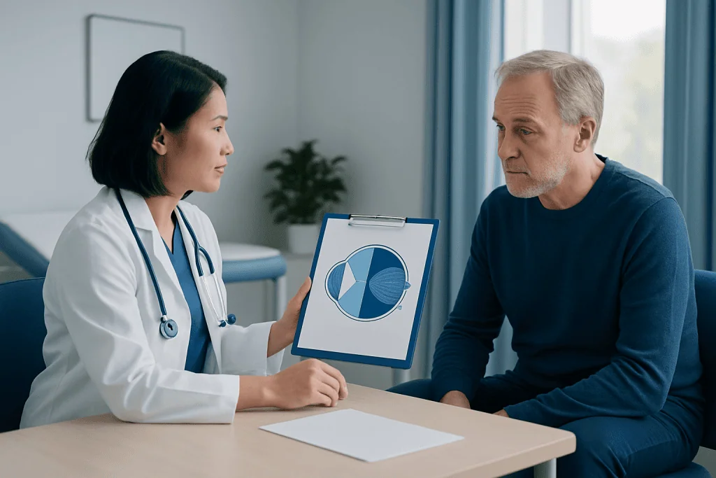
The main symptom of harmonious hemianopia is visual field loss on the same side in both eyes. For example, in a right-sided defect, the right half of the visual scene is missing from each eye. This is different from blurred vision or reduced sharpness, because central visual acuity may remain quite good, especially if the area responsible for sharp central vision has been spared.
People may also describe visual confusion rather than obvious loss. They may say they are “not seeing things on one side,” skipping lines while reading, or being startled by objects that seem to appear suddenly. In some cases, the condition is accompanied by other neurologic symptoms depending on the cause, such as weakness, speech difficulty, headache, dizziness, or changes in attention.
Some patients also develop visual neglect, especially after certain strokes. Neglect is not the same as hemianopia: in neglect, the brain has difficulty attending to one side, while in hemianopia, there is a true visual field defect. A specialist assessment helps separate these problems because management may differ.
Causes and risk factors
Harmonious hemianopia most often occurs when the visual pathways behind the optic chiasm are affected, such as the optic tract, optic radiations, or occipital lobe. The most common cause is stroke, particularly when blood flow to areas of the brain involved in vision is interrupted. This is why sudden onset should never be ignored.
Other possible causes include traumatic brain injury, brain tumors, bleeding in the brain, infections, inflammatory conditions, and less commonly demyelinating disease. In children and younger adults, the causes may be broader and can include congenital or developmental brain conditions. The pattern of visual loss can help doctors estimate where the problem is located.
Risk factors depend on the underlying condition. For stroke-related cases, they include high blood pressure, diabetes, smoking, high cholesterol, heart rhythm disorders, and vascular disease. For tumor-related or inflammatory causes, the risk profile is different, so a careful medical history is important.
Because harmonious hemianopia is often linked to neurologic illness, evaluation may involve both eye specialists and brain specialists. In some patients, further assessment is needed for conditions such as stroke or brain tumor when symptoms or imaging suggest these possibilities.
How doctors diagnose it
Diagnosis starts with listening carefully to the patient’s experience. A doctor may ask when the vision change began, whether it was sudden or gradual, and whether there are other symptoms such as headache, weakness, numbness, trouble speaking, or imbalance. A full eye examination is still important, but the focus often extends beyond the eyes to the nervous system.
Visual field testing is central to diagnosis. This may be done with bedside confrontation testing or more formally with automated perimetry, which maps the missing parts of the field in each eye. The pattern helps determine whether the defect is consistent with harmonious hemianopia and may suggest where the lesion is located.
Brain imaging is usually needed, especially when the condition is new. CT may be used urgently in emergency settings, while MRI often provides more detail about the visual pathways and surrounding brain tissue. Depending on the suspected cause, doctors may also order blood tests, vascular imaging, or heart-related tests. In some cases, advanced MRI imaging or urgent CT scan assessment helps clarify the diagnosis quickly and safely.
Treatment options and rehabilitation
Treatment for harmonious hemianopia focuses first on the underlying cause. If the vision loss is due to stroke, treatment may involve urgent stroke care and later secondary prevention. If a tumor, bleeding, inflammation, or another neurologic condition is responsible, treatment is directed at that condition. Visual recovery may occur partly over time, especially in the early months, but the degree of improvement varies from person to person.
Even when the visual field defect does not fully resolve, rehabilitation can make daily life easier. This may include visual scanning training, reading strategies, occupational therapy, and mobility techniques. Patients are often taught to turn the head and eyes more deliberately toward the affected side and to organize tasks in ways that reduce missed information.
Some people may benefit from prisms or other low-vision aids, depending on their exact pattern of loss and daily needs. If the cause is surgically treatable, management may involve services such as neurosurgery or specialist stroke rehabilitation after acute treatment. The best plan is individualized and often coordinated across neurology, ophthalmology, rehabilitation, and sometimes neurosurgery.
Near the end of the care pathway, ongoing review matters because vision needs can change with recovery. Acibadem International’s multidisciplinary specialists and JCI-accredited hospitals diagnose and treat this condition for international patients, with coordinated assessment when neuro-ophthalmic and neurologic expertise are both needed.
Living with the condition: self-care and practical tips
Adapting to harmonious hemianopia takes time, but many patients develop effective strategies. Keeping commonly used objects in consistent places, reducing clutter at home, improving lighting, and scanning consciously from side to side can make daily routines safer and less tiring. Reading may improve by using larger print, line guides, or structured training methods suggested by a therapist.
Driving rules vary by country and by the severity of the field loss. Patients should not assume it is safe to drive until they have been medically assessed and advised according to local regulations. This can be a difficult adjustment, but discussing alternatives early may reduce stress.
Emotional adjustment is also important. Losing part of the visual field can affect confidence, independence, and work. Support from rehabilitation professionals, family members, and patient education can help people regain routine and make practical plans for home, travel, and employment.
- Practice slow, deliberate scanning toward the affected side
- Use clear pathways and remove tripping hazards at home
- Ask about occupational therapy or vision rehabilitation
- Follow treatment plans for stroke risk factors or other underlying conditions
- Seek advice before returning to driving or hazardous work
When to seek medical care
Sudden visual field loss should be treated as urgent, especially if it develops over minutes or hours. Emergency evaluation is particularly important if it is accompanied by weakness, facial drooping, trouble speaking, severe headache, confusion, or difficulty walking, because these can be signs of a stroke or another acute neurologic problem.
Medical review is also important for vision loss that develops gradually, even if it seems mild. A slower onset does not rule out a significant cause, and assessment can help identify whether the problem relates to the brain, the eyes, or both. New difficulty reading, repeated collisions on one side, or unusual visual disorientation should prompt an appointment with a qualified doctor.
After diagnosis, follow-up matters if symptoms worsen, if new neurologic signs appear, or if daily functioning remains difficult despite treatment. Early specialist input can support both investigation and rehabilitation, helping patients understand what is happening and what next steps are available.
Frequently asked questions
Is harmonious hemianopia the same as blindness in one eye?
No. Harmonious hemianopia affects the same side of the visual field in both eyes, rather than causing total vision loss in one eye. Many people still have normal or near-normal sharpness of vision in the central area, but they miss information on one side.
Can harmonious hemianopia improve over time?
It can improve in some people, especially in the early period after the underlying brain injury or illness. Recovery depends on the cause, the location of the damage, and how extensive it is. Even when the field defect remains, rehabilitation can still help with daily function.
What is the most common cause of harmonious hemianopia?
Stroke is one of the most common causes, particularly in adults. However, trauma, brain tumors, bleeding, inflammation, and other neurologic disorders can also lead to this pattern of visual field loss.
How is harmonious hemianopia tested?
Doctors usually assess it with a visual field test, which maps the areas a person can and cannot see. This is combined with an eye examination and often brain imaging, such as CT or MRI, to identify the cause.
Can a person with harmonious hemianopia drive?
That depends on the severity of the visual field loss and the driving regulations in the country or region where the person lives. Medical assessment is essential before driving, because reduced awareness on one side can create safety risks.
Does this condition always mean there is a serious brain problem?
It does indicate that the visual pathways in the brain may be affected, so it should be properly assessed. Some causes are urgent and need immediate care, while others are more gradual. The key step is timely medical evaluation rather than assuming the problem will go away on its own.
References
- National Eye Institute
- American Academy of Ophthalmology
- American Stroke Association
- National Institute of Neurological Disorders and Stroke
- Royal College of Ophthalmologists
This article is for general information only and is not a substitute for professional medical advice. Please consult a qualified doctor about your individual situation.
Explore treatments in Turkey — costs, top hospitals & a free quote
JCI-accredited · board-certified surgeons · reply within 24h
Add us as a Preferred Source to see more of our trusted health content across Google Search, AI Overviews and Discover.
Check your numbers in seconds
BMI, calories, due date, blood pressure and 30+ more clinical calculators — free, instant, doctor-reviewed ranges.

More from the Health Library
Photographic Memory: An Evidence-Based Guide for Patients

Acupuncture: An Evidence-Based Guide for Patients
Metoprolol Is Killing Me — Explained by Medical Evidence, Not Myths

Herbs for Hair Growth: An Evidence-Based Guide for Patients

Superior Mesenteric Vein — Explained by Medical Evidence, Not Myths