Heart Anatomy: An Evidence-Based Guide for Patients
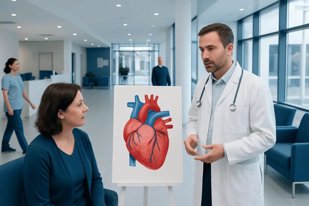
Heart anatomy includes four chambers, four main valves, major blood vessels, coronary arteries, and an electrical conduction system. The right side of the heart sends blood to the lungs, while the left side sends oxygen-rich blood to the body.
Key Takeaways
- Heart anatomy includes four chambers, four main valves, major blood vessels, coronary arteries, and an electrical conduction system.
- The right side of the heart sends blood to the lungs, while the left side sends oxygen-rich blood to the body.
- Heart valves keep blood moving in one direction and help the heart pump efficiently.
- Coronary arteries supply oxygen to the heart muscle itself.
- Common tests such as echocardiography, ECG, CT, and MRI help doctors assess heart structure and function.
- New chest pain, shortness of breath, fainting, or sudden palpitations should be assessed by a medical professional.
Heart anatomy describes how the heart’s chambers, valves, blood vessels, and electrical system work together to move blood efficiently through the lungs and the rest of the body. Understanding these parts can help patients make sense of symptoms, tests, and common heart conditions.
Overview: what heart anatomy means
Heart anatomy is the study of the heart’s structure and how each part supports circulation. In simple terms, the heart is a muscular pump in the chest with four chambers, four valves, major blood vessels, and an electrical system that coordinates each heartbeat. These structures work continuously to move oxygen-poor blood to the lungs and oxygen-rich blood to the body.
For patients, understanding heart anatomy can make medical terms easier to follow during checkups, scans, and treatment discussions. It also helps explain why symptoms such as chest discomfort, breathlessness, swelling, or palpitations can happen when a valve, chamber, artery, or rhythm pathway is not working as expected.
The heart sits slightly left of the center of the chest, behind the breastbone and between the lungs. It is enclosed by a protective sac called the pericardium. The heart wall itself has three main layers: the endocardium lining the inside, the myocardium forming the pumping muscle, and the epicardium covering the outer surface.
The main parts of the heart
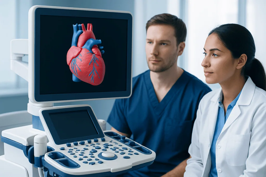
The heart has four chambers. The two upper chambers are the right atrium and left atrium, which receive blood returning to the heart. The two lower chambers are the right ventricle and left ventricle, which pump blood out of the heart. The left ventricle has the thickest muscular wall because it must generate enough force to send blood throughout the entire body.
A wall of tissue called the septum separates the right and left sides of the heart. This separation is important because the right side handles oxygen-poor blood, while the left side handles oxygen-rich blood. When this separation is incomplete, as in some congenital heart defects, blood flow may mix in ways that affect oxygen delivery.
Several large blood vessels connect to the heart. The superior vena cava and inferior vena cava bring oxygen-poor blood from the body into the right atrium. The pulmonary artery carries blood from the right ventricle to the lungs. The pulmonary veins return oxygen-rich blood from the lungs to the left atrium. The aorta then carries oxygen-rich blood from the left ventricle to the body.
The heart also has its own blood supply through the coronary arteries. These arteries branch from the aorta and deliver oxygen and nutrients to the heart muscle. When coronary arteries become narrowed or blocked, the heart muscle may not receive enough blood, which can lead to coronary artery disease.
How blood flows through the heart

Blood follows a precise path through the heart. First, oxygen-poor blood from the body enters the right atrium through the vena cavae. It then passes through the tricuspid valve into the right ventricle. When the right ventricle contracts, blood moves through the pulmonary valve into the pulmonary artery and travels to the lungs.
In the lungs, blood releases carbon dioxide and picks up oxygen. Oxygen-rich blood then returns to the left atrium through the pulmonary veins. From there, it passes through the mitral valve into the left ventricle. When the left ventricle contracts, blood is pushed through the aortic valve into the aorta and distributed to the body.
This flow depends on timing, pressure, and one-way valves. The valves open when pressure moves blood forward and close to prevent backflow. If a valve becomes narrowed or leaky, the heart may have to work harder. Over time, this can contribute to symptoms or complications such as enlargement of a chamber or heart failure.
Doctors often describe circulation as two linked loops: the pulmonary circulation between the heart and lungs, and the systemic circulation between the heart and the rest of the body. These loops run continuously, allowing tissues to receive oxygen and nutrients while waste products are carried away.
Heart valves and the electrical system
The four heart valves are the tricuspid, pulmonary, mitral, and aortic valves. Each valve is made of thin flaps, also called leaflets or cusps, that open and close with each heartbeat. Their role is to keep blood moving in one direction. When a valve does not open fully, it is called stenosis. When it does not close properly, it is called regurgitation or insufficiency.
Valve disease may affect any of the four valves, but problems involving the aortic or mitral valve are especially common in adults. Mild valve changes may cause no symptoms at first, while more advanced disease may lead to fatigue, breathlessness, swelling, dizziness, or a heart murmur detected during examination. Evaluation may include an echocardiogram and, in some cases, heart valve surgery.
The heart also has an internal electrical system that controls rate and rhythm. The sinoatrial node in the right atrium acts as the natural pacemaker, starting each heartbeat. The signal travels through the atria to the atrioventricular node, then down specialized pathways into the ventricles, causing them to contract in a coordinated way.
If this electrical system is disrupted, an irregular heartbeat may occur. Some rhythm changes are harmless, while others need treatment or monitoring. A common example is atrial fibrillation, in which the atria beat in a disorganized way and may increase the risk of stroke in some patients.
How doctors examine heart anatomy
Doctors assess heart anatomy using a combination of physical examination and imaging tests. During a clinic visit, they may listen for murmurs, extra heart sounds, or rhythm changes and look for signs such as leg swelling, bluish skin color, or shortness of breath. Blood pressure, pulse, and oxygen level provide additional clues about how well the heart is working.
Echocardiography is one of the most useful tests for viewing heart structure. It uses ultrasound to show the chambers, valves, wall motion, and blood flow in real time. It can help identify enlargement, valve disease, pumping weakness, fluid around the heart, and many congenital abnormalities. It is often the first imaging test used when structural heart disease is suspected.
An electrocardiogram records the heart’s electrical activity and can help detect rhythm problems, evidence of strain, or signs of a prior heart attack. Chest X-ray may show heart size and fluid in the lungs. Cardiac CT and cardiac MRI can provide more detailed images of anatomy, including the aorta, congenital defects, and the heart muscle itself. In selected cases, doctors may recommend coronary angiography to assess the coronary arteries directly.
Test choice depends on the clinical question. For example, valve disease is often assessed with echocardiography, while coronary artery narrowing may need CT angiography, stress testing, or invasive angiography. A patient’s symptoms, age, medical history, and exam findings help guide the most appropriate evaluation.
Common conditions related to heart anatomy
Many heart conditions are linked to changes in anatomy. Some people are born with structural differences, known as congenital heart defects. Others develop changes over time due to aging, high blood pressure, infection, inflammation, or reduced blood supply to the heart muscle. Structural problems may involve the chambers, valves, vessels, wall thickness, or the heart’s pumping function.
Coronary artery disease affects the vessels that feed the heart muscle. Valve disease affects how blood moves between chambers and out to the lungs or body. Cardiomyopathy changes the heart muscle itself and can affect pumping or relaxation. Enlargement of the heart may happen in response to chronic pressure or volume overload. Some conditions also involve the aorta, the body’s largest artery, which arises from the heart.
Symptoms vary widely and can include chest discomfort, shortness of breath, fatigue, dizziness, fainting, swelling in the legs, or palpitations. However, some structural abnormalities cause no symptoms and are first noticed during a routine exam or imaging test. That is why symptoms are important, but they are not the only way heart problems are found.
Treatment depends on the problem identified. Options may include lifestyle measures, medicines, procedures to improve blood flow, or surgery. For certain rhythm disorders, congenital defects, severe valve disease, or blocked arteries, specialist care is often needed. In some cases, patients may be evaluated for cardiac surgery as part of a broader treatment plan.
Protecting heart health and understanding test results
Knowing basic heart anatomy can help patients understand what doctors mean when they discuss ejection fraction, valve leakage, chamber enlargement, wall motion, or arterial narrowing. For example, the ejection fraction refers to how much blood the left ventricle pumps out with each beat. It is only one part of heart function, but it is commonly reported on echocardiograms and other scans.
Although anatomy itself does not prevent disease, healthy habits can reduce strain on the heart and blood vessels. These include not smoking, staying physically active, eating a balanced diet, maintaining a healthy weight, controlling blood pressure, managing cholesterol and blood sugar, sleeping well, and attending regular medical checkups when advised.
Patients who already have a heart condition may need ongoing monitoring even if they feel well. Follow-up can help detect changes in valve function, chamber size, rhythm, or blood flow before symptoms worsen. A doctor can explain which findings matter most for the individual patient and whether repeat imaging is needed.
Near the end of the care pathway, some people benefit from assessment in specialized centers. Acibadem International’s multidisciplinary specialists and JCI-accredited hospitals diagnose and treat heart conditions for international patients, including structural, vascular, and rhythm-related disorders.
When to seek medical care
Medical advice should be sought promptly for new, persistent, or worsening symptoms that may relate to the heart. These include chest pain or pressure, shortness of breath at rest or with mild activity, fainting, severe dizziness, rapid or irregular heartbeats, swelling in the legs, or unexplained fatigue. Even when symptoms are not caused by heart disease, they deserve proper assessment.
Emergency care is important for severe chest pain, sudden shortness of breath, fainting, new weakness on one side of the body, or symptoms that suggest a heart attack or stroke. Patients should not try to diagnose these situations on their own. Fast evaluation can be critical.
People with known heart disease should also contact their doctor if symptoms change, medicines cause side effects, or blood pressure and pulse readings become concerning. Anyone with a family history of inherited heart problems, a known heart murmur, or an abnormal test may need specialist follow-up even without symptoms.
Frequently asked questions
What is heart anatomy in simple terms?
Heart anatomy refers to the structure of the heart and its related blood vessels. It includes the four chambers, the valves, the coronary arteries, and the electrical system that keeps the heart beating in a coordinated way.
What are the four chambers of the heart?
The heart has two upper chambers called the atria and two lower chambers called the ventricles. The right side sends blood to the lungs, and the left side sends oxygen-rich blood to the rest of the body.
What do heart valves do?
Heart valves open and close to keep blood moving in one direction through the heart. If a valve becomes too narrow or too leaky, blood flow can be disrupted and the heart may have to work harder.
Why are the coronary arteries important?
Coronary arteries supply blood and oxygen to the heart muscle itself. If they become narrowed or blocked, the heart may not get enough oxygen, which can cause chest pain or a heart attack.
Which test best shows heart anatomy?
An echocardiogram is often the first and most useful test for looking at heart structure and function. Depending on the situation, doctors may also use ECG, CT, MRI, or angiography for more detail.
Can someone have a heart problem even if they feel fine?
Yes. Some heart conditions do not cause symptoms early on and are found during a routine examination or imaging test. This is one reason regular checkups and follow-up testing can be important.
References
- American Heart Association
- National Heart, Lung, and Blood Institute
- Centers for Disease Control and Prevention
- Mayo Clinic
- Merck Manual Consumer Version
This article is for general information only and is not a substitute for professional medical advice. Please consult a qualified doctor about your individual situation.
Explore treatments in Turkey — costs, top hospitals & a free quote
JCI-accredited · board-certified surgeons · reply within 24h
Add us as a Preferred Source to see more of our trusted health content across Google Search, AI Overviews and Discover.

More from the Health Library

Nervous System: A Complete Medical Overview

Sinus Massage: What Patients Need to Know

Rales: A Complete Medical Overview

Paraplegic: A Complete Medical Guide for Patients

Weaponized Incompetence: An Evidence-Based Guide for Patients