How Heart Disease Is Diagnosed: ECG, Echocardiogram, Stress Testing, and More

Doctors diagnose heart disease by combining symptoms, examination findings, and one or more heart tests. An ECG records the heart’s electrical activity, while an echocardiogram shows its structure and pumping function.
Key Takeaways
- Doctors diagnose heart disease by combining symptoms, examination findings, and one or more heart tests.
- An ECG records the heart’s electrical activity, while an echocardiogram shows its structure and pumping function.
- Stress tests help reveal problems that may appear only when the heart works harder.
- Blood tests, CT scans, MRI, and coronary angiography may be used when more detail is needed.
- Not every patient needs every test; the testing plan is tailored to the suspected condition.
Heart disease diagnosis usually combines a medical history, physical exam, and targeted tests such as ECG, echocardiography, blood work, and stress testing. The right test depends on a person’s symptoms, age, risk factors, and whether the doctor suspects a problem with heart rhythm, heart muscle, valves, or blood flow.
Overview: How heart disease is diagnosed
Heart disease is a broad term that can include coronary artery disease, heart rhythm disorders, valve problems, heart failure, congenital heart disease, and inflammation affecting the heart. Because these conditions can cause similar symptoms, diagnosis usually happens step by step rather than with a single test. The goal is to identify what part of the heart is affected and how serious the problem may be.
The process often starts with a careful conversation about symptoms such as chest discomfort, shortness of breath, palpitations, dizziness, fatigue, or swelling in the legs. A doctor also asks about medical history, family history, smoking, diabetes, high blood pressure, high cholesterol, and previous infections or surgeries. These details help guide which tests are most useful.
A physical examination can provide important clues. The doctor may listen for a heart murmur, check blood pressure in one or both arms, assess the pulse, and look for signs such as ankle swelling or fluid in the lungs. In many people, this first assessment already suggests whether the main concern is blocked arteries, a valve disorder, an abnormal rhythm, or weakening of the heart muscle.
From there, testing is chosen to answer specific questions. Some tests look at the heart’s electrical signals, while others show its shape, movement, and blood flow. In more complex cases, advanced imaging or catheter-based procedures are used to confirm a diagnosis and plan treatment.
Symptoms and warning signs that may lead to testing
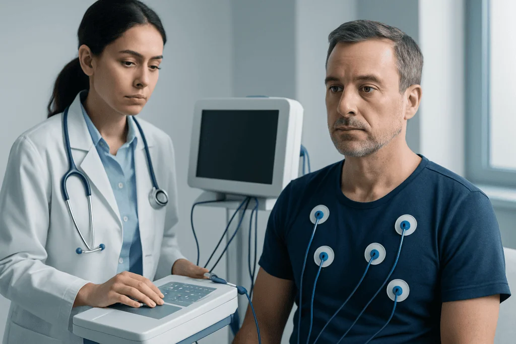
Many people are evaluated for heart disease because of symptoms, but some are tested after an abnormal screening result or because they have strong risk factors. Common symptoms that may prompt investigation include chest pain or pressure, breathlessness during activity or at rest, a racing or irregular heartbeat, fainting, and reduced exercise tolerance. In older adults and people with diabetes, symptoms can sometimes be less typical, such as unusual tiredness, nausea, or discomfort in the jaw, back, or arm.
Doctors also consider how symptoms appear. Chest discomfort brought on by walking and relieved by rest may suggest reduced blood flow to the heart muscle. Sudden palpitations may point to an arrhythmia, while breathlessness with lying flat or swelling in the feet can suggest heart failure. A history of fever, infection, or autoimmune disease may raise concern for inflammation affecting the heart or its lining.
Sometimes heart disease is found before symptoms appear. High blood pressure, elevated cholesterol, a heart murmur heard during a routine exam, or an abnormal test before surgery can lead to further evaluation. Family history also matters, especially when close relatives developed heart disease at a young age or had inherited heart conditions.
- Chest pain, tightness, or pressure
- Shortness of breath
- Palpitations or skipped beats
- Dizziness, fainting, or near-fainting
- Leg swelling or unexplained fatigue
- Reduced stamina with everyday activity
First-line tests: ECG, blood tests, and chest imaging

One of the first and most common tests is the electrocardiogram, or ECG. This quick, painless test records the heart’s electrical activity through sticky electrodes placed on the skin. An ECG can show a fast or irregular rhythm, evidence of a previous heart attack, signs of strain on the heart, or changes that suggest reduced blood flow. However, a normal ECG does not always rule out heart disease, especially if symptoms come and go.
Blood tests are another important part of evaluation. Depending on the situation, doctors may check cardiac biomarkers such as troponin when a heart attack is suspected, as well as cholesterol, blood sugar, kidney function, thyroid function, and markers of infection or inflammation. These tests do not diagnose every heart problem on their own, but they often help clarify risk, urgency, and possible causes.
A chest X-ray may be used to look at the size of the heart and the condition of the lungs. It can sometimes show fluid buildup, an enlarged heart, or other causes of breathlessness that are not primarily cardiac. While it is not the main test for most heart conditions, it can provide useful context during the initial workup.
If symptoms are intermittent, a standard ECG may miss the problem. In that case, doctors may use ambulatory monitoring such as a Holter monitor or event recorder. These devices track heart rhythm over a longer period, making them especially helpful for unexplained palpitations, dizziness, or suspected intermittent arrhythmias.
Echocardiogram and ultrasound-based heart evaluation
An echocardiogram is an ultrasound scan of the heart. It uses sound waves to create moving images, allowing doctors to see the heart chambers, valves, walls, and pumping action in real time. This test is central in the diagnosis of many conditions because it can show whether the heart is enlarged, whether the muscle contracts normally, and whether valves open and close properly.
Echocardiography is especially useful when symptoms suggest heart failure, valve disease, congenital abnormalities, or fluid around the heart. It can estimate how strongly the heart pumps, identify areas that are not moving well after reduced blood flow or a heart attack, and assess pressure changes that may affect the lungs and right side of the heart. It is noninvasive and generally well tolerated.
In some cases, a doctor may recommend a transesophageal echocardiogram, in which a small probe is passed into the esophagus to obtain clearer images from behind the heart. This approach can be helpful when standard images are limited or when there is a need to look closely at valves, blood clots, infection, or structural defects. Stress echocardiography combines ultrasound with exercise or medicine to see how the heart performs under strain.
When a structural heart problem is suspected, an echocardiogram often becomes one of the most informative tests in the whole diagnostic pathway. It may also be used repeatedly over time to monitor known conditions such as valve disease, cardiomyopathy, or heart failure.
Stress testing: checking the heart during exertion
A stress test evaluates how the heart responds when it has to work harder. This can help reveal problems that may not appear while a person is resting. The most familiar type is the exercise treadmill test, in which the patient walks on a treadmill or pedals a stationary bike while heart rhythm, blood pressure, and symptoms are monitored. Doctors may use this test when they suspect reduced blood flow to the heart or when they want to assess exercise tolerance and symptom patterns.
If a person cannot exercise enough because of joint problems, lung disease, or general weakness, medication can be used to simulate the effect of exercise on the heart. Some stress tests are combined with imaging. A stress echocardiogram looks at heart wall motion before and after stress, while nuclear perfusion imaging shows how well blood reaches the heart muscle. These imaging-based tests can provide more detail than an ECG-only stress test.
Stress testing is not right for every patient. The choice depends on the person’s symptoms, baseline ECG, fitness level, and overall likelihood of coronary artery disease. A doctor may also avoid or postpone a stress test if there is concern for an unstable condition, such as ongoing chest pain at rest or severe uncontrolled rhythm disturbance.
Used appropriately, stress testing can help determine whether chest symptoms are likely related to blocked arteries, whether further imaging is needed, and how best to plan treatment. It may be part of the workup for suspected coronary artery disease or follow-up after treatment in selected patients.
Advanced imaging and coronary angiography
When first-line tests do not fully explain symptoms, doctors may recommend advanced imaging. Cardiac CT can provide detailed pictures of the heart and coronary arteries. In some patients, CT coronary angiography helps show whether plaque or narrowing is present in the arteries supplying the heart. This can be useful for people with stable chest pain and an intermediate likelihood of coronary disease.
Cardiac MRI offers high-resolution images of heart structure, function, scarring, inflammation, and tissue characteristics. It may be used when there is concern for cardiomyopathy, myocarditis, congenital abnormalities, or unclear findings on echocardiography. MRI can answer questions that other tests cannot, particularly about the heart muscle itself.
Coronary angiography is a more invasive test, usually performed through a catheter inserted into an artery in the wrist or groin. Contrast dye is injected so doctors can see the coronary arteries directly under X-ray. This test is often recommended when symptoms, blood tests, or noninvasive imaging strongly suggest significant artery blockage, or when immediate treatment may be needed. In the same setting, doctors may proceed to coronary angiography to confirm the diagnosis and guide next steps.
Advanced testing is chosen carefully, balancing how much information is needed with the risks and benefits of each option. For some patients, these tests provide reassurance. For others, they identify a problem that needs medication, close follow-up, or procedures such as coronary artery bypass graft surgery or heart valve surgery.
How doctors choose the right test
There is no single best test for all forms of heart disease. The right approach depends on the clinical question. For example, a person with palpitations may need rhythm monitoring, while someone with a murmur may need an echocardiogram. Chest pain during exercise may lead to stress testing or coronary imaging, whereas sudden severe chest pain and elevated troponin may require urgent hospital-based evaluation.
Doctors also consider age, sex, existing medical conditions, kidney function, pregnancy, previous test results, and ability to exercise. Some tests are excellent for ruling out a disease in low- to intermediate-risk patients, while others are better when there is already a strong suspicion of a significant problem. This is why two people with similar symptoms may receive different testing plans.
It is also common for testing to happen in stages. A normal initial test may be enough if symptoms are mild and risk is low. If symptoms continue or change, more detailed assessment may follow. In other situations, test results are combined to build a clearer picture. For example, an ECG might suggest an abnormality, an echocardiogram might show how it affects heart function, and angiography might explain the underlying cause.
For patients, it can be helpful to ask what each test is looking for, whether preparation is needed, and how the result could change treatment decisions. Understanding the purpose of testing often makes the process less stressful and more meaningful.
After diagnosis: next steps, self-care, and when to seek medical attention
Once heart disease is diagnosed, the next step is to match treatment to the cause and severity of the condition. Some people need lifestyle changes and regular monitoring, while others may need medicines, catheter-based procedures, or surgery. Treatment may focus on improving blood flow, controlling heart rhythm, lowering blood pressure, reducing cholesterol, treating valve disease, or supporting a weakened heart muscle.
Self-care remains important at every stage. Doctors commonly advise stopping smoking, eating a heart-healthy diet, staying physically active within safe limits, managing stress, maintaining a healthy weight, and keeping diabetes, blood pressure, and cholesterol under good control. Taking prescribed medicines consistently and attending follow-up visits are also essential parts of care.
Medical attention should be sought promptly for chest pain that lasts more than a few minutes, severe shortness of breath, fainting, sudden weakness, or a new fast or irregular heartbeat with symptoms. Even when symptoms seem mild, persistent or recurring problems deserve medical review. Early assessment often leads to clearer answers and better treatment planning.
For people seeking coordinated evaluation, Acibadem International’s multidisciplinary specialists and JCI-accredited hospitals diagnose and treat heart conditions for international patients. The best results begin with an accurate diagnosis and a personalized plan based on the individual’s symptoms, risks, and test findings.
Frequently asked questions
What is usually the first test used to diagnose heart disease?
The first steps are usually a medical history, physical examination, blood pressure measurement, and an ECG. Blood tests and sometimes a chest X-ray are also commonly used, depending on the symptoms. These early tests help the doctor decide whether more detailed heart testing is needed.
Can an ECG detect all heart problems?
No. An ECG is very useful, but it may be normal even when heart disease is present, especially if symptoms are intermittent or the problem is structural rather than electrical. That is why doctors often combine it with other tests such as echocardiography, stress testing, or rhythm monitoring.
What does an echocardiogram show?
An echocardiogram shows the heart’s size, shape, pumping function, and valve movement. It can help diagnose heart failure, valve disease, congenital problems, and fluid around the heart. Because it creates moving images, it gives doctors a clear picture of how the heart is working in real time.
Why might a doctor order a stress test?
A stress test helps show how the heart performs during exercise or medication-induced stress. It is often used when chest discomfort or breathlessness may be related to reduced blood flow to the heart. It can also help guide decisions about whether further imaging or treatment is necessary.
Is coronary angiography always necessary?
No. Many patients can be assessed with noninvasive tests such as ECG, echocardiography, stress testing, CT, or MRI. Coronary angiography is usually reserved for situations where there is a strong suspicion of significant artery blockage or when a procedure may be needed.
How long does it take to diagnose heart disease?
It depends on the symptoms and the type of condition suspected. Some urgent problems can be identified within hours in a hospital, while other diagnoses may require several outpatient tests over days or weeks. The timeline is guided by how serious the symptoms are and what each test shows.
References
- World Health Organization
- American Heart Association
- European Society of Cardiology
- National Heart, Lung, and Blood Institute
- Centers for Disease Control and Prevention
This article is for general information only and is not a substitute for professional medical advice. Please consult a qualified doctor about your individual situation.
Heart care in Turkey — expert evaluation and treatment
JCI-accredited · board-certified surgeons · reply within 24h
Add us as a Preferred Source to see more of our trusted health content across Google Search, AI Overviews and Discover.
Check your numbers in seconds
BMI, calories, due date, blood pressure and 30+ more clinical calculators — free, instant, doctor-reviewed ranges.