Interventional Neuroradiology in Turkey: Planning Care From Abroad

Interventional neuroradiology uses catheters and imaging to diagnose and treat some brain and spinal vascular conditions without open surgery. Not every patient or every condition is suitable for this approach; treatment decisions depend on imaging findings, symptoms, and overall health.
Key Takeaways
- Interventional neuroradiology uses catheters and imaging to diagnose and treat some brain and spinal vascular conditions without open surgery.
- Not every patient or every condition is suitable for this approach; treatment decisions depend on imaging findings, symptoms, and overall health.
- Patients traveling from abroad usually need to share prior scans, reports, medicines, and medical history before treatment planning.
- Recovery time may be shorter than with open surgery, but close follow-up and medication guidance remain important.
- Choosing an experienced multidisciplinary center helps coordinate neurology, neurosurgery, imaging, anesthesia, and aftercare.
Interventional neuroradiology in Turkey can help diagnose and treat certain brain, spine, and blood vessel conditions using image-guided, minimally invasive techniques. For people traveling from abroad, understanding evaluation, travel planning, treatment steps, and follow-up can make the process clearer and more manageable.
Overview: What Interventional Neuroradiology Means
Interventional neuroradiology is a medical specialty that treats certain conditions of the brain, spinal cord, head, neck, and blood vessels using imaging guidance and very small instruments. Instead of making a large surgical opening, the doctor typically guides a thin catheter through a blood vessel, often starting from the wrist or groin, and advances it to the area that needs treatment. This approach is often described as minimally invasive.
It is commonly used for problems such as brain aneurysms, arteriovenous malformations, dural arteriovenous fistulas, some kinds of stroke care, narrowing of blood vessels, and other neurovascular conditions. Depending on the diagnosis, treatment may involve coiling, embolization, stenting, thrombectomy, or targeted delivery of medications or devices. The exact procedure is selected only after careful imaging review and specialist evaluation.
For international patients, interventional neuroradiology in Turkey may be considered when a person needs advanced imaging, a second opinion, or a procedure that can potentially reduce recovery time compared with open surgery in selected cases. However, treatment planning is highly individualized. A specialist team needs to review the condition, current symptoms, prior scans, and general health before advising whether travel is appropriate and what care timeline is realistic.
Which Conditions May Be Treated
Interventional neuroradiology is not one single treatment. It is a group of image-guided procedures used for specific diagnoses. Some of the most common conditions include brain aneurysms, arteriovenous malformations, carotid or intracranial artery narrowing, spinal vascular malformations, and certain urgent stroke-related vessel blockages. In some cases, it is also used to help control bleeding or reduce blood flow to a lesion before surgery.
Each condition has its own pattern, risks, and treatment goals. For example, an aneurysm may be treated to reduce the risk of rupture, while a blocked artery during an acute stroke may require urgent clot removal to restore blood flow. An arteriovenous malformation may need embolization alone or as part of a wider plan that also includes surgery or radiosurgery. Because these conditions differ so much, the same procedure is not suitable for everyone.
Before care is planned from abroad, specialists usually need to confirm the diagnosis with good-quality imaging. This may include MRI, CT, CT angiography, MR angiography, or a catheter angiogram. Some patients already have enough imaging from home, while others may need repeat scans after arrival to clarify anatomy and choose the safest strategy.
How Evaluation Works Before Traveling

Planning care from abroad starts with collecting complete medical information. This usually includes the patient’s symptoms, past diagnoses, current medicines, allergies, prior operations, and any history of stroke, bleeding, seizures, high blood pressure, or heart disease. Imaging files are especially important. Whenever possible, patients should provide the original scan images, not only written reports, because specialists often need to review the anatomy directly.
During the remote review process, the medical team may ask practical questions such as when symptoms started, whether they are stable or changing, and whether the condition is urgent. If there are warning signs of an emergency, the person may be advised to seek immediate local medical care rather than travel first. This step helps protect patient safety.
Once the records are reviewed, the team may outline whether the person appears to be a candidate for consultation, further testing, or a procedure. It is important to understand that a final treatment plan may still change after in-person examination and repeat imaging. Patients should also ask about expected length of stay, possible need for a companion, language support, and how follow-up will be managed after returning home.
- Prepare digital copies of scans and reports.
- List all medications, including blood thinners and supplements.
- Share allergy history and previous anesthesia issues.
- Ask whether fasting, lab tests, or medication changes are needed before travel.
Diagnosis and Treatment Planning After Arrival
After arrival, assessment usually begins with a consultation and review of previous records. The doctor may recommend updated imaging if the earlier scans are incomplete, low quality, or no longer reflect the current condition. In many neurovascular cases, detailed vessel imaging is essential before any intervention can be planned safely.
A catheter angiogram, also called cerebral angiography, may be used when more precise information is needed. This test provides highly detailed images of the blood vessels and can guide decisions about whether to proceed with angiography alone or to combine diagnosis and treatment in the same hospital stay. The findings help the team understand the shape of an aneurysm, the source of abnormal blood flow, or the location of a blockage or narrowing.
Interventional neuroradiology care is often planned by a multidisciplinary team. Depending on the diagnosis, this may include neuroradiologists, neurologists, neurosurgery specialists, anesthesiologists, intensive care physicians, and rehabilitation experts. Team-based planning is especially important for complex conditions where catheter-based treatment may need to be combined with surgery, medication, or later monitoring.
Common Procedures and What to Expect
Procedures vary according to the condition. For aneurysms, treatment may involve placing tiny coils, flow-diverting devices, or stents to reduce blood flow into the aneurysm and lower the risk of bleeding in selected cases. For vascular malformations or fistulas, embolization may be used to close abnormal vessels with special materials. For acute ischemic stroke in appropriate patients, clot removal may be performed through stroke treatment pathways when timing and imaging support this approach.
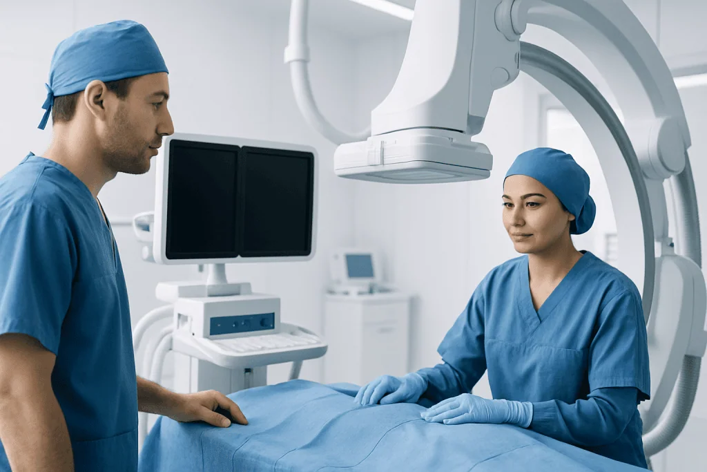
Most procedures are performed in a specialized angiography suite under sedation or general anesthesia, depending on the case. A small catheter is inserted into a blood vessel and guided to the target area using live imaging. Many patients are monitored in a recovery area or intensive care setting afterward, especially if the procedure was complex or urgent.
Although these treatments avoid open surgery, they still carry important risks. Possible complications may include bleeding, clot formation, stroke, vessel injury, contrast reactions, kidney strain in susceptible patients, or device-related problems. The specialist explains the expected benefits, alternatives, and potential risks before treatment, and informed consent is an essential part of the process.
Hospital stay can range from same-day discharge for some diagnostic procedures to several days for more complex treatment. Recovery depends on the condition treated, the procedure performed, and the person’s baseline health. Even when recovery is relatively quick, patients still need clear guidance about medicines, physical activity, wound care, and follow-up scans.
Travel, Recovery, and Follow-Up for International Patients
Patients coming from abroad should think beyond the procedure itself. Travel timing matters, especially for conditions that are urgent, recently symptomatic, or associated with neurological weakness or severe headaches. Air travel may not be appropriate for everyone immediately after treatment, so return plans should remain flexible until the treating doctor confirms it is safe.
Medication management is another key part of recovery. Some procedures require antiplatelet or blood-thinning medicines before or after treatment, while others do not. Patients should understand exactly which medicines to continue, stop, or avoid, and they should bring an updated medication list when traveling. It is also wise to ask for written discharge documents that can be shared with doctors at home.
Follow-up imaging is often necessary to confirm that the treated vessel remains stable or that an aneurysm or malformation has been adequately closed. Depending on the condition, follow-up may involve MRI, CT angiography, or repeat catheter angiography after a defined period. For this reason, patients should ask early whether checks can be done locally after returning home and how results will be reviewed by the treating team.
Near the end of planning, some international patients prefer centers that can coordinate several specialties in one place. Acibadem International’s multidisciplinary specialists and JCI-accredited hospitals diagnose and treat neurovascular conditions for international patients, with care plans shaped by the individual diagnosis, imaging findings, and recovery needs.
How to Prepare and When to Seek Urgent Medical Help
Good preparation can make the experience smoother. Before travel, patients should confirm passport and visa requirements, insurance details if relevant, expected costs, language support, and whether a companion is recommended. Comfortable clothing, a list of emergency contacts, and copies of all medical records can also be helpful. People with chronic conditions such as diabetes, hypertension, or kidney disease should make sure these are discussed in advance, since they may affect imaging contrast use, anesthesia, or recovery plans.
After treatment, patients should follow instructions carefully about hydration, activity, bathing, wound care, and medication use. It is important not to restart or stop blood thinners, antiplatelet medicines, or supplements without direct medical advice. Even if a person feels well, scheduled follow-up should not be skipped, because some neurovascular conditions require imaging surveillance over time.
Urgent medical attention is needed for warning signs such as sudden weakness, numbness, trouble speaking, severe sudden headache, confusion, seizure, fainting, chest pain, shortness of breath, uncontrolled bleeding, or increasing swelling or pain at the catheter entry site. These symptoms do not always mean a serious complication has occurred, but they should be assessed promptly by a qualified doctor or emergency service.
Frequently asked questions
What is interventional neuroradiology used for?
It is used to diagnose and treat certain conditions involving the brain, spine, head, neck, and blood vessels. Examples include aneurysms, vascular malformations, narrowed arteries, and some kinds of stroke-related vessel blockage.
Is interventional neuroradiology the same as brain surgery?
No. It is a minimally invasive, image-guided approach that usually works through a catheter placed in a blood vessel rather than through a large surgical opening. In some cases, however, it may be combined with neurosurgery or used as part of a broader treatment plan.
What records should a patient send before traveling from abroad?
Patients should usually send scan images, scan reports, clinic notes, a medicine list, allergy information, and details of previous treatments or operations. Original imaging files are especially helpful because specialists often need to review them directly.
How long does recovery take after a procedure?
Recovery depends on the diagnosis, the type of procedure, and the patient’s general health. Some people recover quickly after diagnostic angiography, while others need several days in hospital and a longer period of rest and follow-up after more complex treatment.
Can a patient fly home soon after treatment?
Sometimes, but not always. The decision depends on the procedure performed, the patient’s neurological condition, and whether there were any complications. The treating doctor should confirm when air travel is safe.
Are these procedures always safer than open surgery?
Not necessarily. Minimally invasive treatment can offer important advantages in selected cases, but every procedure has risks and not every condition is suitable for a catheter-based approach. The safest option depends on the exact diagnosis, anatomy, and overall health of the patient.
References
- World Health Organization
- American Stroke Association
- Society of NeuroInterventional Surgery
- Radiological Society of North America
- National Institute of Neurological Disorders and Stroke
This article is for general information only and is not a substitute for professional medical advice. Please consult a qualified doctor about your individual situation.
Interventional Neuroradiology in Turkey — costs, top hospitals & a free quote
JCI-accredited · board-certified surgeons · reply within 24h
Add us as a Preferred Source to see more of our trusted health content across Google Search, AI Overviews and Discover.
Check your numbers in seconds
BMI, calories, due date, blood pressure and 30+ more clinical calculators — free, instant, doctor-reviewed ranges.

Neurosurgery
Surgical treatment of the brain, spine and nervous system, including minimally invasive and functional procedures.
70 specialists in this unit