Ischemic Optic Neuropathy: Sudden Vision Loss, Risk Factors, and Next Steps

Ischemic optic neuropathy happens when blood flow to the optic nerve is reduced. Vision loss is often sudden, painless, and usually affects one eye first.
Key Takeaways
- Ischemic optic neuropathy happens when blood flow to the optic nerve is reduced.
- Vision loss is often sudden, painless, and usually affects one eye first.
- There are two main types: non-arteritic and arteritic; the arteritic form is a medical emergency.
- Diagnosis usually involves an eye exam, visual field testing, imaging, and blood tests when needed.
- Treatment depends on the cause and focuses on protecting vision and managing underlying risk factors.
- People with sudden vision changes should seek urgent medical care.
Ischemic optic neuropathy is a condition in which the optic nerve is damaged because its blood supply is reduced. It often causes sudden vision loss in one eye and should be assessed promptly to confirm the diagnosis, look for urgent causes, and guide treatment.
Overview
Ischemic optic neuropathy is a form of optic nerve injury caused by insufficient blood flow. The optic nerve carries visual information from the eye to the brain, so when its circulation is disrupted, vision can be affected quickly. The condition most often appears as sudden vision loss in one eye, although the severity varies from person to person.
Doctors generally divide ischemic optic neuropathy into two major types. Non-arteritic anterior ischemic optic neuropathy, often called NAION, is the more common form and is usually linked to blood vessel risk factors and the structure of the optic nerve. Arteritic anterior ischemic optic neuropathy, often called AION due to giant cell arteritis, is less common but much more urgent because ongoing inflammation in blood vessels can threaten vision in the other eye if treatment is delayed.
Posterior ischemic optic neuropathy can also occur, although it is less common. In this form, the part of the optic nerve behind the eye is affected, so the front of the optic nerve may look normal early on. Because the causes, urgency, and treatment can differ, prompt evaluation by an eye specialist or neuro-ophthalmologist is important.
Symptoms
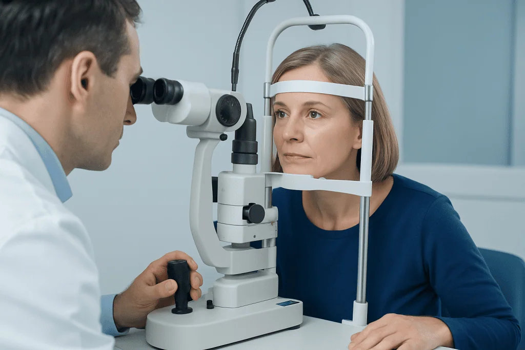
The most common symptom of ischemic optic neuropathy is sudden vision loss in one eye. Some people notice this on waking in the morning, while others become aware of blurred, dim, or patchy vision over hours. Vision changes are often painless, which can make the condition easy to underestimate even though it needs attention.
Loss of vision may affect the upper or lower part of the visual field rather than all vision equally. Colors may seem less vivid, contrast can be reduced, and central vision may or may not be involved. Some people describe a dark shadow, a missing section, or a general sense that vision is not as bright as usual in the affected eye.
Symptoms that suggest the arteritic form need especially urgent care. These may include a new headache, scalp tenderness, jaw pain when chewing, fever, unexplained fatigue, shoulder or hip aching, or weight loss. When sudden vision loss occurs together with these symptoms, doctors consider inflammation of blood vessels an emergency because the second eye may be at risk.
- Sudden blurred or dim vision in one eye
- Partial visual field loss, often top or bottom
- Reduced color vision or contrast sensitivity
- Painless onset in many cases
- Headache, scalp tenderness, or jaw pain in arteritic cases
Causes and Risk Factors

In ischemic optic neuropathy, the immediate problem is reduced circulation to the optic nerve. In non-arteritic cases, this is thought to involve a combination of small blood vessel factors and the anatomy of the optic nerve head. Some people have a crowded optic disc, sometimes described as a “disc at risk,” which may make the nerve more vulnerable when blood flow drops.
Common risk factors include high blood pressure, diabetes, high cholesterol, smoking, and obstructive sleep apnea. Older age also increases risk. Some episodes may be linked to low blood pressure during sleep or other situations that reduce perfusion to the optic nerve, although the exact mechanism is not always clear.
Arteritic ischemic optic neuropathy is usually caused by giant cell arteritis, an inflammatory disease of medium and large arteries. This form tends to affect older adults and requires rapid treatment. Because giant cell arteritis can also reduce blood flow to the brain and other tissues, doctors may evaluate related concerns such as stroke.
Less commonly, optic nerve ischemia may occur after major blood loss, severe anemia, or prolonged surgery, especially in posterior ischemic optic neuropathy. Doctors also consider other causes of optic nerve dysfunction, such as optic neuritis, because several conditions can produce similar visual symptoms but need different treatment.
How Diagnosis Is Made
Diagnosis begins with a careful medical history and a detailed eye examination. The doctor asks when vision changed, whether the loss was sudden or progressive, and whether there are symptoms such as headache, jaw pain, or body aches. Visual acuity, color vision, pupil responses, and visual fields are checked, and the optic nerve is examined after the pupils are dilated.
In anterior ischemic optic neuropathy, the optic nerve head may look swollen on examination, especially early in the condition. Visual field testing helps show the pattern of sight loss. Optical coherence tomography may be used to assess the optic nerve and retina in detail, while fundus photography can document changes over time.
Blood tests are especially important when arteritic ischemic optic neuropathy is suspected. Doctors often request inflammatory markers and may arrange further testing for giant cell arteritis. In some cases, temporal artery ultrasound, biopsy, or both may be considered. Imaging such as MRI may be used when the diagnosis is uncertain or when doctors need to rule out other causes affecting the optic nerve or brain.
Because sudden vision loss has several possible causes, diagnosis also involves excluding conditions such as retinal artery blockage, retinal vein occlusion, compressive lesions, and neurological disease. Depending on the situation, clinicians may use a comprehensive eye examination and selected vascular or neurological tests to build the clearest picture.
Treatment Options
Treatment depends on the type of ischemic optic neuropathy and its underlying cause. In arteritic ischemic optic neuropathy, treatment is urgent and usually begins immediately with corticosteroids to reduce vessel inflammation and protect the other eye. Doctors do not wait for all confirmatory test results if the suspicion is high, because delaying treatment can lead to further irreversible vision loss.
For non-arteritic ischemic optic neuropathy, there is no single treatment that reliably restores lost vision in every case. Care focuses on confirming the diagnosis, managing vascular risk factors, and addressing conditions that may contribute to reduced optic nerve blood flow. This may include treatment of diabetes, blood pressure problems, high cholesterol, and smoking cessation, along with evaluation for sleep apnea when appropriate.
Some patients experience partial spontaneous improvement over time, while others have stable long-term deficits. Follow-up is important to monitor the affected eye, watch for changes in the other eye, and reassess overall health risks. If the diagnostic work-up suggests another cause for visual symptoms, treatment is tailored accordingly.
Because the optic nerve is closely linked to both eye and brain health, management may involve ophthalmologists, neuro-ophthalmologists, neurologists, rheumatologists, and primary care physicians. In selected cases, clinicians may also request neurological rehabilitation or visual support services to help patients adapt to persistent field loss and maintain daily functioning.
Prevention and Self-care
It is not always possible to prevent ischemic optic neuropathy, but reducing vascular risk can support eye and overall health. Managing blood pressure, blood sugar, and cholesterol is an important step. Regular follow-up with a doctor helps keep these factors under control and may lower the chance of further circulation-related problems.
Healthy habits matter. Not smoking, staying physically active, eating a balanced diet, and maintaining a healthy sleep routine can all support blood vessel health. If snoring, daytime sleepiness, or suspected sleep apnea is present, assessment is worthwhile because sleep-disordered breathing has been linked to non-arteritic ischemic optic neuropathy.
People who have had ischemic optic neuropathy in one eye should be especially attentive to any new symptoms in the other eye. They should keep a current list of medications and discuss blood pressure treatment timing, especially if they have episodes of low blood pressure or dizziness. Medication decisions should only be changed with medical advice.
Self-care also includes adapting to any lasting visual change. Better lighting, contrast aids, magnifiers, and organized home layouts can make reading and mobility easier. If additional support is needed, low-vision services may help patients remain independent and confident.
When to See a Doctor
Any sudden change in vision should be treated as urgent. A person should seek prompt medical attention if vision becomes blurred, dim, or partly missing in one or both eyes, even if there is no pain. Early assessment helps doctors identify whether the cause is ischemic optic neuropathy or another condition that also needs rapid care.
Emergency evaluation is especially important when vision loss is accompanied by headache, scalp tenderness, jaw pain with chewing, fever, new muscle aches, or unexplained fatigue. These features can point to giant cell arteritis, which requires immediate treatment to reduce the risk of vision loss in the other eye and other vascular complications.
Ongoing follow-up is also important after diagnosis. Doctors monitor vision, review risk factors, and look for clues to related cardiovascular or inflammatory disease. If there are signs of broader neurological involvement, the care team may assess conditions such as brain tumors or other less common causes of optic nerve dysfunction when clinically appropriate.
For international patients who need coordinated assessment, Acibadem International’s multidisciplinary specialists and JCI-accredited hospitals diagnose and treat ischemic optic neuropathy with ophthalmology, neuro-ophthalmology, neurology, and imaging support as needed.
Frequently asked questions
Is ischemic optic neuropathy the same as a stroke in the eye?
They are not exactly the same, but both involve reduced blood flow. Ischemic optic neuropathy affects the optic nerve, while other circulation problems can affect the retina or brain. Because the symptoms can overlap, urgent evaluation is important.
Can vision return after ischemic optic neuropathy?
Some people have partial improvement, especially in non-arteritic cases, but lost vision may also remain permanent. The amount of recovery varies widely. Early diagnosis is still important because it helps guide treatment and reduce further risk.
What is the difference between arteritic and non-arteritic ischemic optic neuropathy?
Non-arteritic ischemic optic neuropathy is usually related to small vessel circulation problems and risk factors such as diabetes, high blood pressure, or sleep apnea. Arteritic ischemic optic neuropathy is caused by blood vessel inflammation, most often giant cell arteritis. The arteritic form is more urgent because the second eye can be affected quickly without treatment.
Is ischemic optic neuropathy painful?
It is often painless, especially in non-arteritic cases. However, the arteritic form may come with headache, scalp tenderness, or jaw pain rather than pain in the eye itself. Any sudden vision loss, with or without pain, should be assessed promptly.
Who is most at risk for ischemic optic neuropathy?
Risk is higher in older adults and in people with vascular risk factors such as diabetes, high blood pressure, high cholesterol, and smoking. Obstructive sleep apnea is also an important association, particularly in non-arteritic cases. In arteritic cases, giant cell arteritis is the key concern.
How is ischemic optic neuropathy diagnosed?
Doctors use a detailed eye examination, vision testing, and an optic nerve assessment to look for characteristic findings. Blood tests are important when giant cell arteritis is suspected, and imaging may be used when the diagnosis is unclear. The goal is both to confirm the condition and to exclude other serious causes of sudden vision loss.
References
- American Academy of Ophthalmology
- National Eye Institute
- National Institute of Neurological Disorders and Stroke
- Mayo Clinic
- Royal College of Ophthalmologists
This article is for general information only and is not a substitute for professional medical advice. Please consult a qualified doctor about your individual situation.
Explore treatments in Turkey — costs, top hospitals & a free quote
JCI-accredited · board-certified surgeons · reply within 24h
Add us as a Preferred Source to see more of our trusted health content across Google Search, AI Overviews and Discover.
Check your numbers in seconds
BMI, calories, due date, blood pressure and 30+ more clinical calculators — free, instant, doctor-reviewed ranges.